. 2005 Nov 17:5:67.
doi: 10.1186/1471-2148-5-67.

The Enhancer of split and Achaete-Scute complexes of Drosophilids derived from simple ur-complexes preserved in mosquito and honeybee

Affiliations

The Enhancer of split and Achaete-Scute complexes of Drosophilids derived from simple ur-complexes preserved in mosquito and honeybee

Rebekka Schlatter et al. BMC Evol Biol. .

Abstract

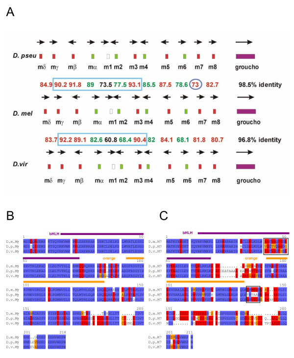
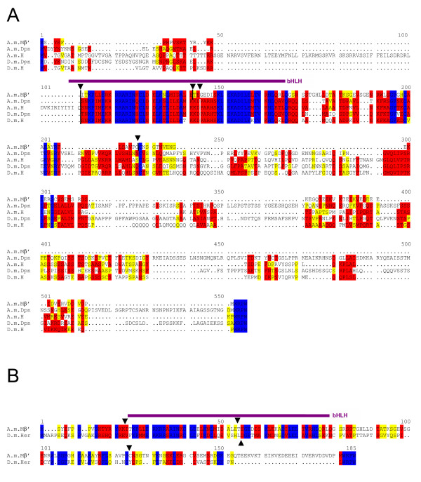
Background: In Drosophila melanogaster the Enhancer of split-Complex [E(spl)-C] consists of seven highly related genes encoding basic helix-loop-helix (bHLH) repressors and intermingled, four genes that belong to the Bearded (Brd) family. Both gene classes are targets of the Notch signalling pathway. The Achaete-Scute-Complex [AS-C] comprises four genes encoding bHLH activators. The question arose how these complexes evolved with regard to gene number in the evolution of insects concentrating on Diptera and the Hymenoptera Apis mellifera.

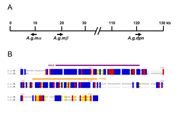
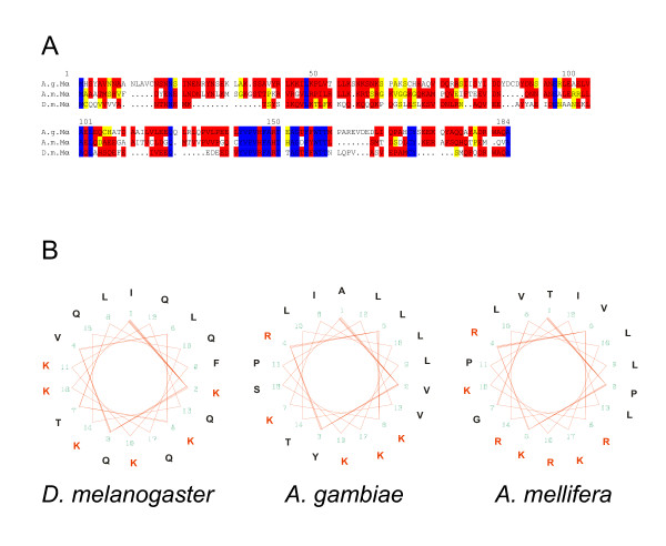
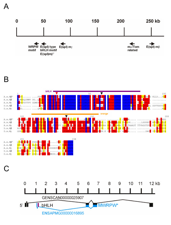
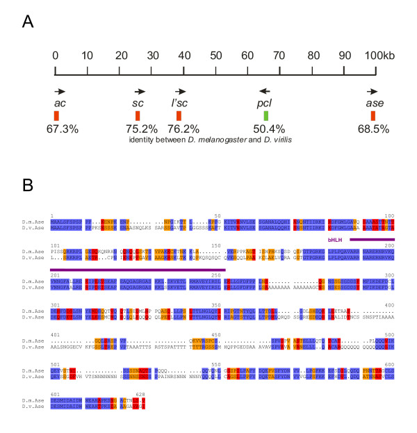
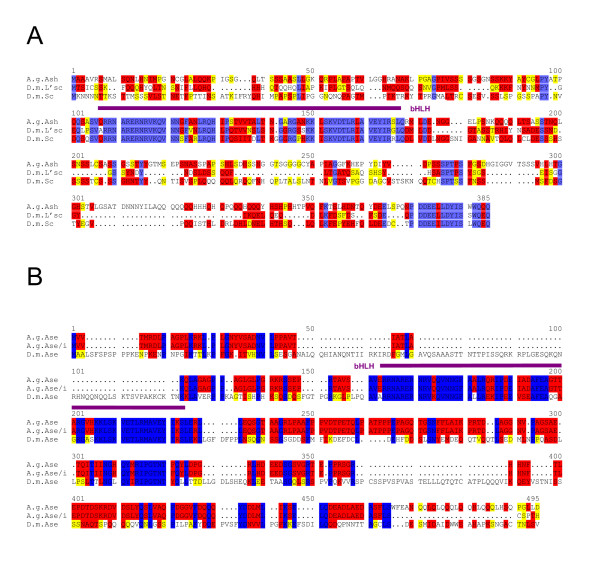
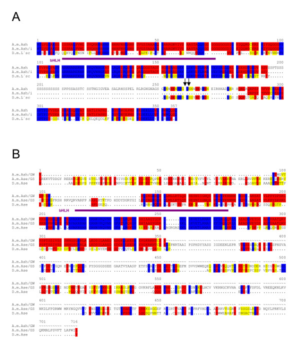
Results: In Drosophilids both gene complexes are highly conserved, spanning roughly 40 million years of evolution. However, in species more diverged like Anopheles or Apis we find dramatic differences. Here, the E(spl)-C consists of one bHLH (mbeta) and one Brd family member (malpha) in a head to head arrangement. Interestingly in Apis but not in Anopheles, there are two more E(spl) bHLH like genes within 250 kb, which may reflect duplication events in the honeybee that occurred independently of that in Diptera. The AS-C may have arisen from a single sc/l'sc like gene which is well conserved in Apis and Anopheles and a second ase like gene that is highly diverged, however, located within 50 kb.

Conclusion: E(spl)-C and AS-C presumably evolved by gene duplication to the nowadays complex composition in Drosophilids in order to govern the accurate expression patterns typical for these highly evolved insects. The ancestral ur-complexes, however, consisted most likely of just two genes: E(spl)-C contains one bHLH member of mbeta type and one Brd family member of malpha type and AS-C contains one sc/l'sc and a highly diverged ase like gene.


Figures

Figure 1
Figure 1
Conservation of the E(spl)-C in Drosophilids. A) The E(spl)-C is highly conserved in Drosophilids with regard to gene number and transcript orientation (arrows). The size of the complex is also almost the same; in D. pseudoobscura (D. pseu) it is only slightly larger than in D. melanogaster (D. mel). The smallest seems to be the D. virilis (D. vir) complex, however, the virilis sequence was not completed at the time. The best identity at protein level is found between the Gro orthologs (purple) followed by the bHLH proteins (red). Interestingly the proteins of the centrally located bHLH genes Mγ, Mβ and M3 (framed blue) are best conserved. Higher identities are seen between the melanogaster and pseudoobscura orthologs than between the ones of D. melanogaster and D. virilis, with the exception of M7 (blue circle). The worst conserved member of the complex is M1. (Numbers give % identity between the proteins). B) Alignment of the Mγ and (C) M7 orthologs. The bHLH (purple) and orange domains are the best conserved parts of the orthologs. The M7 sequences labelled with the black box are unexpectedly better conserved in D. virilis than in D. pseudoobscura compared with D. melanogaster. Identical residues are marked in blue; red shows highly related and yellow similar residues.
Figure 2
Figure 2
The E(spl)-C in mosquito A. gambiae. (A) The E(spl)-C in the mosquito is composed of two putative genes, A.g.mβ and A.g.mα. Approximately 100 kb away a second bHLH coding gene was detected, however, the analysis predicted a close relationship to Deadpan and Hairy. Since a good fitting hairy ortholog is elsewhere in the mosquito genome, the gene is most likely a dpn ortholog. B) Alignment between A.g.Mβ and D.m.Mβ shows good conservation within the bHLH and the orange domains (marked) as well as the WRPW motif at the C-terminus. Identical residues are marked in blue; red shows highly related and yellow similar residues.
Figure 3
Figure 3
Conservation of the Brd-family member Mα. A) Alignment of the presumptive Mα proteins of D. melanogaster, A. mellifera and A. gambiae. Although the alignment reveals not much identity (blue), the postulated features that typify Brd-proteins are present [14]. Red, highly related and yellow, similar residues. B) Mα contains a typcial amphipathic α-helix with high concentration of lysine (K) and arginine (R) residues on one side of the wheel (red).
Figure 4
Figure 4
The E(spl)-C in Apis mellifera. A) A genomic region spanning about 250 kb (GroupUn.159; numbers are correspondingly) contains 3 presumptive E(spl) bHLH genes and one related gene. The gene at position ~250 kb encodes a bHLH protein with best overall identity to D.m.Mβ. This gene is disrupted by an intron inside the bHLH domain (dash and red arrowhead in B). Close by, at position 220 kb a related coding region is found. Based on high similarity to D.m.Mα and its close neighbourhood to A.m.mβ, we named it A.m.mα. A second intronless E(spl) like bHLH coding gene is located at position ~90 kb. We name it A.m.mγ since the encoded protein shows best overall identity to the D.m.Mγ. Approximately 50 kb away we detected sequences encoding an E(spl) like bHLH domain (A.m.Mβ') and a long open reading frame ending with a WRPW motif. The Ensembl honeybee database annotated a gene with five introns spanning 12 kb within this region but missed the respective motifs (GENSCAN00000025907). We propose a different gene structure; see Figure C for details. B) Alignment of the three putative honeybee bHLH proteins (A.m.Mβ', A.m.Mβ, A.m.Mγ) with D. melanogaster Mβ and Mγ proteins is shown. Identical residues are marked in blue; red shows highly related and yellow similar residues. Intron positions are marked with a triangle above and a dash in the respective A. mellifera sequence. C) Structure of the 12 kb GENSCAN00000025907 region. Black shows the Ensembl gene annotation, and blue the new ENSAPMG00000016895 annotation. Purple highlights a second exon that encodes part of the bHLH domain, the open reading frame extends into the predicted intron of GENSCAN00000025907 and terminates with WRPW.
Figure 5
Figure 5
The AS-C of D. virilis. A) The AS-C is highly conserved between D. melanogaster and D. virilis concerning gene number, transcript orientation and overall size. At the protein level (% identity) the best conservation is found between the Sc and L'sc orthologs. However, the conservation rate is lower compared with the E(spl) bHLH proteins (see Fig. 1). The pcl gene, although only 50.4% identical, is found between l'sc and ase. Because of gaps in the genomic virilis sequence, a scheme of the D. melanogaster complex is shown. B) Alignment of the Ase protein orthologs of D. melanogaster and D. virilis. Note the extension of the D.v.Ase protein by repetitive amino acid stretches composed of poly N, poly Q and poly A. The best conservation is found within the bHLH domain (purple). Identical residues are marked in blue; red shows highly related and yellow similar residues.
Figure 6
Figure 6
The AS-C in A. gambiae. A) Alignment of the achaete-scute homologous protein of A. gambiae (A.g.Ash; [36]) with melanogaster L'sc and Sc proteins. Note the high conservation of the bHLH domain (purple) and the very C-terminus. Comparison over the entire length gives a higher identity score to D.m.L'sc, however, the alignment shows also regions that are more similar to D.m.Sc. B) Alignment of the neighbouring bHLH gene product from A. gambiae with D. melanogaster Ase. The alignment is shown to A.g.Ase/i (database predicted version without intron) and A.g.Ase (second intron translated). The second predicted intron comprises almost 2.5 kb and ends with an exon translated into five residues (CSPTH; in A.g.Ase/i). Translation into this intron leads to A.g.Ase that is similar in size and in its terminus to D.m.Ase. Highest conservation is found in the bHLH domain (purple). Identical residues are marked in blue; red shows highly related and yellow similar residues.
Figure 7
Figure 7
The AS-C in A. mellifera. A) Comparison of D.m.L'sc with the predicted A.m.Ash protein. Two forms were compared, without intron sequence, A.m.Ash/i, and with translated intron, A.m.Ash. Arrows mark additional splice consensus sites. B) Within 40 kb of A.m.ash, there is a second potential gene encoding a widely diverged bHLH protein. Two different programs were used for gene prediction that gives A.m.Ase/GS (Chris Burge's Genscan program) and A.m.Ase/GW (GeneWise model); both predicted proteins were aligned with D.m.Ase. Decent conservation is only found in the putative bHLH domains (purple). Identical residues are marked in blue; red shows highly related and yellow similar residues.
Figure 8
Figure 8
Comparison of A.m.Mβ' with Dpn, Hairy and Her proteins. A) Comparison of A.m.Mβ' with A.m.Dpn, A.m.H, D.m.Dpn and D.m.H. The A.m.Mβ' protein belongs to the E(spl)-C, however, has several introns (triangle on top, vertical dashes within sequence). Two of the introns are within the bHLH domain at similar position as in D. melanogaster hairy or dpn genes. The bHLH domain is indicated in purple. B) Alignment of D. melanogaster er Hwith A m.Mβ' protein. Her has only one intron, however, at very similar position as the second intron of A.m.Mβ'. Introns are marked with triangles.

References

    1. Artavanis-Tsakonas S, Rand MR, Lake RJ. Notch signaling: cell fate control and signal integration in development. Science. 1999;284:770–776. doi: 10.1126/science.284.5415.770. - DOI - PubMed
    1. Welshons WJ. Dosage sensitive interactions with split mutations in the presence of an Enhancer of split. Drosophila Inf Service. 1956;30:157–158.
    1. Dawson SR, Turner DL, Weintraub H, Parkhurst SM. Specificity for the hairy/Enhancer of split basic helix-loop-helix (bHLH) proteins maps outside the bHLH domain and suggests two separable modes of transcriptional repression. Mol Cell Biol. 1995;15:6923–6931. - PMC - PubMed
    1. Delidakis C, Artavanis-Tsakonas S. The Enhancer of split locus of Drosophila encodes seven independent helix-loop-helix proteins. Proc Natl Acad Sci USA. 1992;89:8731–8735. - PMC - PubMed
    1. Hartley D, Preiss A, Artavanis-Tsakonas S. A deduced gene product from the Drosophila neurogenic locus, Enhancer of split, shows homology to mammalian G-protein β subunit. Cell. 1988;55:785–795. doi: 10.1016/0092-8674(88)90134-1. - DOI - PubMed

MeSH terms