. 2006 Mar 15;107(6):2400-8.
doi: 10.1182/blood-2005-08-3340. Epub 2005 Nov 17.

Patterns of expression, membrane localization, and effects of ectopic expression suggest a function for MS4a4B, a CD20 homolog in Th1 T cells

Affiliations

Patterns of expression, membrane localization, and effects of ectopic expression suggest a function for MS4a4B, a CD20 homolog in Th1 T cells

Hui Xu et al. Blood. .

Abstract

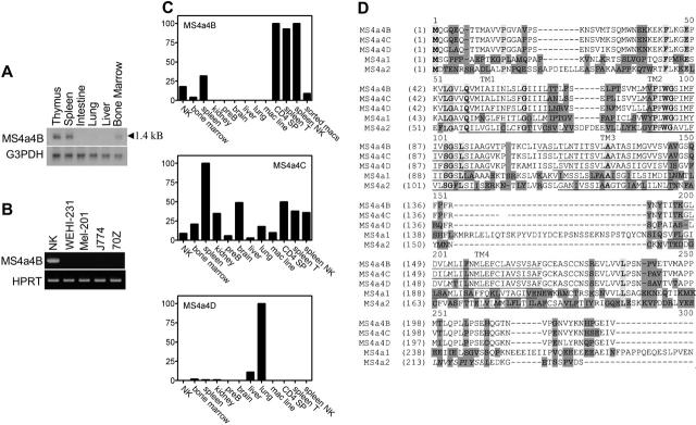
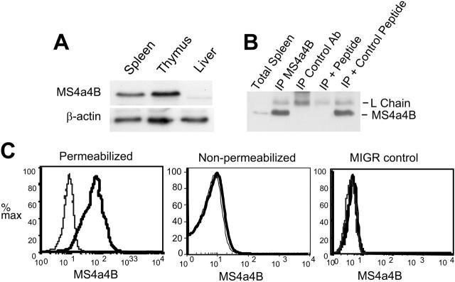
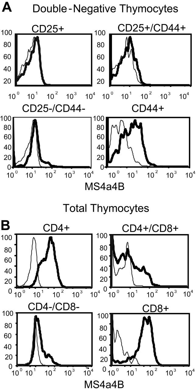
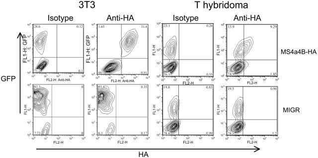
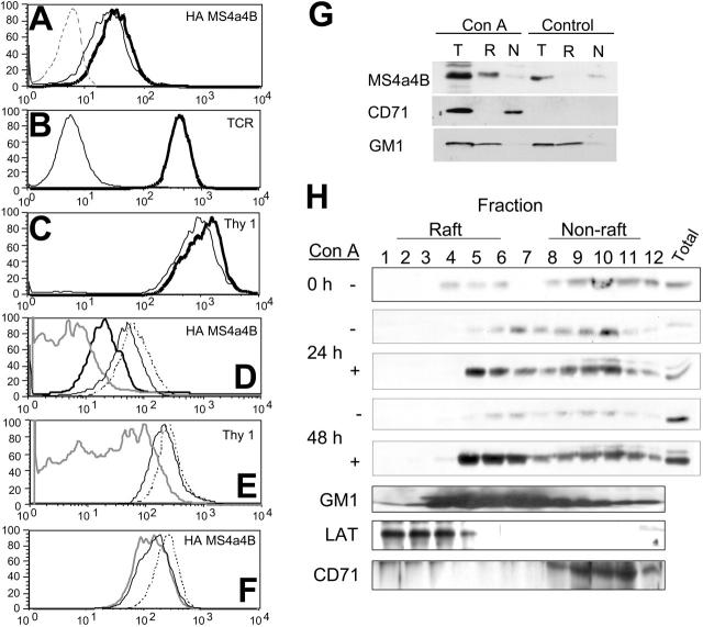
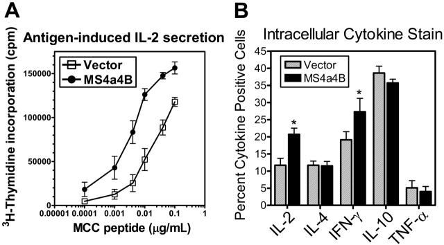
The membrane-spanning 4A (MS4A) family of proteins includes CD20, Fc epsilonRIbeta, and HTm4, whose genes are grouped in a chromosomal location that is associated with increased susceptibility to allergy and atopic asthma. One family member, Chandra/MS4a4B, was reported to be expressed in T helper 1 (Th1) T cells but not Th2 T cells. In the present study, Ms4a4b was isolated in a screen of genes differentially expressed during thymocyte development. MS4a4B was detected in immature CD4- CD8- CD44+ CD25- thymocytes, turned off during further stages of thymocyte development and reexpressed in mature single-positive thymocytes. MS4a4B expression was found in naive CD8+ and CD4+ peripheral T cells and natural killer (NK) cells but not in B cells. MS4a4B is expressed at the cell surface with its C-terminus located in the cytoplasm. When expressed in a T-cell hybridoma by retroviral vector, MS4a4B protein constitutively associated with lipid raft microdomains, whereas in primary T cells endogenous MS4a4B protein became enriched in rafts after T-cell activation. Overexpression of MS4a4B in primary CD4+ T-cell blasts enhanced T-cell receptor (TCR)-induced Th1 cytokine production. These results suggest that MS4a4B expression is tightly regulated during T-cell development and that MS4a4B expression promotes Th1 function and/or differentiation.


Figures

Figure 1.
Figure 1.
MS4a4B mRNA is expressed in mouse tissues and cells. (A) Northern blot: 10 μg RNA was separated on 1% denatured agarose gel and transferred to membrane, probed using Ms4a4b or G3PDH probes, washed, and exposed overnight. The length of the transcript is shown. (B) RT-PCR: samples were normalized by HPRT PCR and then were amplified with the primers specific for Ms4a4b, separated by gel, stained with ethidium bromide, and photographed. Samples were IL-2-expanded NK cells from severe combined immunodeficient (SCID) bone marrow, WEHI-231 B cells, Mel-201 erythroid line, J774 macrophage line, and 70Z-13 pre-B-cell line. (C) Quantitative PCR for expression of Ms4a4b, Ms4a4c, and Ms4a4d was performed using the Light-Cycler system (see “Materials and methods”). Results are in arbitrary units normalized to HPRT, and ribosomal RNA standards were done in parallel. Results shown are representative of 2 experiments. Samples are (1) expanded primary NK cells, (2) bone marrow, (3) whole spleen, (4) kidney, (5) pre-B-cell line 70Z13, (6) brain, (7) liver, (8) lung, (9) macrophage line (J774) FACS-sorted cells, (10) CD4 SP (single-positive thymocytes), (11) spleen T cells (CD3+), (12) spleen NK cells (NK1.1+), and (13) macrophages (Mac1+). (D) Alignments of deduced amino acid sequences for the mouse Ms4a4 subfamily, Ms4a1 (CD20), and Ms4a2 (FcεRIβ). The membrane-spanning domains (TM) are predicted by TMHMM 2.0 program and underlined. Identical sequences are indicated by bold letters in light shade; conservative sequences by unshaded letters; nonhomologous sequences by dark shade. ITAM motif in cytoplasmic domain of MS4a2 is indicated as italic.
Figure 2.
Figure 2.
Anti-MS4a4B antibody recognizes both native and retrovirus-expressed MS4a4B. (A) Western blot of MS4a4B in tissues. Tissue samples from mouse liver, spleen, and thymus were lysed, blotted, and probed with rabbit anti-MS4a4B antibody. Blots were stripped and reprobed with anti-β-actin as a loading control. The position of MS4a4B is shown with an apparent MW of 24 kDa. (B) Lysates from thymus were immunoprecipitated by anti-MS4a4B antibody (or rabbit Ig control)-coated Protein A-Sepharose 4B. The bound proteins were eluted and separated by sodium dodecyl sulfate-polyacrylamide gel electrophoresis (SDS-PAGE) and blotted by anti-MS4a4B antibody as described under “Materials and methods.” Expected positions of MS4a4B protein and Ig light chain are shown. The lanes are as follows: (1) total spleen protein (no immunoprecipitation), (2) IP with MS4a4B, (3) IP with control rabbit IgG, (4) MS4a4B IP blocked with MS4a4B immunizing peptide, (5) MS4a4B IP blocked with control MCC peptide. (C) NIH 3T3 cells were infected by MS4a4B retroviral vector or MIGR (empty vector) control. Staining and flow cytometry were performed as described in “Materials and methods.” The results are shown as fluorescence intensity of MS4a4B (solid heavy line) or rabbit IgG control (light line, shaded) in gated GFP+ populations. Results shown are representative of 2 experiments.
Figure 3.
Figure 3.
MS4a4B protein is expressed in thymocytes. (A) Double-negative thymocytes were analyzed by flow cytometry using 4-color staining. The samples were gated first on the CD4/CD8 double-negative population and subsequently on the 4 subsets of CD44 and CD25 expression, and then analyzed for MS4a4B as shown. Fluorescence intensity of MS4a4B (solid heavy line) or rabbit IgG control (light line) in each selected subset. (B) Total thymocytes were analyzed by flow cytometry as described in “Materials and methods” using 3-color staining. CD4 and CD8 were used for gating thymocyte subpopulations, and anti-MS4a4B (bold line) or Ig as control (light line) was analyzed subsequently as shown. Results shown are representative of 2 separate experiments.
Figure 4.
Figure 4.
MS4a4B protein is detected by FACS analysis. Cells were isolated, stained for surface markers where indicated, fixed and permeabilized, and stained with anti-MS4a4B (solid line) or rabbit Ig (light line with shading) as control. (A) Spleen cells stained for the indicated lineage markers. (B) Staining results for T-cell clones (5cc7, T32, AE7, CTLL-2, HT-2) and hybridomas (58α and α28) are shown. (C) Staining results for Mac1+ cells, freshly isolated from spleen, analyzed immediately (fresh), or expanded in vitro with MCSF (expanded) for 7 days before analysis. Results shown are representative of 3 separate experiments.
Figure 5.
Figure 5.
MS4a4B is expressed on the cell surface. NIH 3T3 cells or 58α T-hybridoma cells were infected with retrovirus encoding MS4a4B with an extracellular HA epitope tag or with an MIGR vector control. Viral transduction was detected by expression of green fluorescent protein (GFP). Cells were stained with anti-HA mAb and analyzed by flow cytometry. The inset numbers represent the percent of viable (propidium iodide-negative) cells in each quadrant. Results shown are representative of at least 5 separate experiments.
Figure 6.
Figure 6.
MS4a4B is located in lipid rafts. (A-F) T-hybridoma cells expressing the MS4a4B-HA tag were stained with antibodies before FACS analysis as indicated. (A) HA staining of cells untreated (bold line) or treated with Triton X-100 (TX; light line) compared with control vector-infected cells (dashed line). (B) TCR staining of cells untreated (bold line) or TX treated (light line). (C) Thy-1-stained cells untreated (bold line) or TX treated (light line). (D) HA staining for cells untreated (dash line), treated with TX (light line), treated with 10 mM MbCD (bold line), treated with 10 mM MbCD and subsequently TX (shaded line). (E) Thy-1 staining of cells treated as in panel G (MbCD alone not shown). (F) HA staining of cells untreated (dashed line) or treated with sphingomyelinase, 5 U/mL (light line), 10 U/mL (shaded line). (G-H) MS4a4B translocates to lipid rafts in activated spleen cells. Spleen cells were cultured for the indicated times in the presence or absence of concanavalin A (ConA). Cells lysates were separated on discontinuous sucrose gradients, fractionated and selected raft (R) and non-raft (N) fractions were pooled (G), or all fractions were analyzed separately (H). A corresponding amount of the total lysate was reserved before fractionation for comparative purposes (T). Fractions were separated by PAGE and immunoblotted with antibodies to MS4a4B, LAT, or transferrin receptor (CD71) or blotted with labeled cholera toxin B (to detect raft glycoprotein GM1). Results shown are representative of 3 separate experiments.
Figure 7.
Figure 7.
MS4a4B augments IL-2 production from stimulated T cells. (A) T-cell hybridoma cells (α3A3) were infected with retrovirus encoding MS4a4B or with an MIGR vector control. GFP+ infected cells were FACS sorted and stimulated with graded amounts of antigen (moth cytochrome C [MCC] peptide) presented by antigen-presenting cells (APCs). Supernatants were harvested after 24 hours, and IL-2 production was measured by CTLL-2 bioassay. The data are representative of 4 independent experiments, and points are averages and SDs of quadruplicate wells. (B) Primary CD4+ mouse T-cell blasts were infected with retrovirus-encoding MS4a4B or with a MIGR vector control. Cells were restimulated with PMA and ionomycin in the presence of monensin for 4 hours, and cytokine production was determined by intracellular cytokine staining in GFP+ cells using flow cytometry. The data represent the percentage of GFP+ cells that were cytokine positive. The results shown are the averages plus SE of 4 separate experiments. *Significantly different from vector control (P < .05).

Similar articles

Cited by

References

    1. Ishibashi K, Suzuki M, Sasaki S, Imai M. Identification of a new multigene four-transmembrane family (MS4A) related to CD20, HTm4 and beta subunit of the high-affinity IgE receptor. Gene. 2001;264: 87-93. - PubMed
    1. Liang Y, Tedder TF. Identification of a CD20-, FcepsilonRIbeta-, and HTm4-related gene family: sixteen new MS4A family members expressed in human and mouse. Genomics. 2001;72: 119-127. - PubMed
    1. Liang Y, Buckley TR, Tu L, Langdon SD, Tedder TF. Structural organization of the human MS4A gene cluster on Chromosome 11q12. Immunogenetics. 2001;53: 357-368. - PubMed
    1. Collee JM, ten Kate LP, de Vries HG, et al. Allele sharing on chromosome 11q13 in sibs with asthma and atopy [letter]. Lancet. 1993;342: 936. - PubMed
    1. Cookson WO, Sharp PA, Faux JA, Hopkin JM. Linkage between immunoglobulin E responses underlying asthma and rhinitis and chromosome 11q. Lancet. 1989;1: 1292-1295. - PubMed

Publication types