

Neutrophils, lymphocytes, and monocytes exhibit diverse behaviors in transendothelial and subendothelial migrations under coculture with smooth muscle cells in disturbed flow
- PMID: 16293605
- PMCID: PMC1895706
- DOI: 10.1182/blood-2005-08-3137
Neutrophils, lymphocytes, and monocytes exhibit diverse behaviors in transendothelial and subendothelial migrations under coculture with smooth muscle cells in disturbed flow
Abstract
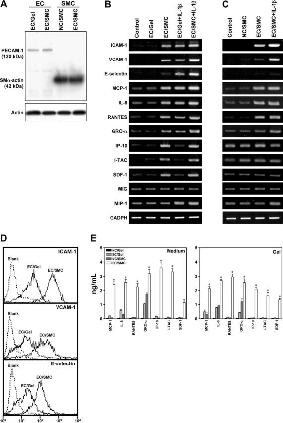
Atherosclerosis develops at regions of the arterial tree exposed to disturbed flow. The early stage of atherogenesis involves the adhesion of leukocytes (white blood cells [WBCs]) to and their transmigration across endothelial cells (ECs), which are located in close proximity to smooth muscle cells (SMCs). We investigated the effects of EC/SMC coculture and disturbed flow on the adhesion and transmigration of 3 types of WBCs (neutrophils, peripheral blood lymphocytes [PBLs], and monocytes) using our vertical-step flow (VSF) chamber, in which ECs were cocultured with SMCs in collagen gels. Such coculture significantly increased the adhesion and transmigration of neutrophils, PBLs, and monocytes under VSF, particularly in the reattachment area, where the rolling velocity of WBCs and their transmigration time were decreased, as compared with the other areas. Neutrophils, PBLs, and monocytes showed different subendothelial migration patterns under VSF. Their movements were more random and shorter in distance in the reattachment area. Coculture of ECs and SMCs induced their expressions of adhesion molecules and chemokines, which contributed to the increased WBC adhesion and transmigration. Our findings provide insights into the mechanisms of WBC interaction with the vessel wall (composed of ECs and SMCs) under the complex flow environments found in regions of prevalence for atherogenesis.
Figures


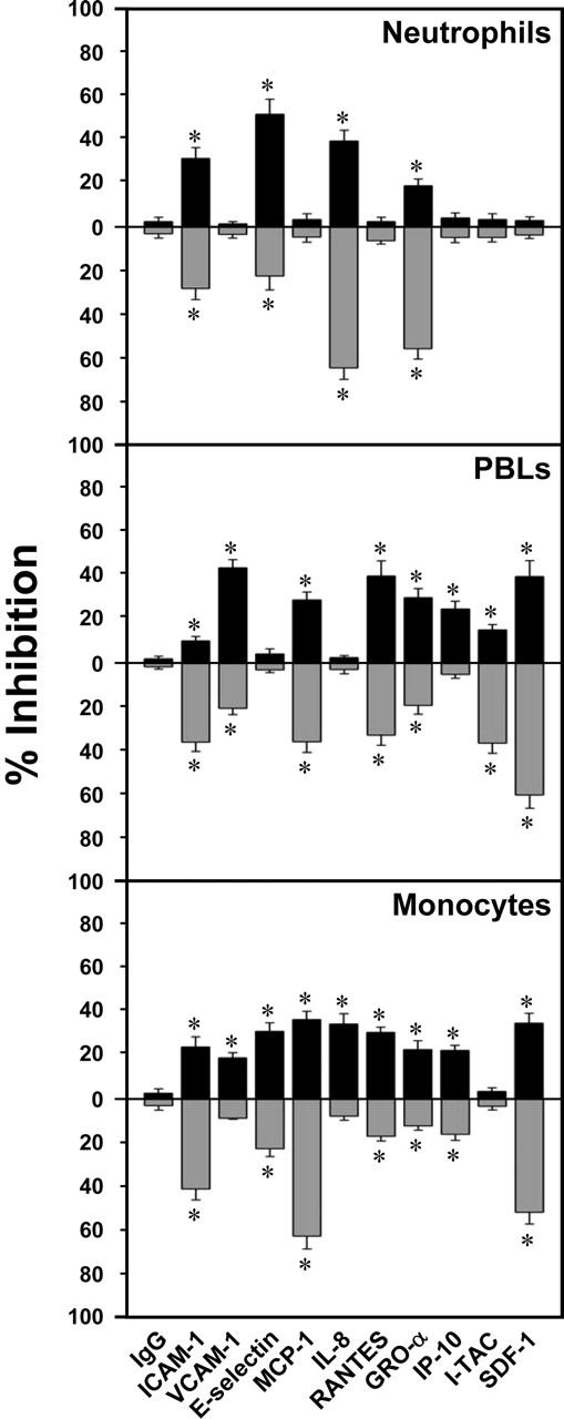
 ) WBCs with antibody treatment (CE) and without antibody treatment (CC): % inhibition = 100 [(CC - CE)/CC]. Data are expressed as mean ± SEM, n = 4. *P < .05 versus EC/SMC treated with control IgG or without antibody treatment.
Similar articles
-
Fluid shear stress suppresses ICAM-1-mediated transendothelial migration of leukocytes in coculture model.Biochem Biophys Res Commun. 2018 Jul 20;502(3):403-408. doi: 10.1016/j.bbrc.2018.05.182. Epub 2018 May 31. Biochem Biophys Res Commun. 2018. PMID: 29852173
-
Shear stress inhibits adhesion molecule expression in vascular endothelial cells induced by coculture with smooth muscle cells.Blood. 2003 Apr 1;101(7):2667-74. doi: 10.1182/blood-2002-08-2560. Epub 2002 Dec 5. Blood. 2003. PMID: 12468429
-
Development of an endothelial-smooth muscle cell coculture model using phenotype-controlled smooth muscle cells.Ann Biomed Eng. 2011 Nov;39(11):2750-8. doi: 10.1007/s10439-011-0372-8. Epub 2011 Aug 3. Ann Biomed Eng. 2011. PMID: 21811870
-
Subendothelial resistin enhances monocyte transmigration in a co-culture of human endothelial and smooth muscle cells by mechanisms involving fractalkine, MCP-1 and activation of TLR4 and Gi/o proteins signaling.Int J Biochem Cell Biol. 2014 May;50:29-37. doi: 10.1016/j.biocel.2014.01.022. Epub 2014 Feb 6. Int J Biochem Cell Biol. 2014. PMID: 24508784 Review.
-
Leukocyte-endothelial cell interactions.Semin Hematol. 1993 Oct;30(4 Suppl 4):45-53; discussion 54-5. Semin Hematol. 1993. PMID: 8303310 Review.
Cited by
-
Upregulation of inflammatory gene transcripts in periosteum of chronic migraineurs: Implications for extracranial origin of headache.Ann Neurol. 2016 Jun;79(6):1000-13. doi: 10.1002/ana.24665. Epub 2016 May 5. Ann Neurol. 2016. PMID: 27091721 Free PMC article.
-
Flow shear stress and atherosclerosis: a matter of site specificity.Antioxid Redox Signal. 2011 Sep 1;15(5):1405-14. doi: 10.1089/ars.2010.3679. Epub 2011 Apr 8. Antioxid Redox Signal. 2011. PMID: 21050140 Free PMC article. Review.
-
Single-cell imaging of mechanotransduction in endothelial cells.Prog Mol Biol Transl Sci. 2014;126:25-51. doi: 10.1016/B978-0-12-394624-9.00002-6. Prog Mol Biol Transl Sci. 2014. PMID: 25081613 Free PMC article. Review.
-
Endothelial cell behaviour within a microfluidic mimic of the flow channels of a modular tissue engineered construct.Biomed Microdevices. 2011 Feb;13(1):69-87. doi: 10.1007/s10544-010-9472-8. Biomed Microdevices. 2011. PMID: 20842530 Free PMC article.
-
Neutrophil adhesion on endothelial cells in a novel asymmetric stenosis model: effect of wall shear stress gradients.Ann Biomed Eng. 2010 Sep;38(9):2791-804. doi: 10.1007/s10439-010-0032-4. Epub 2010 Apr 13. Ann Biomed Eng. 2010. PMID: 20387119 Free PMC article.
References
-
- Ku DN, Giddens DP, Zarins CK, Glagov S. Pulsatile flow and atherosclerosis in the human carotid bifurcation. Positive correlation between plaque location and low oscillating shear stress. Arteriosclerosis. 1985;5: 293-302. - PubMed
-
- Ross R. The pathogenesis of atherosclerosis: a perspective for the 1990s. Nature. 1993;362: 801-809. - PubMed
-
- Skilbeck C, Westwood SM, Walker PG, David T, Nash GB. Population of the vessel wall by leukocytes binding to P-selectin in a model of disturbed arterial flow. Arterioscler Thromb Vasc Biol. 2001; 21: 1294-1300. - PubMed
-
- Chiu JJ, Chen CN, Lee PL, et al. Analysis of the effect of disturbed flow on monocytic adhesion to endothelial cells. J Biomech. 2003;36: 1883-1895. - PubMed
Publication types
MeSH terms
Substances
Grants and funding
LinkOut - more resources
Full Text Sources
