. 2006 Jan 15;570(Pt 2):407-20.
doi: 10.1113/jphysiol.2005.098848. Epub 2005 Nov 17.

Distinct rhythm generators for inspiration and expiration in the juvenile rat

Affiliations

Distinct rhythm generators for inspiration and expiration in the juvenile rat

Wiktor A Janczewski et al. J Physiol. .

Abstract

Inspiration and active expiration are commonly viewed as antagonistic phases of a unitary oscillator that generates respiratory rhythm. This view conflicts with observations we report here in juvenile rats, where by administration of fentanyl, a selective mu-opiate agonist, and induction of lung reflexes, we separately manipulated the frequency of inspirations and expirations. Moreover, completely transecting the brainstem at the caudal end of the facial nucleus abolished active expirations, while rhythmic inspirations continued. We hypothesize that inspiration and expiration are generated by coupled, anatomically separate rhythm generators, one generating active expiration located close to the facial nucleus in the region of the retrotrapezoid nucleus/parafacial respiratory group, the other generating inspiration located more caudally in the preBötzinger Complex.


Figures

Figure 1
Figure 1
Transition from control to quantal pattern of breathing in an 11-day-old vagotomized rat after fentanyl Top three traces, recordings of tidal volume (VT), genioglossus muscle electromyographic (EMGGG) and abdominal muscle electromyographic (EMGABD) activity. Bottom, timing bars indicating occurrence of inspiratory airflow (inspiration) and expiratory motor activity (EMGABD). A, control recording had all normal (I-E) cycles (i.e. cycles with inspiration and expiration). B, onset of quantal breathing as an occasional sequence of inspiration–expiration–expiration, i.e. E-only cycle (cycle without inspiration), is observed at 9 min after an initial dose of fentanyl (20 μg kg−1s.c.). C, at 4 min after a supplemental dose of fentanyl (10 μg kg−1), there are more E-only cycles. Note split EMGABD bursts in I-E cycles, and single EMGABD burst in E-only cycles.
Figure 2
Figure 2
Quantal pattern of breathing in an 8-day-old vagotomized rat given fentanyl A, intervals between successive EMGABD bursts (circles) and inspirations (squares) for 220 consecutive respiratory cycles: 120 I-E cycles (mean duration of I-E cycles formula image) and 100 E-only cycles (mean duration of E-only cycles formula image
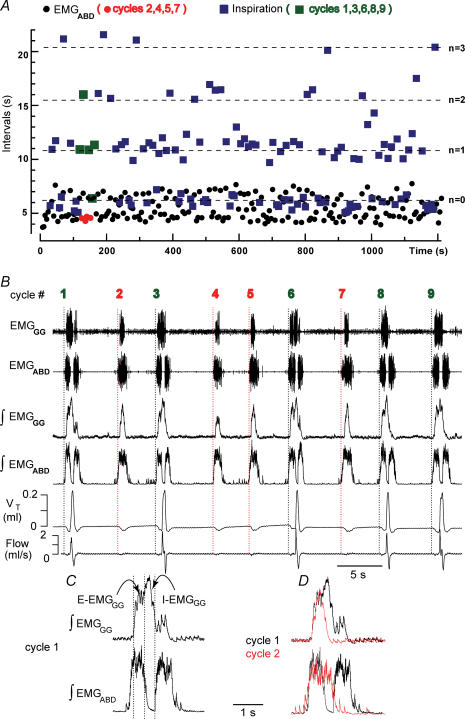
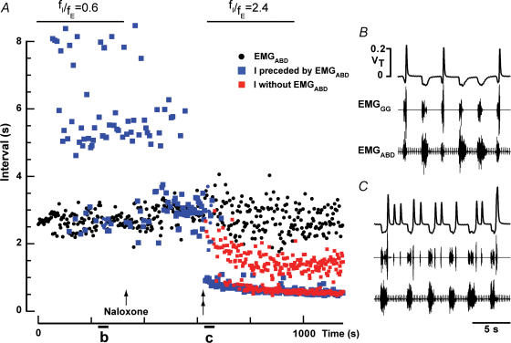
). B, recording of epoch shown in A (∼(150 s, 190 s); green squares represent normal, i.e. I-E cycles (1, 3, 6, 8, 9), and red circles represent E-only cycles (2, 4, 5, 7). Traces from top to bottom: cycle number, EMGGG, EMGABD, their integrals (∫EMGGG and ∫EMGABD), VT and respiratory flow. Vertical dotted lines mark the onset of each EMGABD burst. E-only cycles differed from I-E cycles; see C, D and text. C, ∫EMGGG and ∫EMGABD on an expanded time scale to show pattern of bursting in an I-E cycle (cycle 1); note that ∫EMGGG has two distinct components (I-EMGGG and E-EMGGG) and can have no delay between them or be separate, e.g. Fig. 3, paired vertical lines. EMGABD activity has a two-burst structure. D, comparison of an I-E cycle (cycle 1) and an E-only cycle (cycle 2, red).
Figure 3
Figure 3
Response to continuous positive airway pressure (CPAP) in an 11-day-old rat with intact vagi and transected pons given fentanyl Top traces, VT (continuous line) with superimposed resting lung volume (dashed line), EMGGG and EMGABD. Paired vertical lines above the VT trace indicate I-E cycles where termination of an I-EMGGG burst and onset of subsequent E-EMGGG burst were separate; this is consistent with distinct sources of these bursts. Bottom, timing bars (see Fig. 1). Initially, inspiratory frequency/expiratory frequency (fI/fE) = 0.7, i.e. on average 3 of 10 cycles were E-only. With CPAP = 3 cmH2O, fI/fE= 0, as I-E cycles were completely suppressed. Note that during CPAP the amplitude and duration of the expiratory efforts increased, and VT generated by ‘passive’ inflations matched that of active inspirations.
Figure 4
Figure 4
Rhythmic lung inflations increased fE and rhythmic lung deflations increased fI in a 9-day-old rat with intact vagi given fentanyl in which all cycles were E-only Traces, EMGABD, EMGGG and tracheal pressure (TP). A and B, Breuer-Hering inflation/deflation reflexes before administration of fentanyl. A, inflation reflex triggered by brief lung inflations (upward deflections of TP trace) leads to apnoea and briefly inhibits EMGABD activity. B, brief lung deflation triggers an extra inspiration, and sustained lung deflation increases fI from 92 to 208 min−1. C and D, after administration of fentanyl in a dose that eliminated inspiratory activity, i.e, all cycles were E-only. C, brief lung inflations applied shortly after the beginning of each EMGABD burst increased fE from 11 to 29 bursts min−1 and shortened EMGABD bursts from 2.2 to 0.4 s. D, brief lung deflations triggered inspirations (indicated by further downward deflections of TP trace) and terminated ongoing EMGABD bursts, e.g. D-1. Note that fE remained fairly constant despite shortening of EMGABD bursts. D-1, the first deflation in D on an expanded time scale. Note that EMGABD burst was not terminated until the onset of inspiration.
Figure 5
Figure 5
Response to lung deflation in an 11-day-old rat with intact vagi and transected pons given fentanyl Traces, tracheal pressure (TP), respiratory flow, EMGABD, EMGGG. Bottom, timing bars (see Fig. 1). Arrows mark onset of EMGABD bursts. Negative pressure of −1.5 cmH2O did not change fE but did increase fI, increasing fI/fE from 0.7 to 1.0. Negative pressure of −3 cmH2O recruited additional inspirations (triangles/dotted lines). fI increased leading to fI/fE= 2.5. The frequency of I activity locked to EMGABD bursts was similar to that of preceding I-E cycles, and was clearly different from the rhythm of inspirations ‘in between’ EMGABD bursts (triangles). Note that during lung deflation, EMGABD bursts became shorter.
Figure 6
Figure 6
Naloxone increased fI/fE in an 11-day-old rat with intact vagi given fentanyl A, intervals between successive EMGABD bursts (black circles), and between inspirations preceded (blue squares) or not preceded (red squares) by an EMGABD burst. A small dose of naloxone (0.03 mg kg−1s.c.) was injected (arrow) and antagonized fentanyl-induced changes in breathing apparent after ∼300 s (double arrow). Note that naloxone had little effect on fE, but increased fI to the point where some inspirations (red squares) occurred in between E-bursts. B, quantal breathing after fentanyl but before naloxone. C, ∼300 s after injection of naloxone, ectopic inspiratory bursts were induced.
Figure 7
Figure 7
Transection rostral to the facial nucleus did not affect breathing pattern, whereas transection at the caudal end of the facial nucleus eliminated EMGABD bursts (but not inspirations) in vagotomized rats A, breathing after transection at the level just rostral to the VII nucleus (dotted line, ‘rostral-t’ in C) in rats that spontaneously recovered from quantal pattern of breathing. B, breathing after transection marked as ‘caudal-t’ in C. Only this most caudal transection completely eliminated EMGABD bursts. Tonic EMGABD activity, inhibited during inspirations, could still be observed. A–1 and B–2, expanded time-scale of flow and VT during the first cycles of traces in A and B. C, sagittal representation of medulla and pons showing the levels of the most rostral (‘rostral-t’) and caudal (‘caudal-t’) transections. Intermediate transections were also made (see text). C-1 and C-2, medullary sections (40 μm thick) of remaining medulla cut at the same angle as ‘caudal-t’ transection. The first section (C-1), showing the caudal end of the VII nucleus, was modestly damaged, but the next section (C-2), showing rostral end of BötC, was intact in every rat (n = 5). V, motor trigeminal nucleus; VII, facial nucleus; LRN, lateral reticular nucleus; preBötC, preBötzinger Complex; BötC, Bötzinger Complex; pFRG, parafacial respiratory group; RTN, retrotrapezoid nucleus; SO, superior olivary complex; Ac, ambiguus nucleus compact.

Comment in

References

    1. Alheid GF, Gray PA, Jiang MC, Feldman JL, McCrimmon DR. Parvalbumin in respiratory neurons of the ventrolateral medulla of the adult rat. J Neurocytol. 2002;31:693–717. - PubMed
    1. Ballanyi K. Neuromodulation of the perinatal respiratory network. Curr Neuropharmacol. 2004;2:221–243. - PubMed
    1. Ballanyi K, Onimaru H, Homma I. Respiratory network function in the isolated brainstem–spinal cord of newborn rats. Prog Neurobiol. 1999;59:583–634. - PubMed
    1. Bianchi AL, Denavit-Saubie M, Champagnat J. Central control of breathing in mammals: neuronal circuitry, membrane properties, and neurotransmitters. Physiol Rev. 1995;75:1–45. - PubMed
    1. Borday C, Wrobel L, Fortin G, Champagnat J, Thaeron-Antono C, Thoby-Brisson M. Developmental gene control of brainstem function: views from the embryo. Prog Biophys Mol Biol. 2004;84:89–106. - PubMed

Publication types

MeSH terms