Comparative Study
. 2005 Nov 29;102(48):17412-7.
doi: 10.1073/pnas.0508698102. Epub 2005 Nov 17.

Visualization of a primary anti-tumor immune response by positron emission tomography

Affiliations
Comparative Study

Visualization of a primary anti-tumor immune response by positron emission tomography

Chengyi J Shu et al. Proc Natl Acad Sci U S A. .

Abstract

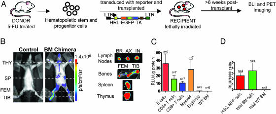
Current methodologies that monitor immune responses rely on invasive techniques that sample tissues at a given point in time. New technologies are needed to elucidate the temporal patterns of immune responses and the spatial distribution of immune cells on a whole-body scale. We describe a noninvasive, quantitative, and tomographic approach to visualize a primary anti-tumor immune response by using positron emission tomography (PET). Bone marrow chimeric mice were generated by engraftment of hematopoietic stem and progenitor cells transduced with a trifusion reporter gene encoding synthetic Renilla luciferase (hRluc), EGFP, and Herpes virus thymidine kinase (sr39TK). Mice were challenged with the Moloney murine sarcoma and leukemia virus complex (M-MSV/M-MuLV), and the induced immune response was monitored by using PET. Hematopoietic cells were visualized by using 9-[4-[(18)F]fluoro-3-(hydroxymethyl)butyl]guanine ([(18)F]FHBG), a radioactive substrate specific for the sr39TK PET reporter protein. Immune cell localization and expansion were seen at the tumor and draining lymph nodes (DLNs). 2-[(18)F]fluoro-2-deoxy-D-glucose ([(18)F]FDG), which is sequestered in metabolically active cells, was used to follow tumor growth and regression. Elevated glucose metabolism was also seen in activated lymphocytes in the DLNs by using the [(18)F]FDG probe. When M-MSV/M-MuLV-challenged mice were treated with the immunosuppressive drug dexamethasone, activation and expansion of immune cell populations in the DLNs could no longer be detected with PET imaging. The method we describe can be used to kinetically measure the induction and therapeutic modulations of cell-mediated immune responses.


Figures

Fig. 1.
Fig. 1.
Trifusion reporter is stably expressed in the hematopoietic cell compartment of BM chimeras. (A) Schematic of the generation of BM chimeric mice. The trifusion reporter construct is composed of a ubiquitin promoter driving expression of an optical bioluminescence reporter gene Renilla luciferase (hrl), a green fluorescent protein marker (egfp), and a PET reporter mutant herpes simplex virus type 1 thymidine kinase (tk). (B) (Left) Renilla luciferase expression in control and BM chimeric mice was imaged by using a charge-coupled device (CCD) camera. (Right) Organs were imaged ex vivo to assess tissue origin of bioluminescence signal. THY, thymus; SP, spleen; FEM, femurus; TIB, tibia; BR, brachial; AX, axillary; IN, inguinal. p/s/cm2/sr, photons·s-1·cm-2·steridian-1. The results are representative of 40 BM chimeric mice. (C) B cells (B220+), T cells (CD4+ and CD8+), myeloid cells (CD11b+), and erythroid cells (Ter119+) were purified from lymphoid organs of chimeras. (D) HSCs and multipotent progenitor (MPP) (c-kit+Thy1.2-/loLin-Sca-1+) cells were sorted by FACS. (C and D) Cells were lysed, and Renilla luciferase activity was assayed. BM cells from C57BL/6 mice were used as a control. RLU, relative light units.
Fig. 2.
Fig. 2.
Whole-body [18F]FDG PET imaging of metabolically active tumor cells and immune cells. (A) Whole-body tomographic computed tomography (CT) image of untreated mouse. Coronal, transverse, and sagittal planes are shown. BM chimeric mice (B) and CB-17SCID/SCID mice (C) were injected i.p. with [18F]FDG on day 13 post tumor challenge and imaged as described in Materials and Methods. Whole-body coronal, transverse, and sagittal PET sections (1.6-mm thickness) are shown. TUM, tumor; H, heart; BL, bladder; SP, spleen; KID, kidney.
Fig. 3.
Fig. 3.
Alterations in immune cell localization and activation with or without DEX treatment can be detected by [18F]FHBG and [18F]FDG PET imaging. (A) Schematic diagram of imaging schedule and DEX treatment. Mice were injected i.v with [18F]FHBG and imaged on day 8, 10, and 14 (B) and i.p with [18F]FDG and imaged on day 9, 13, and 15 (F). The coronal section displayed is centered at the tumor (TUM) and DLNs (1.6-mm thickness). (B and F) Shown are untreated mice (No DEX) (Left) and DEX-treated mice (DEX) (Right). Representative of a total of 10 animals performed in three separate experiments. ROIs were drawn to quantify [18F]FHBG (C) and [18F]FDG (G) uptake in the TUM and DLN on different days post tumor challenge. The quantification of [18F]FHBG and [18F]FDG signals was calculated as a ratio of %ID/g at region of interest to the %ID/g at a background region. Immediately after whole-body imaging, each organ was isolated day 14 post challenge to assess organ size to the [18F]FHBG (D) and [18F]FDG (H) signal. (H) Naive unchallenged C57BL/6 mouse lymph nodes were used as a control. Representative of three animals from each group. CON MUS, contralateral muscle; CON LNs, contralateral LNs. (E) Picture of the DLNs and CON LNs from untreated (No DEX) and DEX-treated (DEX) mice.
Fig. 4.
Fig. 4.
Histological analysis of tumor sections detects myeloid cell populations in DEX-treated tumors. Shown are stainings of tumor sections from untreated (No DEX) and DEX-treated (DEX) animals with α-CD3 and α-CD11b. (Magnification: ×200.)

References

    1. Butcher, E. C., Williams, M., Youngman, K., Rott, L. & Briskin, M. (1999) Adv. Immunol. 72, 209-253. - PubMed
    1. Therasse, P., Arbuck, S. G., Eisenhauer, E. A., Wanders, J., Kaplan, R. S., Rubinstein, L., Verweij, J., Van Glabbeke, M., van Oosterom, A. T., Christian, M. C. & Gwyther, S. G. (2000) J. Natl. Cancer Inst. 92, 205-216. - PubMed
    1. Iparraguirre, A. & Weninger, W. (2003) Int. Arch. Allergy Immunol. 132, 277-293. - PubMed
    1. Shah, K., Jacobs, A., Breakefield, X. O. & Weissleder, R. (2004) Gene Ther. 11, 1175-1187. - PubMed
    1. Gambhir, S. S. (2002) Nat. Rev. Cancer 2, 683-693. - PubMed

Publication types

MeSH terms

LinkOut - more resources