Comparative Study
. 2005 Nov 29;102(48):17531-6.
doi: 10.1073/pnas.0508418102. Epub 2005 Nov 17.

MINISEED3 (MINI3), a WRKY family gene, and HAIKU2 (IKU2), a leucine-rich repeat (LRR) KINASE gene, are regulators of seed size in Arabidopsis

Affiliations
Comparative Study

MINISEED3 (MINI3), a WRKY family gene, and HAIKU2 (IKU2), a leucine-rich repeat (LRR) KINASE gene, are regulators of seed size in Arabidopsis

Ming Luo et al. Proc Natl Acad Sci U S A. .

Abstract

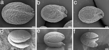
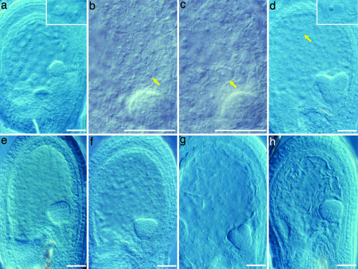
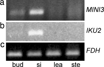
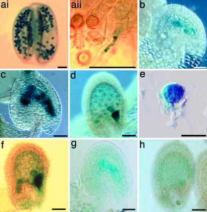
We have identified mutant alleles of two sporophytically acting genes, HAIKU2 (IKU2) and MINISEED3 (MINI3). Homozygotes of these alleles produce a small seed phenotype associated with reduced growth and early cellularization of the endosperm. This phenotype is similar to that described for another seed size gene, IKU1. MINI3 encodes WRKY10, a WRKY class transcription factor. MINI3 promoter::GUS fusions show the gene is expressed in pollen and in the developing endosperm from the two nuclei stage at approximately 12 hr postfertilization to endosperm cellularization at approximately 96 hr. MINI3 is also expressed in the globular embryo but not in the late heart stage of embryo development. The early endosperm expression of MINI3 is independent of its parent of origin. IKU2 encodes a leucine-rich repeat (LRR) KINASE (At3g19700). IKU2::GUS has a similar expression pattern to that of MINI3. The patterns of expression of the two genes and their similar phenotypes indicate they may operate in the same genetic pathway. Additionally, we found that both MINI3 and IKU2 showed decreased expression in the iku1-1 mutant. IKU2 expression was reduced in a mini3-1 background, whereas MINI3 expression was unaltered in the iku2-3 mutant. These data suggest the successive action of the three genes IKU1, IKU2, and MINI3 in the same pathway of seed development.


Figures

Fig. 1.
Fig. 1.
Phenotypes of mini3-1 and iku2-3. (a) Ler dry seed, (b) mini3-1 dry seed, (c) iku2-3 dry seed, (d) mature Ler embryo, (e) mature mini3-1 embryo, and (f) mature iku2-3 embryo. (Scale bars, 0.05 mm.)
Fig. 2.
Fig. 2.
Cytological phenotypes of mini3-1 and iku2-3 developing seeds. (a) Seventy-two-hour-old Ler seed with globular stage embryo and uncellularized endosperm. (Inset) Free nuclei. (b) Seventy-two-hour-old mini3-1 seed with globular stage embryo and cellularized peripheral endosperm. Arrow shows cell walls of endosperm. Embryo is not in focus. (c) Seventy-two-hour-old iku2-3 seed with globular-stage embryo and cellularized peripheral endosperm. Arrow shows cell walls of endosperm. Embryo is not in focus (d) Ninety-six-hour-old Ler seed with heart stage embryo and cellularized endosperm. (Inset) Cell walls of endosperm. (e) Ninety-six-hour-old mini3-1 seed with triangular embryo and cellularized endosperm. (f) Ninety-six-hour-old iku2-3 seed with triangular embryo and cellularized endosperm. (g) Ninety-six-hour-old fis2 seed with early-heart stage embryo and uncellularized endosperm. (h) Ninety-six-hour-old fis2/mini3-1 seed with late globular stage embryo and uncellularized endosperm. (Scale bars, 0.05 mm.)
Fig. 3.
Fig. 3.
Expression of MINI3 and IKU2. cDNA from flower buds prepollination (bud), young siliques (0- to 3-day fertilized siliques) (si), cauline leaves (lea), and bolt stem without cauline leaves (ste) was used for PCR amplification. (a) RT-PCR of MINI3. Bands are seen in bud and si. (b) RT-PCR of IKU2. Bands are seen in si. (c) RT-PCR of FDH. All tissues give bands.
Fig. 4.
Fig. 4.
MINI3::GUS and IKU2::GUS activity. (ai) Mature pollen showing GUS activity in homozygous MINI3::GUS transgenic plants. (aii) MINI3::GUS pollen germination on Ler stigma, showing a blue pollen tube. (b) MINI3::GUS developing seed showing GUS activity in two endosperm nuclei 12 hr postfertilization. (c) MINI3::GUS developing seed showing GUS activity in eight endosperm nuclei 24 hr postfertilization. (d) MINI3::GUS developing seed showing GUS activity in endosperm nuclei, globular embryo, and embryo surrounding region (ESR) in the seed 72 hr postfertilization. (e) MINI3::GUS developing seed showing GUS activity in a triangular embryo 96 hr postfertilization. (f)Ler developing seed showing paternal MINI3::GUS activity in endosperm nuclei, embryo, and ESR 72 hr post-fertilization. (g and h) IKU2::GUS activity in developing seeds 12 and 48 hr postfertilization. (Scale bars, 0.05 mm.)
Fig. 5.
Fig. 5.
Expression of MINI3 and IKU2 in mutant siliques. (a) RT-PCR of IKU2. IKU2 is repressed in mutant siliques of iku1-1, iku2-3, and mini3-1. (b) RT-PCR of MINI3. MINI3 is repressed only in iku1-1 mutant siliques. (c) RT-PCR of FDH. (d) Total RNA loading. (e) IKU1, MINI3, and IKU2 operate in a single pathway.
Fig. 6.
Fig. 6.
Autorepression of MINI3 and MINI3::GUS activity in fie-2 autonomous seeds. (a and b)Ler ovule showing higher GUS activity when pollinated with mini3-1 (a) than with MINI3 (b). (c and d) mini-3 ovule showing higher GUS activity when pollinated with mini3-1 pollen (c) than with MINI3 pollen (d). (e) A fie-2 autonomous seed showing MINI3::GUS activity in two endosperm nuclei 72 hr postemasculation. (f) fie-2 autonomous seed showing MINI3::GUS activity in endosperm nuclei 96-hr postemasculation. (Scale bars, 0.05 mm.) b and d represent separate experiments with different staining periods and exposures, and thus the intensity of GUS in these figures is not directly comparable.

Similar articles

Cited by

References

    1. Faure, J. E., Rotman, N., Fortuné, P. & Dumas, C. (2002) Plant J. 30, 481-488. - PubMed
    1. Boisnard-Lorig, C., Colon-Carmona, A., Bauch, M., Hodge, S., Doerner, P., Bancharel, E., Dumas, C., Haseloff, J. & Berger, F. (2001) Plant Cell 13, 495-509. - PMC - PubMed
    1. Olsen, O. A. (2001) Annu. Rev. Plant Physiol. Plant Mol. Biol. 52, 233-267. - PubMed
    1. Sorensen, M. B., Mayer, U., Lukowitz, W., Robert, H., Chambrier, P., Jurgens, G., Somerville, C., Lepiniec, L. & Berger, F. (2002) Development (Cambridge, U.K.) 129, 5567-5576. - PubMed
    1. Golden, T. A., Schauer, S. E., Lang, J. D., Pien, S., Mushegian, A. R., Grossniklaus, U., Meinke, D. W. & Ray, A. (2002) Plant Physiol. 130, 808-822. - PMC - PubMed

Publication types