. 2005 Dec;115(12):3494-505.
doi: 10.1172/JCI26052. Epub 2005 Nov 17.

Timp3 deficiency in insulin receptor-haploinsufficient mice promotes diabetes and vascular inflammation via increased TNF-alpha

Affiliations

Timp3 deficiency in insulin receptor-haploinsufficient mice promotes diabetes and vascular inflammation via increased TNF-alpha

Massimo Federici et al. J Clin Invest. 2005 Dec.

Abstract

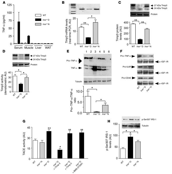
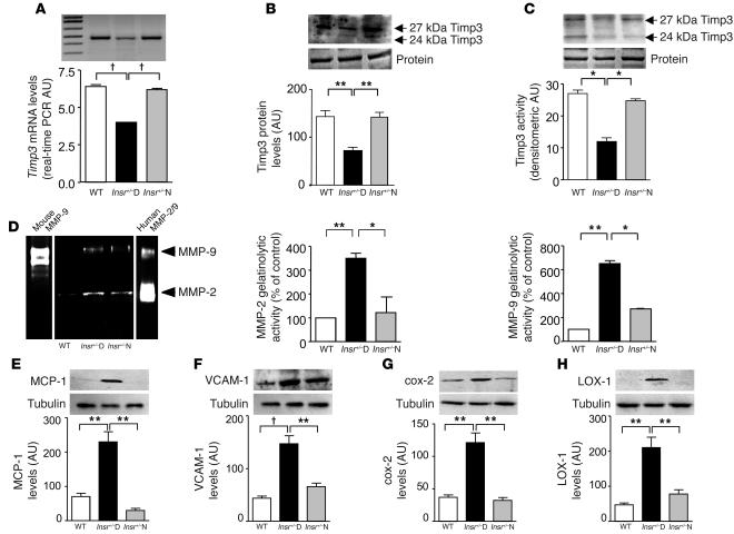
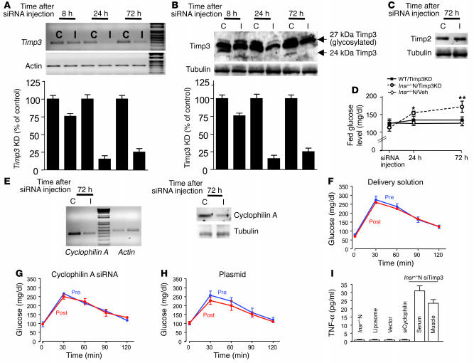
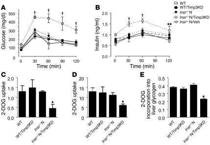
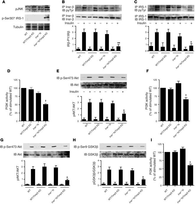
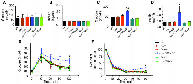
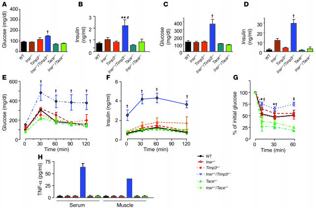
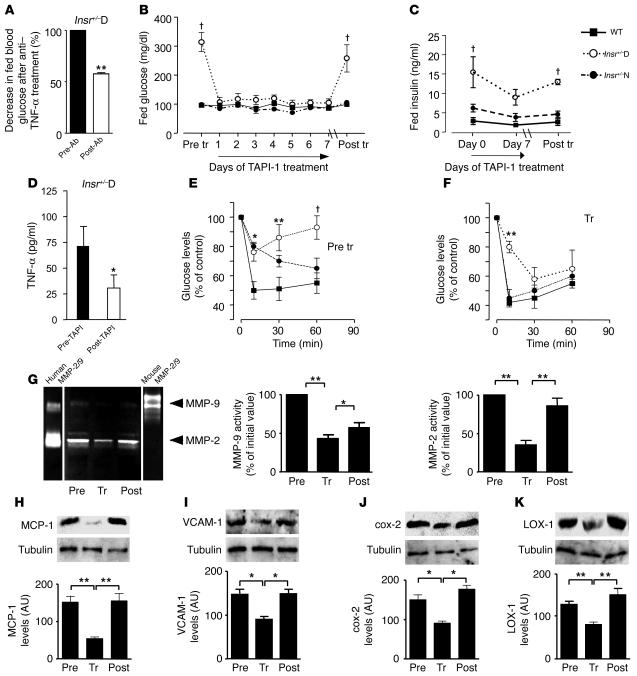
Activation of inflammatory pathways may contribute to the beginning and the progression of both atherosclerosis and type 2 diabetes. Here we report a novel interaction between insulin action and control of inflammation, resulting in glucose intolerance and vascular inflammation and amenable to therapeutic modulation. In insulin receptor heterozygous (Insr+/-) mice, we identified the deficiency of tissue inhibitor of metalloproteinase 3 (Timp3, an inhibitor of both TNF-alpha-converting enzyme [TACE] and MMPs) as a common bond between glucose intolerance and vascular inflammation. Among Insr+/- mice, those that develop diabetes have reduced Timp3 and increased TACE activity. Unchecked TACE activity causes an increase in levels of soluble TNF-alpha, which subsequently promotes diabetes and vascular inflammation. Double heterozygous Insr+/-Timp3+/- mice develop mild hyperglycemia and hyperinsulinemia at 3 months and overt glucose intolerance and hyperinsulinemia at 6 months. A therapeutic role for Timp3/TACE modulation is supported by the observation that pharmacological inhibition of TACE led to marked reduction of hyperglycemia and vascular inflammation in Insr+/- diabetic mice, as well as by the observation of increased insulin sensitivity in Tace+/- mice compared with WT mice. Our results suggest that an interplay between reduced insulin action and unchecked TACE activity promotes diabetes and vascular inflammation.


Figures

Figure 1
Figure 1
Timp3 expression and activity in skeletal muscle of Insr+/–mice. (A) Quantification of TNF-α in Insr+/–D, Insr+/–N, and WT littermates. (B) Real-time RT-PCR analysis of Timp3 mRNA expression in Insr+/–D, Insr+/–N, and WT mice. **P < 0.01; P < 0.001 by 1-way ANOVA. (C) Western blot analysis of Timp3 protein expression in Insr+/–D, Insr+/–N, and WT mice (equal loading was confirmed by silver staining). **P < 0.01 by 1-way ANOVA. (D) Comparison of Timp3 activity, measured by reverse zymography in Insr+/–D, Insr+/–N, and WT mice (equal loading was confirmed by silver staining). *P < 0.05 by 1-way ANOVA. (E) Pro–TNF-α to TNF-α conversion in Insr+/–D (lanes 3 and 4), Insr+/–N (lanes 5 and 6), and WT (lanes 1 and 2) mice. *P < 0.05 by 1-way ANOVA. (F) Shedding of ProTNF-α, pro–IL-6R, and pro-CD40 in Insr+/–D, Insr+/–N, and WT mice. Gel loading was normalized by anti–IGF-1R. (G) TACE activity measured by a fluorimetric assay in Insr+/–D, Insr+/–N, and WT mice in the presence or absence of recombinant Timp3 (rTimp-3) (100 mM), recombinant Timp2 (rTimp-2) (100 mM), and specific MMP inhibitor BMS-275291 (1 μM). **P < 0.01 versus WT; #P < 0.01 versus Insr+/–D by 1-way ANOVA. (H) IRS-1 phosphorylation on Ser307 in Insr+/–D, Insr+/–N, and WT. *P < 0.05 by 1-way ANOVA. Data from 3 separate experiments are summarized in the bar graphs and expressed as mean ± SD.
Figure 2
Figure 2
Decreased Timp3 activity and increased inflammation in aortas of Insr+/–D mice. (A) Real-time RT-PCR analysis of Timp3 mRNA expression in Insr+/–D, Insr+/–N, and WT mice. P < 0.001 by 1-way ANOVA. (B) Western blot analysis of Timp3 protein expression in Insr+/–D, Insr+/–N, and WT mice (equal loading was confirmed by silver staining). **P < 0.01 by 1-way ANOVA. (C) Comparison of Timp3 activity, measured by reverse zymography in Insr+/–D, Insr+/–N, and WT mice (equal loading was confirmed by silver staining). *P < 0.05 by 1-way ANOVA. (D) MMP measured by zymography in Insr+/–D, Insr+/–N, and WT mice. Mouse MMP-9 and human MMP-2/9 were used as standards. **P < 0.01, *P < 0.05 by 1-way ANOVA. Analysis by Western blotting of inflammatory markers, MCP-1 (E), VCAM-1 (F), cox-2 (G), LOX-1 (H), and tubulin as control in Insr+/–D, Insr+/–N, and WT mice. **P < 0.01, P < 0.001 by 1-way ANOVA. Data from 3 separate experiments are summarized in the bar graphs and expressed as mean ± SD.
Figure 3
Figure 3
Effect of Timp3 KD in skeletal muscle. siRNA-mediated inhibition of Timp3 mRNA (A) and protein (B) expression in mice injected with insert 3 containing vector at 8 hours, 24 hours, and 72 hours after injection. I, muscle injected with insert; C, contralateral muscle injected only with vehicle. Data from 3 separate experiments are summarized in the bar graphs and expressed as mean ± SD. (C) Effect of Timp3 KD on Timp2 protein levels. (D) Fed glucose levels in Insr+/–N/Timp3KD mice (open circles), WT/Timp3KD (filled squares), and Insr+/–N mice injected with vehicle (Insr+/–N/Veh; open diamonds). Data are expressed as mean ± SD (n = 6 per group). *P < 0.05, **P < 0.01. (E) Effect of cyclophilin A siRNA on cyclophilin A mRNA and protein level in Insr+/–N mice. (FH) Intraperitoneal glucose tolerance tests before (Pre) and after (Post) injection of cyclophilin A (G), delivery solution (F), or empty plasmid (H) (n = 3 per group). (I) TNF-α levels in Insr+/–N mice injected with siRNA reagents and in Insr+/–N/cyclophilin A KD and Insr+/–N/Timp3KD mice. Data are expressed as mean ± SD (n = 3–6 per group).
Figure 4
Figure 4
Effect of Timp3 KD on glucose metabolism. Glucose (A) and insulin (B) levels during an intraperitoneal glucose tolerance test in WT mice (open squares), WT/Timp3KD (filled squares), Insr+/–N (filled circles), Insr+/–N/Timp3KD mice (open circles), and Insr+/–N/Veh (open diamonds) mice (n = 6 per group). Data are expressed as mean ± SD. **P < 0.01, P < 0.001 by 2-way ANOVA for Insr+/–N/Timp3KD compared with other groups. 2-DOG uptake into triceps (C) and perigonadal WAT (D) during a glucose tolerance test. (E) Glucose uptake into liver, as estimated by 2-DOG incorporation into glycogen (n = 6 per group). Data in CE represent moles/milligram protein per minute and are expressed as mean ± SD. *P < 0.05 compared with WT by 1-way ANOVA for Insr+/–N/Timp3KD compared with other groups.
Figure 5
Figure 5
Effect of Timp3 KD on insulin signaling in WT and Insr+/– mice. (A) TNF-α signaling in skeletal muscle measured by phosphorylation state of JNK-1/2 and of Ser307 in IRS1. After an overnight fast, 6-month-old WT, WT/Timp3KD, Insr+/– and Insr+/–/Timp3KD mice were injected with either saline (–) or 25 mU/kg of insulin (Ins). Skeletal muscle (BE), livers (FH), and WAT (I) were collected 5–10 minutes later. Protein extracts were prepared, immunoprecipitated with antibodies against Insr (B) and IRS-1 (C), and after gel separation were immunoblotted with antibodies specific for phosphotyrosine (pyTyr) (PY20) and normalized by reblotting with specific antibodies. PI3K activity (D, F, and I) was measured in phosphotyrosine immunoprecipitates by ELISA assay. The levels of total AKT1/2 and activated AKT (p-Ser473) (E and G), total glycogen synthase kinase 3β (GSK3β) and inactivated GSK3β (p-Ser9) (H) were determined by immunoblotting of the original lysates. Band intensities were quantified by densitometry and expressed as mean ± SD (n = 3 mice per group). *P < 0.05, **P < 0.01, P < 0.001 by 1-way ANOVA for Insr+/–N/Timp3KD compared with other groups.
Figure 6
Figure 6
Glucose homeostasis in Insr+/–Timp3+/–, Insr+/–Tace+/–, Insr+/–, Timp3+/–, Tace+/–, and WT littermates at 3 months. Fasted glucose (A), fasted insulin (B), fed glucose (C), and fed insulin (D) levels in mice (n = 8–10 per group). Data are expressed as mean ± SD. P < 0.001 for Insr+/–Timp3+/– versus Timp3+/–, Tace+/–, and WT mice by 1-way ANOVA; #P < 0.01 for Insr+/–Timp3+/– versus Insr+/– and Insr+/–Tace+/– mice; *P < 0.05 for Insr+/–Timp3+/– versus all the other genotypes by 1-way ANOVA. (E) Glucose levels after intraperitoneal glucose load. Data are expressed as mean ± SD (n = 5 per group). *P < 0.05 for Insr+/–Timp3+/– versus other genotypes by 2-way ANOVA. (F) Insulin tolerance test (0.75 U kg–1). Data are expressed as mean ± SD (n = 5 per group). *P < 0.05 for Insr+/–/Timp3+/– versus other genotypes by 2-way ANOVA.
Figure 7
Figure 7
Glucose homeostasis in Insr+/–Timp3+/–, Insr+/–Tace+/–, Insr+/–, Timp3+/–, Tace+/–, and WT littermates at 6 months. Fasted glucose (A), fasted insulin (B), fed glucose (C), and fed insulin (D) levels (n = 8–10 per group). Data are expressed mean ± SD. P < 0.001 for Insr+/–/Timp3+/– versus other genotypes by 1-way ANOVA; **P < 0.01 for Insr+/–Timp3+/– versus Timp3+/–, Tace+/–, and WT; #P < 0.05, Insr+/–Timp3+/– versus Insr+/– and Insr+/–Tace+/– by 1-way ANOVA. Glucose (E) and insulin (F) levels during an intraperitoneal glucose tolerance test. Data are expressed as mean ± SD (n = 5 per group). P < 0.001, Insr+/–Timp3+/– versus other genotypes by 2-way ANOVA. (G) Insulin tolerance test (0.75 U kg–1). Data are expressed as mean ± SD (n = 5 per group). P < 0.001, Insr+/–Timp3+/– versus Timp3+/–, Tace+/–, and WT; *P < 0.05, Insr+/–/Timp3+/– versus Insr+/– and Insr+/–Tace+/– by 2-way ANOVA at 10 and 30 minutes; and P < 0.001, Insr+/–Timp3+/– versus Insr+/–Tace+/–, Insr+/–, Timp3+/–, Tace+/–, and WT by 2-way ANOVA at 60 minutes. (H) Quantification of TNF-α in Insr+/–Timp3+/–, Insr+/–Tace+/–, Insr+/–, Timp3+/–, Tace+/–, and WT littermates.
Figure 8
Figure 8
Glucose homeostasis in Insr+/–D mice is improved by inhibition of Tace/MMP activity. (A) Fed glucose levels were measured before (Pre-Ab) and 24 hours after (Post-Ab) anti–TNF-α antibody injection in Insr+/–D (n = 6). **P < 0.01 by 1-way ANOVA. (B) Fed glucose levels were measured before and on day 7 of TAPI-1 treatment and 14 days after discontinuing TAPI-1 in Insr+/–D, Insr+/–N, and WT mice at 6 months of age (n = 12 per group). P < 0.001 by 1-way ANOVA. (C) Fed insulin levels before, on day 7 of TAPI-1 treatment, and day 14 after discontinuing TAPI-1 in Insr+/–D, Insr+/–N, and WT mice at 6 months of age (n = 12 per group). P < 0.001 by 1-way ANOVA. (D) TNF-α levels before and after TAPI-1 treatment (7 days) in Insr+/–D mice. *P < 0.05. (E and F) Insulin tolerance test (0.75 U kg–1) in Insr+/–D (open circles), Insr+/–N (filled circles), and WT (filled squares) mice at 6 months of age (n = 6 per group) before and at the end of TAPI-1 treatment. *P < 0.05, **P < 0.01, P < 0.001 by 2-way ANOVA. (G) MMP-9 and MMP-2 measured by zymography (n = 3 per group). Mouse MMP-9 and human MMP-2/9 were used as standards. MCP-1 (H), VCAM-1 (I), cox-2 (J), and LOX-1 (K) by Western blotting. Gel loading was normalized by tubulin. Data from 3 separate experiments are summarized in the bar graphs and expressed as mean ± SD. *P < 0.05, **P < 0.01 by 1-way ANOVA.

References

    1. Warram JH, Martin BC, Krolewski AS, Soeldner JS, Kahn CR. Slow glucose removal rate and hyperinsulinemia precede the development of type II diabetes in the offspring of diabetic parents. Ann. Intern. Med. 1990;113:909–915. - PubMed
    1. Rewers M, et al. Insulin sensitivity, insulinemia, and coronary artery disease: the Insulin Resistance Atherosclerosis Study. Diabetes Care. 2004;27:781–787. - PubMed
    1. Stern MP. Diabetes and cardiovascular disease. The “common soil” hypothesis. Diabetes. 1995;44:369–374. - PubMed
    1. Ginsberg HN. Insulin resistance and cardiovascular disease. J. Clin. Invest. 2000;106:453–458. - PMC - PubMed
    1. Festa A, et al. Chronic subclinical inflammation as part of the insulin resistance syndrome: the Insulin Resistance Atherosclerosis Study (IRAS) Circulation. 2000;102:42–47. - PubMed

Publication types

MeSH terms