

Ultrasonic drug delivery--a general review
- PMID: 16296719
- PMCID: PMC1361256
- DOI: 10.1517/17425247.1.1.37
Ultrasonic drug delivery--a general review
Abstract
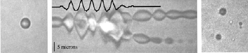
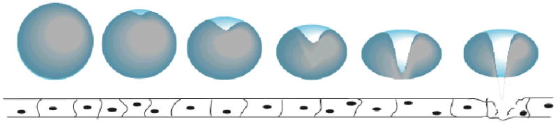
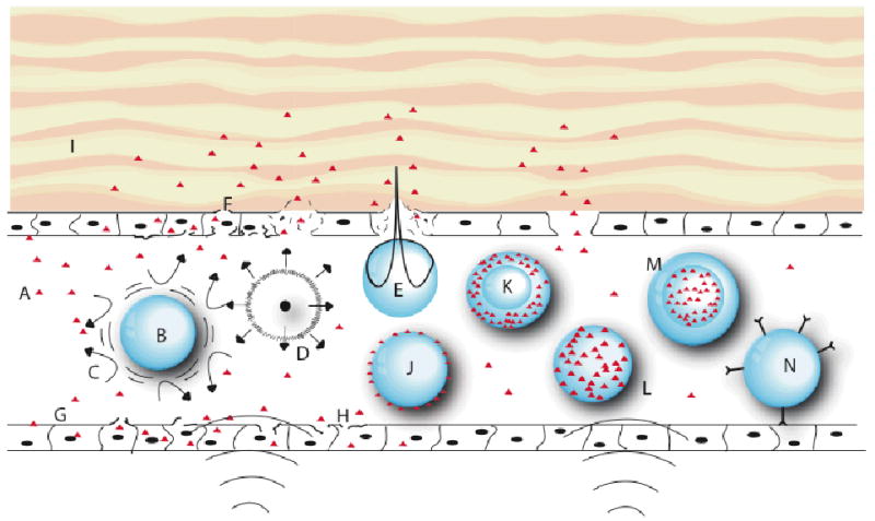
Ultrasound has an ever-increasing role in the delivery of therapeutic agents, including genetic material, protein and chemotherapeutic agents. Cavitating gas bodies, such as microbubbles, are the mediators through which the energy of relatively non-interactive pressure waves is concentrated to produce forces that permeabilise cell membranes and disrupt the vesicles that carry drugs. Thus, the presence of microbubbles enormously enhances ultrasonic delivery of genetic material, proteins and smaller chemical agents. Numerous reports show that the most efficient delivery of genetic material occurs in the presence of cavitating microbubbles. Attaching the DNA directly to the microbubbles, or to gas-containing liposomes, enhances gene uptake even further. Ultrasonic-enhanced gene delivery has been studied in various tissues, including cardiac, vascular, skeletal muscle, tumour and even fetal tissue. Ultrasonic-assisted delivery of proteins has found most application in transdermal transport of insulin. Cavitation events reversibly disrupt the structure of the stratus corneum to allow transport of these large molecules. Other hormones and small proteins could also be delivered transdermally. Small chemotherapeutic molecules are delivered in research settings from micelles and liposomes exposed to ultrasound. Cavitation appears to play two roles: it disrupts the structure of the carrier vesicle and releases the drug; and makes cell membranes and capillaries more permeable to drugs. There remains a need to better understand the physics of cavitation of microbubbles and the impact that such cavitation has on cells and drug-carrying vesicles.
Figures
Similar articles
-
Recent Patents and Formulation of Nanopharmaceuticals Using Ultrasonication Technique.Recent Pat Nanotechnol. 2018;12(2):86-100. doi: 10.2174/1872210511666171120100649. Recent Pat Nanotechnol. 2018. PMID: 29165099
-
Microbubbles in ultrasound-triggered drug and gene delivery.Adv Drug Deliv Rev. 2008 Jun 30;60(10):1153-66. doi: 10.1016/j.addr.2008.03.005. Epub 2008 Apr 3. Adv Drug Deliv Rev. 2008. PMID: 18486268 Free PMC article. Review.
-
Micelles and nanoparticles for ultrasonic drug and gene delivery.Adv Drug Deliv Rev. 2008 Jun 30;60(10):1137-52. doi: 10.1016/j.addr.2008.03.008. Epub 2008 Apr 4. Adv Drug Deliv Rev. 2008. PMID: 18486269 Free PMC article. Review.
-
[Key problems in using ultrasonic cavitating bubble as a gene or drug vector].Sheng Wu Yi Xue Gong Cheng Xue Za Zhi. 2009 Oct;26(5):1129-32. Sheng Wu Yi Xue Gong Cheng Xue Za Zhi. 2009. PMID: 19947504 Review. Chinese.
-
Ultrasound-induced cavitation: applications in drug and gene delivery.Expert Opin Drug Deliv. 2006 Nov;3(6):713-26. doi: 10.1517/17425247.3.6.713. Expert Opin Drug Deliv. 2006. PMID: 17076594 Review.
Cited by
-
Nanoparticle-based materials in anticancer drug delivery: Current and future prospects.Heliyon. 2023 Oct 20;9(11):e21227. doi: 10.1016/j.heliyon.2023.e21227. eCollection 2023 Nov. Heliyon. 2023. PMID: 37954330 Free PMC article. Review.
-
In vitro suppression of oral squamous cell carcinoma growth by ultrasound-mediated delivery of curcumin microemulsions.Int J Nanomedicine. 2012;7:941-51. doi: 10.2147/IJN.S28510. Epub 2012 Feb 21. Int J Nanomedicine. 2012. PMID: 22393291 Free PMC article.
-
Current Challenges in Microcapsule Designs and Microencapsulation Processes: A Review.ACS Appl Mater Interfaces. 2024 Aug 7;16(31):40326-40355. doi: 10.1021/acsami.4c02462. Epub 2024 Jul 23. ACS Appl Mater Interfaces. 2024. PMID: 39042830 Free PMC article. Review.
-
Ultrasound enhanced methanol penetration of zebrafish (Danio rerio) embryos measured by permittivity changes using impedance spectroscopy.Eur Biophys J. 2008 Jul;37(6):1039-44. doi: 10.1007/s00249-007-0229-0. Epub 2007 Nov 6. Eur Biophys J. 2008. PMID: 17985127
-
Pentagalloyl Glucose and Its Functional Role in Vascular Health: Biomechanics and Drug-Delivery Characteristics.Ann Biomed Eng. 2019 Jan;47(1):39-59. doi: 10.1007/s10439-018-02145-5. Epub 2018 Oct 8. Ann Biomed Eng. 2019. PMID: 30298373 Free PMC article. Review.
References
-
- DRAPER DO, CASTEL JC, CASTEL D. Rate of Temperature Increase in Human Muscle During 1 MHz and 3 MHz Continuous Ultrasound. J Orthop Sports Phys Ther. 1995;22(4):142–150. - PubMed
-
- TACKER JR, ANDERSON RU. Delivery of Antitumor Drug to Bladder Cancer by Use of Phase Transition Liposomes and Hyperthermia. Journal of Urology. 1982;127:1211–121214. - PubMed
-
- KENNEDY JE, TER HAARGR, CRANSTON D. High intensity focused ultrasound: surgery of the future? Br J Radiol. 2003;76(909):590–599. - PubMed
-
- MADERSBACHER S, MARBERGER M. High-energy shockwaves and extracorporeal high-intensity focused ultrasound. J Endourol. 2003;17(8):667–672. - PubMed
-
- HUBER PE, JENNE JW, RASTERT R, et al. A New Noninvasive Approach in Breast Cancer Therapy Using Magnetic Resonance Imaging-guided Focused Ultrasound Surgery. Cancer Res. 2001;61:8441–8447. * Seminal paper regarding high intensity ultrasound for tumor destruction. - PubMed
Publication types
MeSH terms
Substances
Grants and funding
LinkOut - more resources
Full Text Sources
Other Literature Sources
Medical