. 2006 Feb 20;345(2):468-81.
doi: 10.1016/j.virol.2005.10.011. Epub 2005 Nov 17.

Phosphorylation of the VP16 transcriptional activator protein during herpes simplex virus infection and mutational analysis of putative phosphorylation sites

Affiliations

Phosphorylation of the VP16 transcriptional activator protein during herpes simplex virus infection and mutational analysis of putative phosphorylation sites

Søren Ottosen et al. Virology. .

Abstract

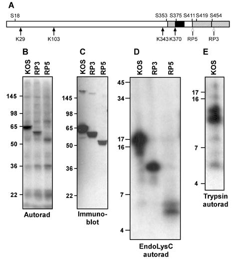
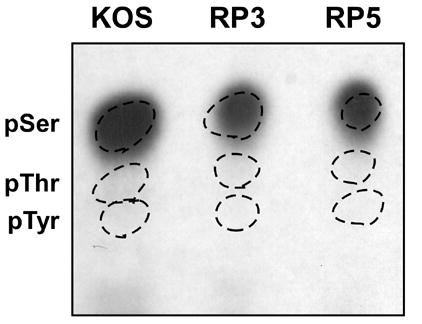
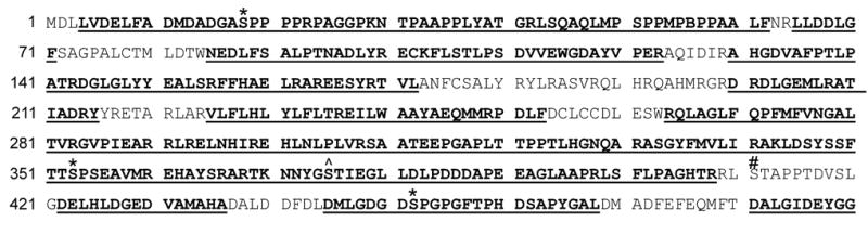
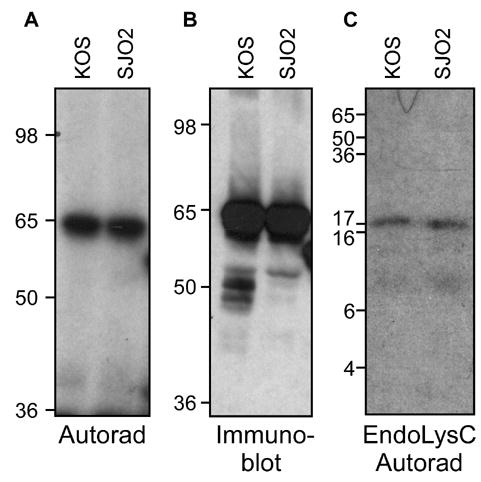
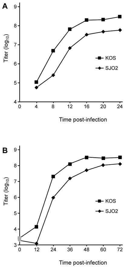
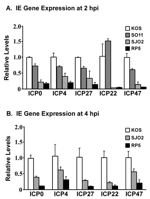
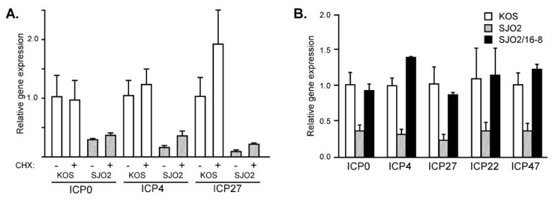
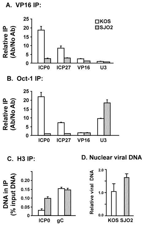
VP16 is a virion phosphoprotein of herpes simplex virus and a transcriptional activator of the viral immediate-early (IE) genes. We identified four novel VP16 phosphorylation sites (Ser18, Ser353, Ser411, and Ser452) at late times in infection but found no evidence of phosphorylation of Ser375, a residue reportedly phosphorylated when VP16 is expressed from a transfected plasmid. A virus carrying a Ser375Ala mutation of VP16 was viable in cell culture but with a slow growth rate. The association of the mutant VP16 protein with IE gene promoters and subsequent IE gene expression was markedly reduced during infection, consistent with prior transfection and in vitro results. Surprisingly, the association of Oct-1 with IE promoters was also diminished during infection by the mutant strain. We propose that Ser375 is important for the interaction of VP16 with Oct-1, and that the interaction is required to enable both proteins to bind to IE promoters.


Figures

Figure 1
Figure 1
Mapping VP16 phosphorylation sites using deletion mutants and peptide mapping. A. Schematic diagram of VP16. The two subregions of the C-terminal activation domain are denoted by hatching. Regions implicated in interaction with Oct-1 and HCF-1 are indicated by black and grey boxes, respectively. The four lysine residues in VP16 (K) and some of the serine residues (S) are indicated. Open arrows mark the truncations of the VP16 open reading frame in the viral strains RP3 and RP5. B. Autoradiogram of material precipitated using a VP16-specific monoclonal antibody (LP1) from HeLa cells infected by virus strains KOS, RP3 and RP5 and radiolabeled with [32P]-orthophosphate from 1.5 to 8 hour post-infection. The relative positions of protein molecular weight standards following separation on a 10% SDS-PAGE gel are indicated (in kDa). C. Immunoblot of a gel in parallel to that shown in panel B, probed with a VP16-specific polyclonal antibody (C8). D. Autoradiogram of radiolabeled VP16 fragments following digestion by lysyl endopeptidase (LysC), separated on a 16% polyacrylamide gel. E. Autoradiogram of radiolabeled VP16 fragments following digestion by trypsin and separation on a 16% polyacrylamide gel.
Figure 2
Figure 2
Phosphoamino acid analysis of VP16 proteins radiolabeled from 1.5 to 8 hours following infection with virus strains KOS, RP3 and RP5. The VP16 protein was isolated from infected cell lysates by immunoprecipitation with monoclonal antibody LP1 followed by SDS-PAGE. Proteins were transferred to a PVDF membrane and the band corresponding to VP16 was excised. Proteins were hydrolyzed in 6 N HCl for 1 hour at 100° C. Samples were mixed with unlabeled phosphoserine, phosphothreonine and phosphotyrosine standards and separated by thin-layer electrophoresis on a cellulose plate at pH 2.5. Phosphoamino acid standards were visualized using ninhydrin (dotted circles) and radiolabeled amino acids were detected by autoradiography.
Figure 3
Figure 3
Peptide sequences identified following LC-MS/MS analysis of VP16 proteins isolated at 8 hour post-infection with HSV-1 strain KOS. VP16 was isolated by immunoprecipitation and SDS-PAGE and then digested with trypsin or endoproteinase AspN. The entire VP16 amino acid sequence is shown. Boldface and underlined type indicates residues within peptide fragments identified following μLC-MS/MS. Asterisks indicate Ser residues found to be phosphorylated. A carat (^) indicates Ser375, for which no phosphorylation was observed. A hatch (#) indicates Ser411, the phosphorylation of which was implicated by peptide mapping experiments but not confirmed by μLC-MS/MS.
Figure 4
Figure 4
Phosphorylation of a Ser375Ala mutant of VP16. Virus strain SJO2 was constructed with an alanine codon replacing serine codon 375. HeLa cells infected with KOS or SJO2 were radiolabeled and the VP16 proteins were immunoprecipitated and separated by SDS-PAGE as described in Fig. 1. A. Autoradiogram, as described in Fig. 1. B. Immunoblot using VP16-specific antiserum C8. C. Autoradiogram of gel-isolated VP16 proteins subjected to digestion by LysC.
Figure 5
Figure 5
Growth curves of SJO2 and KOS. A. Vero cells were infected at high multiplicity (10 pfu/cell) with KOS or SJO2. At 4 h intervals, infected cells were lysed into the growth medium by scraping and sonication. Following centrifugation to remove cell debris, aliquots of the resulting supernatant were titered on Vero cells. B. Vero cells were infected with KOS or SJO2 at low multiplicity (0.01 pfu/cell) and harvested at 12 h intervals. Graphs represent the average of two biological replicates, which showed a range of less than 20% at every timepoint.
Figure 6
Figure 6
Immediate-early gene expression following infection by HSV-1 strains KOS, SJO2 and RP5. HeLa cells were infected at a multiplicity of 1 pfu/cell (KOS) or a corresponding number of virions (SJO2, RP5). RNA was harvested at 2 hpi (panel A) or 4 hpi (panel B). IE gene expression was assayed by quantitative real-time PCR following reverse transcriptase reactions. Expression levels were normalized to 18S rRNAs amplified in parallel Q-PCR reactions and are expressed relative to the levels observed in KOS infection. Error bars represent the range of two biological replicates each measured in duplicate PCRs.
Figure 7
Figure 7
Immediate-early gene expression in cells infected with HSV-1 strains KOS and SJO2. A. HeLa cells were infected with KOS (open bars) or SJO2 (hatched bars) in the presence or absence of cycloheximide (CHX). Total cellular RNA was harvested at 2 hpi and IE mRNA levels were assayed by quantitative real-time PCR as in Fig. 6. B. HeLa cells were infected with KOS (open bars), SJO2 (hatched bars), or SJO2 virions produced in 16-8 cells (filled bars). IE mRNA levels were assayed as in Fig. 6.
Figure 8
Figure 8
Association of VP16 and Oct-1 with IE promoters during infection by HSV-1 strains KOS and SJO2. At 2 h post-infection, proteins and nucleic acids were crosslinked by formaldehyde. Nuclei were isolated and sonicated to shear DNA to small fragments (predominantly 300–500 bp). Immunoprecipitation by antibodies specific to VP16 (panel A), Oct-1 (panel B), or H3 (panel C) was followed by quantitative real-time PCR to detect DNA fragments corresponding to IE gene promoters (ICP0 and ICP27), a late gene promoter (VP16 or gC) and a cellular promoter with an Oct-1 binding site (U3 snRNA). For panels A and B, the abundance of a given DNA in the IP pellet is indicated relative to its abundance in a control precipitation without antibody. For panel C, the abundance of a given DNA in the IP pellet is indicated relative to its abundance in input DNA (prior to IP). The amounts of viral DNA in nuclear DNA preparations from infected cells were assessed using quantitative PCR with primers specific for the VP16 gene (panel D), normalized to cellular DNA fragments from the U3 and IFN-β promoters and expressed relative to levels in wildtype-infected cells.
Figure 9
Figure 9
Phosphorylation of a Ser411Ala mutant of VP16. Virus strain DG1 contains a VP16 gene fused to sequences encoding green-fluorescent protein (GFP). SO11 was constructed with an alanine codon replacing serine codon 411, in the DG1 genetic background. HeLa cells infected with KOS, DG1 or SO11 were radiolabeled and the VP16 proteins were immunoprecipitated and separated by SDS-PAGE as described in Fig. 1. A. Autoradiogram, as described in Fig. 1. B. Immunoblot using VP16-specific antiserum C8.

References

    1. Ace CI, McKee TA, Ryan JM, Cameron JM, Preston CM. Construction and characterization of a herpes simplex virus type 1 mutant unable to transinduce immediate-early gene expression. J Virol. 1989;63:2260–2269. - PMC - PubMed
    1. Babb R, Huang CC, Aufiero DJ, Herr W. DNA recognition by the herpes simplex virus transactivator VP16: a novel DNA-binding structure. Mol Cell Biol. 2001;21:4700–12. - PMC - PubMed
    1. Berger SL, Pina B, Silverman N, Marcus GA, Agapite J, Regier JL, Triezenberg SJ, Guarente L. Genetic isolation of ADA2 - a potential transcriptional adaptor required for function of certain acidic activation domains. Cell. 1992;70:251–265. - PubMed
    1. Cress A, Triezenberg SJ. Nucleotide and deduced amino acid sequences of the gene encoding virion protein 16 of herpes simplex virus type 2. Gene. 1991a;103:235–8. - PubMed
    1. Cress WD, Triezenberg SJ. Critical structural elements of the VP16 transcriptional activation domain. Science. 1991b;251:87–90. - PubMed

Publication types

MeSH terms

Substances

LinkOut - more resources