. 2005 Dec;139(4):1692-703.
doi: 10.1104/pp.105.069765. Epub 2005 Nov 18.

Effects of brefeldin A on pollen germination and tube growth. Antagonistic effects on endocytosis and secretion

Affiliations

Effects of brefeldin A on pollen germination and tube growth. Antagonistic effects on endocytosis and secretion

Qinli Wang et al. Plant Physiol. 2005 Dec.

Abstract

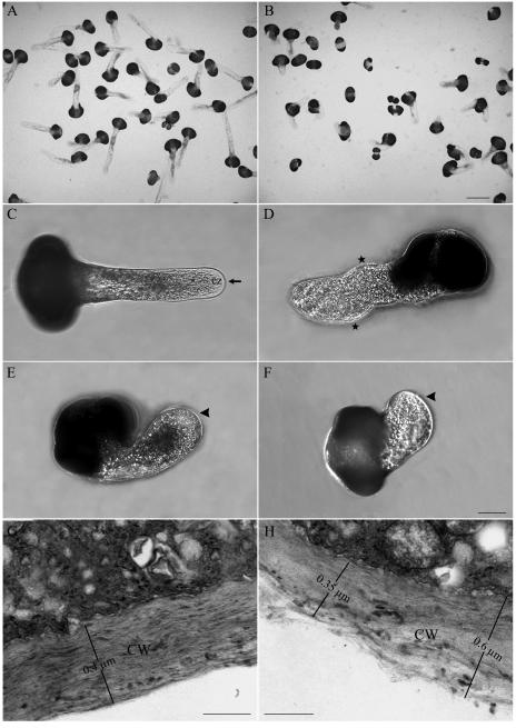
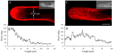
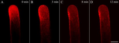
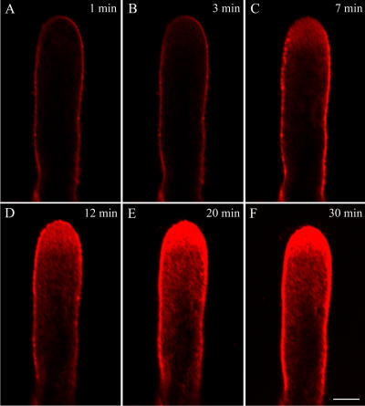
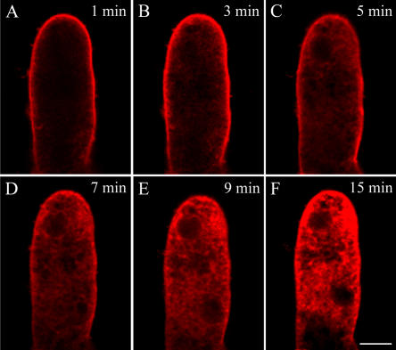
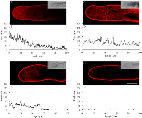
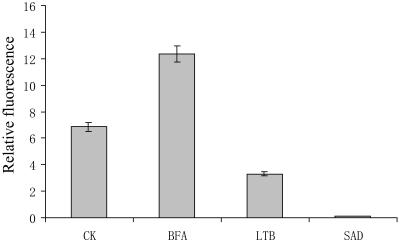
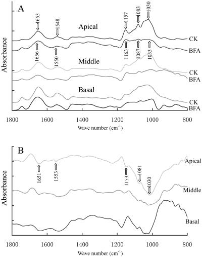
We assessed the effects of brefeldin A (BFA) on pollen tube development in Picea meyeri using fluorescent marker FM4-64 as a membrane-inserted endocytic/recycling marker, together with ultrastructural studies and Fourier transform infrared analysis of cell walls. BFA inhibited pollen germination and pollen tube growth, causing morphological changes in a dose-dependent manner, and pollen tube tip growth recovered after transferring into BFA-free medium. FM4-64 labeling showed typical bright apical staining in normally growing P. meyeri pollen tubes; this apical staining pattern differed from the V-formation pattern found in angiosperm pollen tubes. Confocal microscopy revealed that exocytosis was greatly inhibited in the presence of BFA. In contrast, the overall uptake of FM4-64 dye was about 2-fold that in the control after BFA (5 microg mL(-1)) treatment, revealing that BFA stimulated endocytosis in a manner opposite to the induced changes in exocytosis. Transmission electron microscopic observation showed that the number of secretory vesicles at the apical zone dramatically decreased, together with the disappearance of paramural bodies, while the number of vacuoles and other larger organelles increased. An acid phosphatase assay confirmed that the addition of BFA significantly inhibited secretory pathways. Importantly, Fourier transform infrared microspectroscopy documented significant changes in the cell wall composition of pollen tubes growing in the presence of BFA. These results suggest that enhanced endocytosis, together with inhibited secretion, is responsible for the retarded growth of pollen tubes induced by BFA.


Figures

Figure 1.
Figure 1.
Effects of BFA on pollen germination and pollen tube morphology. A, Healthy P. meyeri pollen tubes cultured in standard medium for 36 h, showing good germination and many long pollen tubes with normal shape. B, P. meyeri pollen tubes cultured in medium containing 5 μg mL−1 BFA for 36 h, showing poor germination and a few short tubes with morphological abnormalities. C, Micrograph of a control P. meyeri pollen tube cultured for 20 h, showing a regularly shaped pollen tube of constant diameter and a clear zone at apical region. D to F, Typical examples of pollen tubes in the presence of BFA for 20 h. D, Pollen tube incubated in 1 μg mL−1 BFA; stars indicate changes in growth direction (wavy pattern). E, Pollen tube incubated in 5 μg mL−1 BFA, showing a swelled tube tip (arrowhead). F, Most of the pollen grains cultured in 8 μg mL−1 BFA, showing aberrant protrusions, which were not regarded as germinated. G, Details of cell wall structure in control pollen tube. The cell wall was uniform, compact, and 0.4 μm thick. H, Details of cell wall structure in BFA-treated pollen tube. The cell wall became more loosely packed, showing the cell wall thickness from 0.35 to 0.6 μm in the presence of BFA. cz, Clear zone; CW, cell wall. Bars in A and B = 100 μm; in C to F = 50 μm; and in G and H = 0.3 μm.
Figure 2.
Figure 2.
Confocal images of FM4-64 staining in pollen tubes of P. meyeri. A, A median focal plane confocal optical section, showing a typical FM4-64 staining pattern in a growing pollen tube; the bright field at one-third size appears as an insert. B, Pixel values along a central transect through the fluorescence image in A. C, A median focal plane confocal optical section, showing a dispersed and disrupted FM4-64 staining pattern in BFA-treated (5 μg mL−1) pollen tube; the bright field at one-third size appears as an insert. D, Pixel values along a central transect through the fluorescence image in C.
Figure 3.
Figure 3.
Time course of disruption in a normal typical FM4-64 staining induced by 5 μg mL−1 BFA. BFA was applied directly to growing pollen tubes on thin gel layers in 70 μL of 115% to 120% liquid medium. A, A typical FM4-64 distribution pattern in a normal growing pollen tube of P. meyeri. B, The changes of FM4-64 staining at 3 min after the addition of BFA, showing the FM4-64 fluorescence tended to be scattered. C and D, The changed FM4-64 staining distribution with increasing time, showing FM4-64 fluorescence became more dispersed and almost distributed in the whole pollen tube after 12 min of BFA treatment. Bar = 25 μm.
Figure 4.
Figure 4.
FM4-64-uptake time course in a growing P. meyeri pollen tube. A to F, Median confocal fluorescence images at increasing times after addition of FM4-64 (2 μm in 115% standard medium). To avoid osmotic perturbation, pollen tubes were pretreated with 115% medium before dye application. The rapid uptake suggests an extremely high rate of endocytosis and membrane traffic; the bright region suggests there is an accumulation of secretory vesicles in the apical zone. Bar = 25 μm.
Figure 5.
Figure 5.
Time course of changes in dye distribution of FM4-64-loaded P. meyeri pollen tubes treated with 5 μg mL−1 BFA. A to F, Confocal fluorescence images of a BFA-treated pollen tube at different times, showing BFA treatment did not inhibit the internalization of FM4-64 dye through endocytosis but the dispersed FM4-64 distributing pattern indicating BFA inhibited secretory vesicle accumulation at tube tip. Bar = 25 μm.
Figure 6.
Figure 6.
Confocal images showing uptake of 2 μm FM4-64 into control and inhibitor-treated pollen tubes of P. meyeri within 12 min. All the pixel values did not include the peripheral region. A, A median focal plane confocal optical section of a control pollen tube; the bright field at one-third size appears as an insert. B, Pixel values along a central transect through the fluorescence image in A. C, A median focal plane confocal optical section of a pollen tube pretreated with 5 μg mL−1 BFA for 60 min; the bright field at one-third size appears as an insert. D, Pixel values along a central transect through the fluorescence image in C, showing that BFA stimulated the internalization of FM4-64 dye. E, A median focal plane confocal optical section of a pollen tube pretreated with 1 μm latrunculin B for 60 min; the bright field at one-third size appears as an insert. F, Pixel values along a central transect through the fluorescence image in E, showing latrunculin B inhibited the internalization of FM4-64 dye. G, A median focal plane confocal optical section of a pollen tube pretreated with 500 μm sodium azide for 60 min; the bright field at one-third size appears as an insert. H, Pixel values along a central transect through the fluorescence image in G, showing sodium azide blocked internalization of FM4-64 dye almost entirely. Bar = 25 μm.
Figure 7.
Figure 7.
FM4-64 internalization in control and inhibitor-treated pollen tubes showing that dye uptake is stimulated by BFA (5 μg mL−1) inhibited by latrunculin B (1 μm) and sodium azide (500 μm). Cell-associated FM4-64 fluorescence was quantified after a 12-min internalization of the dye and normalized to control pollen tubes labeling. Data shown are means ± sd and are representative of three experiments, each containing three individual measurements. CK, Control; LTB, latrunculin B; SAD, sodium azide.
Figure 8.
Figure 8.
Electron micrographs in control and BFA-treated (5 μg mL−1) pollen tubes. A, Tip region of a control pollen tube, showing the apical clear zone. B, Tip region of a BFA-treated pollen tube, showing the apical clear zone was occupied by many organelles, e.g. mitochondria and vacuoles. C, Magnified picture of a control pollen tube tip region, from where many vesicles could be observed; some of them were fusing with, or releasing from, the plasma membrane. D, Tip region of a BFA-treated pollen tube. No PBs and secretory vesicles could be observed, but many other types of vesicles and large organelles, such as mitochondria, vacuoles, and lipid bodies, appeared. E, Another apical zone in control pollen tube, indicating a typical PB (arrow). F, Golgi apparatus in control pollen tubes, showing the Golgi stacks contain four flattened cisternae with a distinct cis-trans polarity with numerous vesicles attached to them. G, Abnormal arrangements of the Golgi stacks in BFA-treated pollen tubes, indicating the disassembly of the Golgi apparatus accompanied by vesiculation of trans-side (arrow), less Golgi-derived vesicles, as well as the ER dilations (arrow). H, Another abnormal arrangement of Golgi apparatus induced by BFA treatment. Golgi cisternae appeared abnormally stacked and curved, engulfing some large vesicles. Bar = 2 μm in A and B; 1 μm in C and D; 0.5 μm in E; and 0.2 μm in F, G, and H. SV, Secretory vesicles; M, mitochondrion; V, vacuole; L, lipid body; G, Golgi apparatus; PB, paramural body.
Figure 9.
Figure 9.
A typical inhibitor experiment showing the effects of BFA on acPase activity (A), germination frequency (B), and mean tube length (C). Pollen grains were incubated in germination medium. After 12 h, BFA (5 μg mL−1) was added to one-half of the pollen culture (), whereas the other half served as a control (). All three parameters were determined every 6 h. The data points represent means with sds of three independent experiments.
Figure 10.
Figure 10.
FTIR spectra obtained from the apical region, middle region, and basal region of P. meyeri pollen tubes cultured for 20 h. A, FTIR spectra obtained from the tip, middle, and basal regions of pollen tubes cultured in standard medium (CK) or in medium containing 5 μg mL−1 BFA (BFA) revealed that BFA treatment induced displacements of the peaks or changes of absorbance. B, Difference spectra generated by digital subtraction of spectra CK from spectra BFA, showing that the content of proteins and polysaccharides decreased after BFA treatment and that the reduction in polysaccharide was most obvious in apical region.

References

    1. Baluška F, Hlavacka A, Šamaj J, Palme K, Robinson DG, Matoh T, McCurdy DW, Menzel D, Volkmann D (2002) F-actin-dependent endocytosis of cell wall pectins in meristematic root cells. Insights from Brefeldin A-induced compartments. Plant Physiol 130: 422–431 - PMC - PubMed
    1. Baluška F, Liners F, Hlavacka A, Schlicht M, Van Cutsem P, McCurdy D, Menzel D (2005) Cell wall pectins and xyloglucans are internalized into dividing root cells and accumulate within cell plates during cytokinesis. Protoplasma 225: 141–155 - PubMed
    1. Battey NH, James NC, Greenland AJ, Brownlee C (1999) Exocytosis and endocytosis. Plant Cell 11: 643–659 - PMC - PubMed
    1. Bolte S, Talbot C, Boutte Y, Catrice O, Read ND, Satiat-Jeunemaitre B (2004) FM-dyes as experimental probes for dissecting vesicle trafficking in living plant cells. J Microsc 214: 159–173 - PubMed
    1. Camacho L, Malhó R (2003) Endo/exocytosis in the pollen tube apex is differentially regulated by Ca2+ and GTPases. J Exp Bot 54: 83–92 - PubMed

Publication types

MeSH terms

LinkOut - more resources