. 2005 Dec;139(4):1677-91.
doi: 10.1104/pp.105.067140. Epub 2005 Nov 18.

At5PTase13 modulates cotyledon vein development through regulating auxin homeostasis

Affiliations

At5PTase13 modulates cotyledon vein development through regulating auxin homeostasis

Wen-Hui Lin et al. Plant Physiol. 2005 Dec.

Abstract

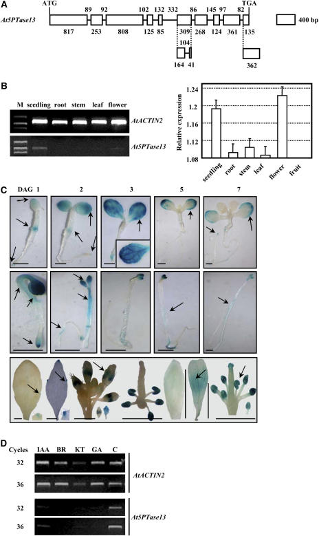
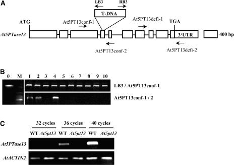
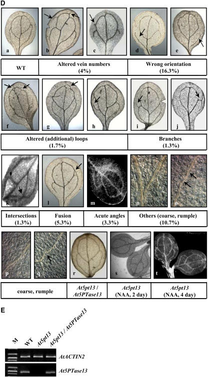
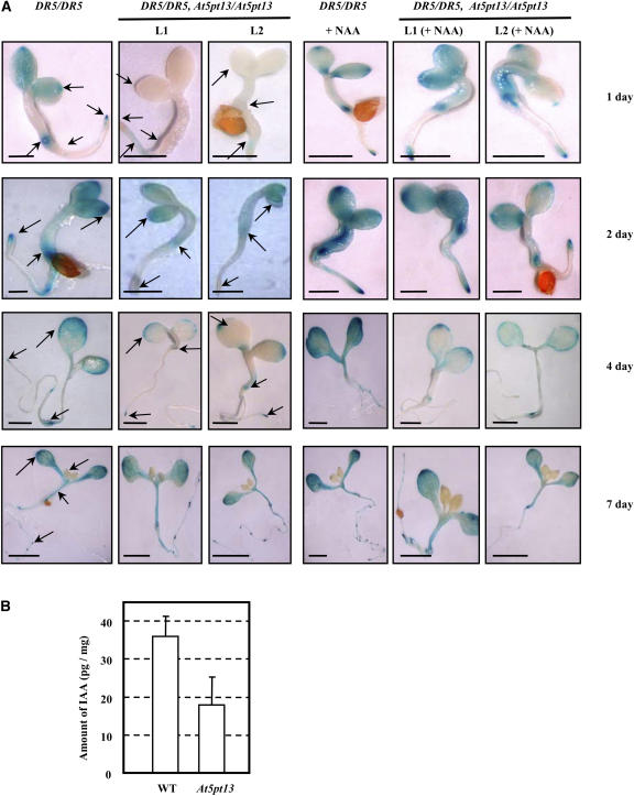
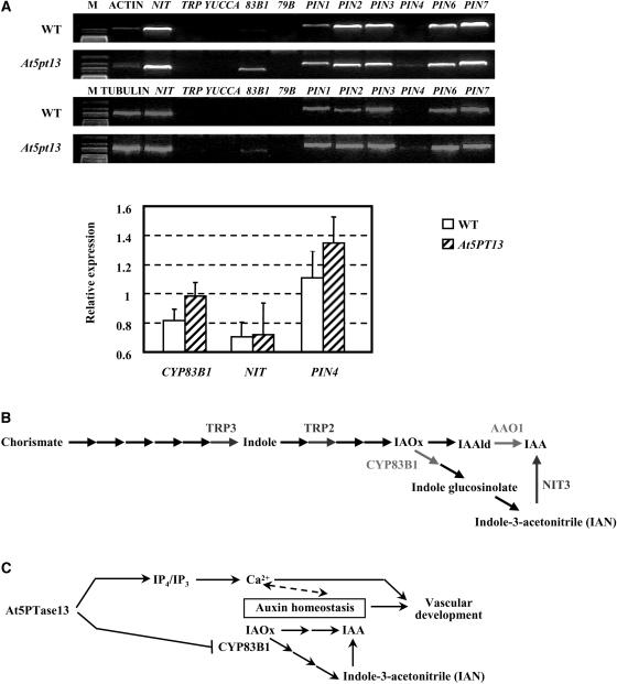
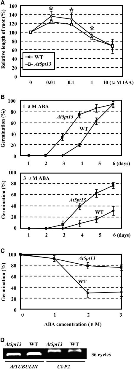
Phosphatidylinositol signaling pathway and the relevant metabolites are known to be critical to the modulation of different aspects of plant growth, development, and stress responses. Inositol polyphosphate 5-phosphatase is a key enzyme involved in phosphatidylinositol metabolism and is encoded by an At5PTase gene family in Arabidopsis thaliana. A previous study shows that At5PTase11 mediates cotyledon vascular development probably through the regulation of intracellular calcium levels. In this study, we provide evidence that At5PTase13 modulates the development of cotyledon veins through its regulation of auxin homeostasis. A T-DNA insertional knockout mutant, At5pt13-1, showed a defect in development of the cotyledon vein, which was rescued completely by exogenous auxin and in part by brassinolide, a steroid hormone. Furthermore, the mutant had reduced auxin content and altered auxin accumulation in seedlings revealed by the DR5:beta-glucuronidase fusion construct in seedlings. In addition, microarray analysis shows that the transcription of key genes responsible for auxin biosynthesis and transport was altered in At5pt13-1. The At5pt13-1 mutant was also less sensitive to auxin inhibition of root elongation. These results suggest that At5PTase13 regulates the homeostasis of auxin, a key hormone controlling vascular development in plants.


Figures

Figure 1.
Figure 1.
Structure and expression of At5PTase13. A, Gene organization of At5PTase13. Numbers represent the sizes of the exons and introns. The computational predictions for the sixth (one 104-bp intron was predicted) and last (the prediction is 227 bp longer) exons were incorrect (bottom section). B, Semiquantitative RT-PCR analysis shows that At5PTase13 is mainly expressed in young seedlings, with weak transcripts in flowers and almost no expression in roots, stems, and leaves (left section). Further analysis via quantitative real-time RT-PCR confirmed the relatively higher expression of At5PTase13 in young seedlings and flowers, while lower expression in fruit, roots, stems, and leaves (right section). Data presented are compared with AtACTIN2 and shown as percentage. Bars indicate sd. C, Promoter-reporter (GUS) fusion studies demonstrate the At5PTase13 expression in the tips of cotyledons, root tip, and hypocotyls-root juncture at day 1 after germination, and then expand to the whole cotyledon. At days 3 and 5, the expression was concentrated at the pinnacle of cotyledons. Later (days 5–7, when the first two pairs of true leaves appeared), the expression of At5PTase13 decreased and was focused in cotyledon margin and root tip. At5PTase13 exhibited stronger expression in seedlings grown in darkness (middle section) and is also detected in inflorescence leaves, petals, and pollen grains, but weakly in sepal and rosette leaves (bottom section). Arrows show the positions of At5PTase13 expression. A few expression patterns are highlighted in rectangles. Bar = 1 mm. D, Semiquantitative RT-PCR analysis revealed that At5PTase13 was down-regulated following treatment with different plant hormones. Seven-day-old seedlings were treated with 100 μm auxin (IAA), cytokinin (Kinetin), GA (GA3), or 1 μm 24-eBL for 8 h.
Figure 2.
Figure 2.
Identification and analysis of At5PTase13 deficiency mutants. A, A single T-DNA insertion in the fourth exon of At5PTase13 in the At5pt13-1 mutant. The positions of primers used to confirm T-DNA (At5PT13conf-1 and 2) in the At5PTase13 gene and deficiency of At5PTase13 (At5PT13defi-1 and 2) are indicated. B, Confirmation of T-DNA insertion (left section) and identification of homozygous lines (right section). T-DNA insertion was confirmed (lane 0) by PCR amplification using primers At5PT13conf-1 (located in the 5′ flanking region) and LB3. Homozygous lines were further identified via PCR amplification through primers At5PT13conf-1 and 2, which are located in the 5′ and 3′ flanked regions and show a positive signal only in the case of heterozygous lines (lanes 1, 2, and 4). C, Semiquantitative RT-PCR analysis with primers At5PT13defi-1 and 2 confirmed the absence of At5PTase13 expression in At5pt13-1. RNA was extracted from 4-d-old seedlings, and PCR amplifications were performed for 32, 36, and 40 cycles. Arabidopsis actin coding gene was employed as a positive internal control. D, Growth of At5pt13-1 seedlings. Compared to the cotyledon veins of wild-type seedlings (a, 4-d, mid-vein, and distal secondary vein developed), those of At5pt13-1 showed various abnormal patterns (b–q), including altered numbers of veins (b and c), improper orientation (d and e), additional loops (f–h), branches (i and j), intersections of the distal and proximal secondary veins (k), fusion of the distal and proximal secondary veins at one side (l), acute angles (m), and coarse (o and q, comparing to that of wild-type plants, n and p). The abnormalities are highlighted by arrows. Vascular tissues were observed using differential interference contrast microscopy. Expression of At5PTase13 in At5pt13-1 rescued the abnormal cotyledon vein development (r), as did the application of exogenous NAA (0.1 μm; s, 2-d-old, and t, 4-d-old seedlings). E, Expression of At5PTase13 was detected after transformation with the construct harboring At5PTase13 under its own promoter (pBI101-P-At5PTase13). RNAs were extracted from 4-d-old seedlings and PCR amplification was performed for 36 cycles.
Figure 2.
Figure 2.
Identification and analysis of At5PTase13 deficiency mutants. A, A single T-DNA insertion in the fourth exon of At5PTase13 in the At5pt13-1 mutant. The positions of primers used to confirm T-DNA (At5PT13conf-1 and 2) in the At5PTase13 gene and deficiency of At5PTase13 (At5PT13defi-1 and 2) are indicated. B, Confirmation of T-DNA insertion (left section) and identification of homozygous lines (right section). T-DNA insertion was confirmed (lane 0) by PCR amplification using primers At5PT13conf-1 (located in the 5′ flanking region) and LB3. Homozygous lines were further identified via PCR amplification through primers At5PT13conf-1 and 2, which are located in the 5′ and 3′ flanked regions and show a positive signal only in the case of heterozygous lines (lanes 1, 2, and 4). C, Semiquantitative RT-PCR analysis with primers At5PT13defi-1 and 2 confirmed the absence of At5PTase13 expression in At5pt13-1. RNA was extracted from 4-d-old seedlings, and PCR amplifications were performed for 32, 36, and 40 cycles. Arabidopsis actin coding gene was employed as a positive internal control. D, Growth of At5pt13-1 seedlings. Compared to the cotyledon veins of wild-type seedlings (a, 4-d, mid-vein, and distal secondary vein developed), those of At5pt13-1 showed various abnormal patterns (b–q), including altered numbers of veins (b and c), improper orientation (d and e), additional loops (f–h), branches (i and j), intersections of the distal and proximal secondary veins (k), fusion of the distal and proximal secondary veins at one side (l), acute angles (m), and coarse (o and q, comparing to that of wild-type plants, n and p). The abnormalities are highlighted by arrows. Vascular tissues were observed using differential interference contrast microscopy. Expression of At5PTase13 in At5pt13-1 rescued the abnormal cotyledon vein development (r), as did the application of exogenous NAA (0.1 μm; s, 2-d-old, and t, 4-d-old seedlings). E, Expression of At5PTase13 was detected after transformation with the construct harboring At5PTase13 under its own promoter (pBI101-P-At5PTase13). RNAs were extracted from 4-d-old seedlings and PCR amplification was performed for 36 cycles.
Figure 3.
Figure 3.
Altered auxin accumulation and distribution in At5PTase13-deficient plants. A, GUS activity detection of homozygous DR5:GUS/At5pt13-1 cross offsprings implies altered auxin levels and distribution in At5pt13-1 seedlings grown at 1, 2, and 4 d (two independent homozygous lines, L1 and 2) after germination. Adscititious NAA in low concentrations could enhance auxin levels in both DR5:GUS/At5pt13-1 and control plants. Bar = 4 mm. B, Quantitative assay of free auxin content in wild-type and At5pt13-1 seedlings. Four-day-old seedlings were used for ELISA analysis. The measurements were repeated four times and bars indicate sd.
Figure 4.
Figure 4.
The At5pt13 knockout mutation alters the expression of auxin biosynthesis- and transport-related genes. A, Semiquantitative RT-PCR analysis indicates increased expression of CYP83B1 and PIN4 in At5pt13-1. RNA was extracted from 4-d-old seedlings and PCR amplifications were performed using Arabidopsis actin or tubulin (locus nos. At3g18780 and At5g62690), respectively, as internal positive controls (bottom section). Further analysis via quantitative real-time RT-PCR confirmed the unaltered expression of NIT1 and stimulated expression of CYP83B1 and PIN4 (bottom section). Data presented are compared with AtACTIN2 and shown as percentage of the AtACTIN2 expression. Bars indicate sd. B, Summary of the genes involved in auxin homeostasis with altered transcripts by microarray analysis. TRP3, TRP2, and NIT3 were suppressed, and CYP83B1 and AAO1 were enhanced, under At5PTase13 deficiency. C, The hypothesized model of how At5PTase13 is involved in the cotyledon vein development through regulating auxin homeostasis and Ins(1,3,4,5)P4-related Ca2+. In normal conditions, At5PTase13 suppresses CYP83B1 and keeps higher IAA/indole-3-acetonitrile (IAN) homeostasis. In At5pt13-1, release of CYP83B1 leads to more IAN in vivo and lower IAA/IAN homeostasis. Ins(1,3,4,5)P4-related Ca2+ may interact with auxin homeostasis to modulate the vascular development.
Figure 5.
Figure 5.
At5pt13-1 is less sensitive to auxin and ABA. A, Lengths of the primary roots of 7-d-old wild-type and At5pt13-1 seedlings on the medium supplemented with gradient auxin (0, 0.01, 0.1, 1, or 10 μm) were measured and the relative ratios of growth promoting and restraining were calculated. Error bars indicate sd, and the asterisk indicates the significant difference (P < 0.01) by one-tailed Student's t test. B, Seed germination for both wild type and At5pt13-1 was assayed on the medium supplemented with 1 μm (top section) or 3 μm ABA (bottom section) over a 6-d period. Error bars indicate sd. C, ABA dosage effects on seed germination for both wild type and At5pt13-1. Squares, At5pt13-1; circles, wild type. Error bars indicate sd. D, Semiquantitative RT-PCR analysis indicates no changes in CVP2 transcript levels in At5pt13-1. RNA was extracted from 4-d-old seedlings and PCR amplifications were performed for 36 cycles using tubulin (locus no. At5g62690) as internal positive control.

References

    1. Alfandari D, Darribere T (1994) A simple PCR method for screening cDNA libraries. PCR Methods Appl 4: 46–49 - PubMed
    1. Bak S, Tax FE, Feldmann KA, Galbraith DW, Feyereisen R (2001) CYP83B1, a cytochrome P450 at the metabolic branch point in auxin and indole glucosinolate biosynthesis in Arabidopsis. Plant Cell 13: 101–111 - PMC - PubMed
    1. Bartel B, LeClere S, Magidin M, Zolman BK (2001) Inputs to the active indole-3-acetic acid pool: de novo synthesis, conjugate hydrolysis, and indole-3-butyric acid β-oxidation. J Plant Growth Regul 20: 198–216
    1. Berdy SE, Kudla J, Gruissem W, Gillaspy GE (2001) Molecular characterization of At5PTase1, an inositol phosphatase capable of terminating inositol trisphosphate signaling. Plant Physiol 126: 801–810 - PMC - PubMed
    1. Burnette RN, Gunesekera BM, Gillaspy GE (2003) An Arabidopsis inositol 5-phosphatase gain-of-function alters abscisic acid signaling. Plant Physiol 132: 1011–1019 - PMC - PubMed

Publication types

MeSH terms

Associated data