. 2005 Dec;73(12):8209-18.
doi: 10.1128/IAI.73.12.8209-8218.2005.

Anaplasma phagocytophilum delay of neutrophil apoptosis through the p38 mitogen-activated protein kinase signal pathway

Affiliations

Anaplasma phagocytophilum delay of neutrophil apoptosis through the p38 mitogen-activated protein kinase signal pathway

Kyoung-Seong Choi et al. Infect Immun. 2005 Dec.

Abstract

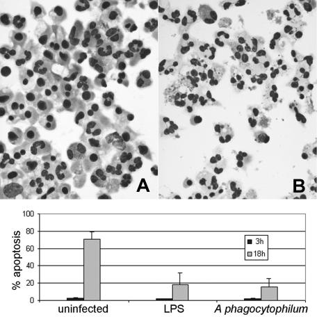
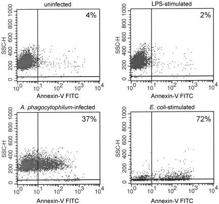
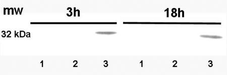
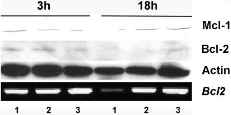
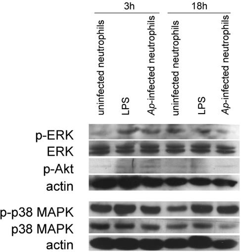
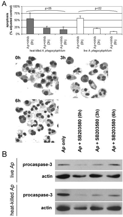
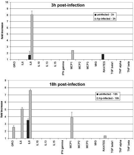
Human granulocytic anaplasmosis is caused by the obligate intracellular bacterium Anaplasma phagocytophilum. The bacterium avoids host innate defenses in part by infecting, surviving in, and propagating in neutrophils, as well as by inhibiting neutrophil apoptosis. However, the mechanisms of A. phagocytophilum survival in neutrophils and the inhibition of spontaneous apoptosis are not well understood. In this study, we demonstrated that antiapoptotic Mcl-1 protein (Bcl-2 family) expression is maintained and that inhibition of procaspase-3 processing occurs in A. phagocytophilum-infected human neutrophils. An evaluation of p38 mitogen-activated protein kinase (MAPK) showed evidence of increased phosphorylation with infection. Moreover, antagonism of p38 MAPK by the inhibitor SB203580 reversed apoptosis inhibition in live or heat-killed A. phagocytophilum-infected neutrophils. A role for the autocrine or paracrine production of antiapoptotic interleukin 8 (IL-8) expressed with A. phagocytophilum infection was excluded by the use of IL-8-, IL-8R1 (CXCR1)-, and IL-8R2 (CXCR2)-blocking antibodies. As previously demonstrated, the antiapoptotic effect was initially mediated by exposure to A. phagocytophilum components in heat-killed bacteria. However, an important role for active infection is demonstrated by the additional delay in apoptosis with intracellular growth and the refractory abrogation of this response by the p38 MAPK inhibitor 3 to 6 h after neutrophil infection. These results suggest that the initial activation of the p38 MAPK pathway leading to A. phagocytophilum-delayed neutrophil apoptosis is bypassed with active intracellular infection. Moreover, active intracellular infection contributes more to the overall delay in apoptosis than do components of heat-killed A. phagocytophilum alone.


Figures

FIG. 1.
FIG. 1.
Morphological appearance of human neutrophils incubated in the presence and absence of A. phagocytophilum. Infected and uninfected neutrophils were cultivated in vitro for 18 h, and cytocentrifuged preparations were Romanowsky stained. A, Uninfected neutrophils exhibit morphological features of apoptosis. B, A. phagocytophilum-infected neutrophils show only a small proportion of cells with an apoptotic morphology.
FIG. 2.
FIG. 2.
Annexin-V apoptosis assays of A. phagocytophilum-infected neutrophils. Neutrophils were cultivated with or without LPS or cell-free A. phagocytophilum; cells were examined by flow cytometry. Annexin-V staining analyzed at 3 h shows early expression of phosphatidyl serine, a membrane marker correlated with apoptosis.
FIG. 3.
FIG. 3.
Inhibition of procaspase-3 processing in A. phagocytophilum-infected neutrophils. Apoptosis results with procaspase-3 cleavage into active caspase-3 in neutrophils after 3 and 18 h of exposure to medium only (lane 1) or LPS (lane 2), compared with A. phagocytophilum infection (lane 3). A representative immunoblot from five replicated experiments is shown.
FIG. 4.
FIG. 4.
Mcl-1 and Bcl-2 expression and bcl-2 transcription are maintained in A. phagocytophilum-infected neutrophils. Neutrophils cultured for 3 and 18 h in medium only (lane 1) rapidly lose Mcl-1 and Bcl-2 expression and bcl-2 family transcription (lane 1) compared to stimulation by LPS (lane 2) or infection with A. phagocytophilum (lane 3). Representative immunoblots and RT-PCRs from three experiments are shown.
FIG. 5.
FIG. 5.
p38 MAPK, but not ERK or Akt, are phosphorylated in A. phagocytophilum (Ap)-infected neutrophils. p38 MAPK was consistently found phosphorylated with A. phagocytophilum infection of neutrophils or LPS controls. Increased phosphorylation of neither ERK nor Akt was detected. The results are representative of three independent experiments. p-ERK, p-Akt, and P-p38, phosphorylated ERK, Akt, and p38, respectively.
FIG. 6.
FIG. 6.
p38 MAPK inhibitor SB203580 reverses the inhibition of neutrophil apoptosis by A. phagocytophilum. A, Apoptotic cells were identified by morphology upon light microscopy (Romanowsky stains; original magnification, ×260), and the percentages of apoptotic cells were calculated; 100% was set at the level of uninfected neutrophils, and 0% was set at the level of untreated A. phagocytophilum (Ap)-infected neutrophils. Note that the antiapoptotic effect was reversed when infected cells were treated with inhibitor at 0 h, regardless of infection with live bacteria or exposure to heat-killed bacteria. In contrast, apoptosis inhibition was reversed in neutrophils with live A. phagocytophilum to a significantly lower degree when p38 MAPK inhibitor was added at 3 or 6 h postinfection (P < 0.02), whereas neutrophils exposed to heat-killed bacteria showed no increased antiapoptotic effect with inhibitor added at these times (P > 0.05). B, As anticipated from changes in morphological apoptosis, p38 MAPK inhibitor promoted procaspase-3 cleavage at 0 h, but less so at 3 and 6 h.
FIG. 7.
FIG. 7.
A. phagocytophilum-infected neutrophils express IL-8, CCL, and CXCL chemokines. Neutrophils were incubated for 3 h and 18 h in medium alone or with A. phagocytophilum. IL-8, CCL, CXCL chemokine, and cytokine expression in culture supernatants was detected by solid-phase antibody capture and chemiluminescence; results are expressed as increases (n-fold) above background (medium alone). GRO, growth-related oncogene; MIG, monokine induced by gamma interferon; TGF, transforming growth factor.

References

    1. Aga, E., D. M. Katschinski, G. van Zandbergen, H. Laufs, B. Hansen, K. Muller, W. Solbach, and T. Laskay. 2002. Inhibition of the spontaneous apoptosis of neutrophil granulocytes by the intracellular parasite Leishmania major. J. Immunol. 169:898-905. - PubMed
    1. Akgul, C., D. A. Moulding, and S. W. Edwards. 2001. Molecular control of neutrophil apoptosis. FEBS Lett. 487:318-322. - PubMed
    1. Alvarado-Kristensson, M., F. Melander, K. Leandersson, L. Ronnstrand, C. Wernstedt, and T. Andersson. 2004. p38-MAPK signals survival by phosphorylation of caspase-8 and caspase-3 in human neutrophils. J. Exp. Med. 199:449-458. - PMC - PubMed
    1. Alvarado-Kristensson, M., M. I. Porn-Ares, S. Grethe, D. Smith, L. Zheng, and T. Andersson. 2002. p38 mitogen-activated protein kinase and phosphatidylinositol 3-kinase activities have opposite effects on human neutrophil apoptosis. FASEB J. 16:129-131. - PubMed
    1. Aoshiba, K., S. Yasui, M. Hayashi, J. Tamaoki, and A. Nagai. 1999. Role of p38-mitogen-activated protein kinase in spontaneous apoptosis of human neutrophils. J. Immunol. 162:1692-1700. - PubMed

Publication types

MeSH terms