. 2005 Dec;73(12):8275-81.
doi: 10.1128/IAI.73.12.8275-8281.2005.

Direct inhibition of T-lymphocyte activation by anthrax toxins in vivo

Affiliations

Direct inhibition of T-lymphocyte activation by anthrax toxins in vivo

Jason E Comer et al. Infect Immun. 2005 Dec.

Abstract

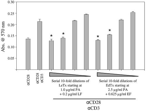
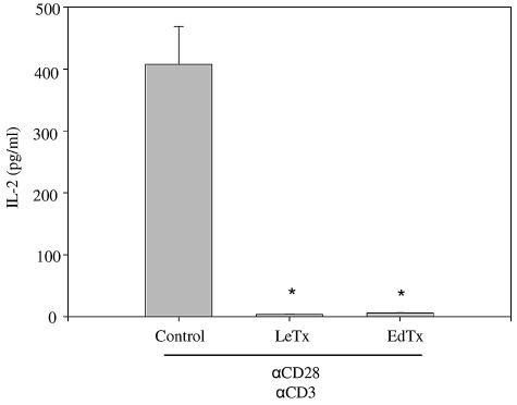
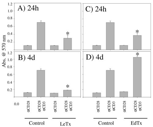
The causative agent of anthrax, Bacillus anthracis, produces two toxins that contribute in part to its virulence. Lethal toxin is a metalloprotease that cleaves upstream mitogen-activated protein kinase kinases. Edema toxin is a calmodulin-dependent adenylate cyclase. Previous studies demonstrated that the anthrax toxins are important immunomodulators that promote immune evasion of the bacterium by suppressing activation of macrophages and dendritic cells. Here we showed that injection of sublethal doses of either lethal or edema toxin into mice directly inhibited the subsequent activation of T lymphocytes by T-cell receptor-mediated stimulation. Lymphocytes were isolated from toxin-injected mice after 1 or 4 days and stimulated with antibodies against CD3 and CD28. Treatment with either toxin inhibited the proliferation of T cells. Injection of lethal toxin also potently inhibited cytokine secretion by stimulated T cells. The effects of edema toxin on cytokine secretion were more complex and were dependent on the length of time between the injection of edema toxin and the isolation of lymphocytes. Treatment with lethal toxin blocked multiple kinase signaling pathways important for T-cell receptor-mediated activation of T cells. Phosphorylation of the extracellular signal-regulated kinase and the stress-activated kinase p38 was significantly decreased. In addition, phosphorylation of the serine/threonine kinase AKT and of glycogen synthase kinase 3 was inhibited in T cells from lethal toxin-injected mice. Thus, anthrax toxins directly act on T lymphocytes in a mouse model. These findings are important for future anthrax vaccine development and treatment.


Figures

FIG. 1.
FIG. 1.
Proliferation of CD4+ T cells in the presence of LeTx or EdTx. CD4+ T cells were isolated from female BALB/c mice and stimulated with anti-CD3 (αCD3) and anti-CD28 (αCD28) antibodies in the presence of 10-fold serial dilutions of LeTx (starting at 1 μg/ml PA and 0.2 μg/ml of LF) or EdTx (starting at 2.5 μg/ml of PA and 0.625 μg/ml of EF). After 48 h of incubation at 37°C and 5% CO2, cellular proliferation was measured using the MTT cell proliferation assay described above. Bars represent means of triplicates ± standard errors. The data are from one experiment representative of three independent repetitions. Asterisks denote a statistically significant difference between untreated and toxin-treated cells (P < 0.05 by Dunnett's test). Abs., absorbance.
FIG. 2.
FIG. 2.
Secretion of IL-2 by CD4+ T cells in the presence of LeTx or EdTx. Isolated CD4+ T cells were incubated with anti-CD28 (αCD28) and anti-CD3 (αCD3) in the presence of LeTx (1 μg/ml PA and 0.2 μg/ml of LF) or EdTx (2.5 μg/ml of PA and 0.625 μg/ml of EF). After 48 h of incubation at 37°C and 5% CO2, IL-2 was measured in culture supernatants. Bars represent means of triplicates ± standard errors. The data are from one experiment representative of three independent repetitions. Asterisks denote a statistically significant difference between untreated and toxin-treated cells (P < 0.05 by the Tukey test).
FIG. 3.
FIG. 3.
Cellular proliferation of T cells isolated from mice injected with LeTx or EdTx. Female BALB/c mice were injected with 100 μg of PA and 7.5 μg of LF or EF. Total lymphocytes were isolated after 24 h (A and C) or 4 days (B and D). T cells were incubated with anti-CD28 (αCD28) and anti-CD3 (αCD3) antibodies for 48 h at 37°C and 5% CO2. Proliferation was measured using the MTT cell proliferation assay described above. Bars represent means of triplicates ± standard errors. The data are from one experiment representative of three independent repetitions. Asterisks indicate a statistically significant difference between stimulated cells from untreated and toxin-injected mice (P < 0.05 by Student's t test).
FIG. 4.
FIG. 4.
(A) Phosphorylation of proteins defining various signaling pathways in T-cell receptor-stimulated T cells isolated from toxin-injected mice. Mice were injected with 100 μg of PA and 7.5 μg of LF or EF, and lymphocytes were isolated 24 h later. The T cells were stimulated with anti-CD3 antibody and cross-linked with a secondary antibody for 5 min. The cells were lysed, and phosphorylation (p-) of ERK 1 and 2, p38, ATF-2, AKT, GSK-3α and β, and JNK was measured. Values are given as relative fluorescence units, and bars represent means of duplicates ± standard errors. The data are from one experiment representative of three independent repetitions. Asterisks indicate a statistically significant difference (P < 0.05 by the Tukey test). (B) The presence of equal amounts of protein in the samples was ascertained by Western blotting with antibodies against beta-actin as a probe.

References

    1. Aandahl, E. M., W. J. Moretto, P. A. Haslett, T. Vang, T. Bryn, K. Tasken, and D. F. Nixon. 2002. Inhibition of antigen-specific T cell proliferation and cytokine production by protein kinase A type I. J. Immunol. 169:802-808. - PubMed
    1. Agrawal, A., J. Lingappa, S. H. Leppla, S. Agrawal, A. Jabbar, C. Quinn, and B. Pulendran. 2003. Impairment of dendritic cells and adaptive immunity by anthrax lethal toxin. Nature 424:329-334. - PubMed
    1. Bradley, K. A., J. Mogridge, M. Mourez, R. J. Collier, and J. A. Young. 2001. Identification of the cellular receptor for anthrax toxin. Nature 414:225-229. - PubMed
    1. Chang, L., and M. Karin. 2001. Mammalian MAP kinase signalling cascades. Nature 410:37-40. - PubMed
    1. Collier, R. J. 1999. Mechanism of membrane translocation by anthrax toxin: insertion and pore formation by protective antigen. J. Appl. Microbiol. 87:283. - PubMed

Publication types

LinkOut - more resources