. 2006 Jan;147(2):175-82.
doi: 10.1038/sj.bjp.0706472.

Inflammatory events in a vascular remodeling model induced by surgical injury to the rat carotid artery

Affiliations

Inflammatory events in a vascular remodeling model induced by surgical injury to the rat carotid artery

Barbara Rinaldi et al. Br J Pharmacol. 2006 Jan.

Abstract

1.--The aim of our study was to gain insight into the molecular and cellular mechanisms of the inflammatory response to arterial injury in a rat experimental model. 2.--Rats (five for each experimental time) were subjected to brief clamping and longitudinal incision of a carotid artery and monitored for 30 days. Subsequently, Nuclear Factor-kappaB (NF-kappaB) expression was measured by electrophoretic mobility shift assay. Heat shock protein (HSP) 27, HSP47 and HSP70 were evaluated by Western blot. Morphological changes of the vessel wall were investigated by light and electron microscopy. 3.--In injured rat carotid artery NF-kappaB activity started immediately upon injury, and peaked between 2 and 3 weeks later. Western blot showed a significant increase of HSP47 and HSP70 7 days after injury. At 2 weeks postinjury, HSP27 expression peaked. Light microscopy showed a neointima formation, discontinuity of the media layer and a rich infiltrate. Among infiltrating cells electron microscopy identified dendritic-like cells in contact with lymphocytes. 4.--Our model of surgical injury induces a significant inflammatory process characterized by enhanced NF-kappaB activity and HSPs hyperexpression. Dendritic-like cells were for the first time identified as a novel component of tissue repair consequent to acute arterial injury.


Figures

Figure 1
Figure 1
Kinetic analysis of NF-κB activation and characterization of NF-κB complex. Representative EMSA (a) as well as densitometric analysis (b) show NF-κB/DNA binding activity in nuclear extracts from uninjured (NT) and injured (T) carotid arteries. Nuclear extracts from NT and T carotid arteries were prepared as described in the text and incubated with 32P-labeled NF-κB probe. Data in (a) are from a single experiment (from three to five carotids) and representative of three separate experiments. Data in (b) are expressed as mean±s.e.m. of three experiments. °P<0.01 vs NT carotids at time zero; *P<0.01 vs NT carotid for each time point. Characterization of NF-κB/DNA complex in nuclear extracts of injured rat carotid arteries at 24 h (c). In competition reaction nuclear extracts were incubated with radiolabeled NF-κB probe in the absence or presence of: identical but unlabeled oligonucleotides (W.T., 50 ×), mutated non-functional NF-κB probe (Mut., 50 ×) or unlabeled oligonucleotide containing the consensus sequence for Sp-1 (Sp-1, 50 ×). In supershift experiments nuclear extracts were incubated with antibodies against p50, p65, p50+p65, 15 min before incubation with radiolabeled NF-κB probe. Data illustrated are from a single experiment and are representative of three separate experiments.
Figure 2
Figure 2
Kinetic analysis of HSP70 expression in injured (T) and uninjured (NT) carotids. The samples obtained from carotids at different times after injury or from NTs were analyzed by Western blot assay using specific monoclonal antibody as indicated in Material and methods. Results are expressed in arbitrary densitometric units as compared to the protein expression level in NT. (a) Representative Western blot for HSP 70 expression. (b) The relative amount of HSP70 was quantified by scanning densitometry and normalized to that of β-actin. Each column represents the mean±s.e.m. of five independent experiments for each time point. *P<0.05 vs NT.
Figure 3
Figure 3
Kinetic analysis of HSP27 expression in injured (T) and uninjured (NT) carotids. The experiments were performed as described in Figure 2. (a) Representative Western blot for HSP 27 expression. (b) Each column represents the mean±s.e.m. of five independent experiments for each time point. *P<0.05 vs NT.
Figure 4
Figure 4
Kinetic analysis of HSP47 expression in injured (T) and uninjured (NT) carotids. The experiments were performed as described in Figure 2. (a) Representative Western blot for HSP 47 expression. (b) Each column represents the mean±s.e.m. of five independent experiments for each time point. *P<0.05 vs NT; #P<0.01 vs NT.
Figure 5
Figure 5
Light microscopy of a vessel wall 7 days after injury. Epoxy resin section, stained with alkaline toluidine blue, shows media discontinuity, neointima formation and cell infiltration which starting from the neointima goes through media and adventitia layers. The cells within the squared inset has been observed at the electron microscopy (see Figure 6). Field is representative of three independent experiments. L=vessel lumen, Bar=100 μm.
Figure 6
Figure 6
The picture shows the ultrastructure of the border between media and adventitia layers (squared insert of Figure 5) of a carotid artery 7 days after injury: dendritic-like cells (D), organized in a mesh and in contact with lymphocytes (L), are observed in the inner part of the neointima, within the media and adventitia. Field is representative of three independent experiments. Bar=5 μm.
Figure 7
Figure 7
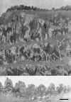
Untreated carotid artery of a rat 7 days after injury, with normal appearance at electron microscopy. The mature endothelium with its thin basement membrane attached to the inner elastic lamina is enlarged in the bottom panel. Field is representative of three independent experiments. Bar=7 μm for the top panel and 2 μm for the bottom panel.
Figure 8
Figure 8
A cluster of dendritic cells expressing MHC-II membrane antigens is shown deep in the neointima of a carotid artery 7 days upon injury. Field is representative of three independent experiments. Immunofluorescence, Bar=20 μm.

References

    1. BAILEY A.S., FLEMING W.H. Converging roads: evidence for an adult hemangioblast. Exp. Hematol. 2003;31:987–993. - PubMed
    1. BHATT D.L. Inflammation and Restenosis: is there a link. Am. Heart J. 2004;147:945–947. - PubMed
    1. CERCEK B., YAMASHITA M., DIMAYUGA P., ZHU J., FISHBEIN M.C., KAUL S., SHAH P.K., NILSSON J., REGNSTROM J. Nuclear factor-kappaB activity and arterial response to balloon injury. Atherosclerosis. 1997;131:59–66. - PubMed
    1. CHEN Y., ROSS B.M., CURRIE R.W. Heat shock treatment protects against angiotensin II-induced hypertension and inflammation in aorta. Cell Stress Chaperones. 2004;9:99–107. - PMC - PubMed
    1. CIRRINCIONE C., PIMPINELLI N., ORLANDO L., ROMAGNOLI P. Lamina propria dendritic cells express activation markers and contact lymphocytes in chronic adult periodontitis. J. Periodontol. 2002;73:46–53. - PubMed

Publication types

MeSH terms