. 2005 Nov;1(5):e67.
doi: 10.1371/journal.pgen.0010067. Epub 2005 Nov 18.

All paired up with no place to go: pairing, synapsis, and DSB formation in a balancer heterozygote

Affiliations

All paired up with no place to go: pairing, synapsis, and DSB formation in a balancer heterozygote

Wei J Gong et al. PLoS Genet. 2005 Nov.

Abstract

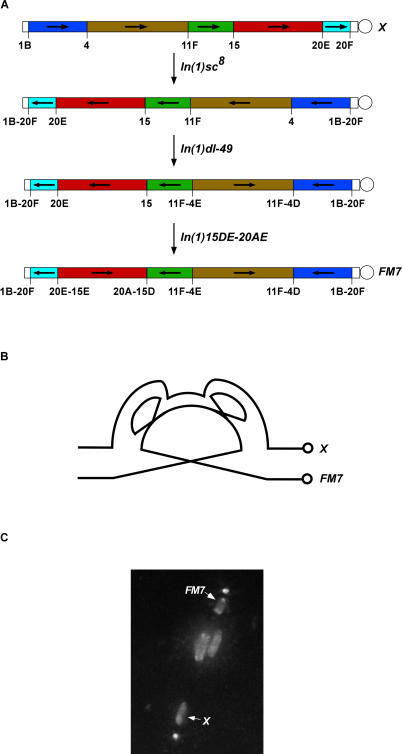
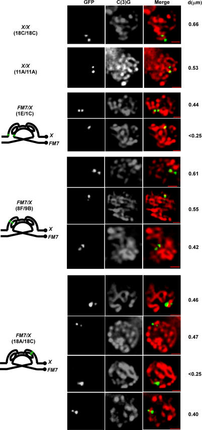
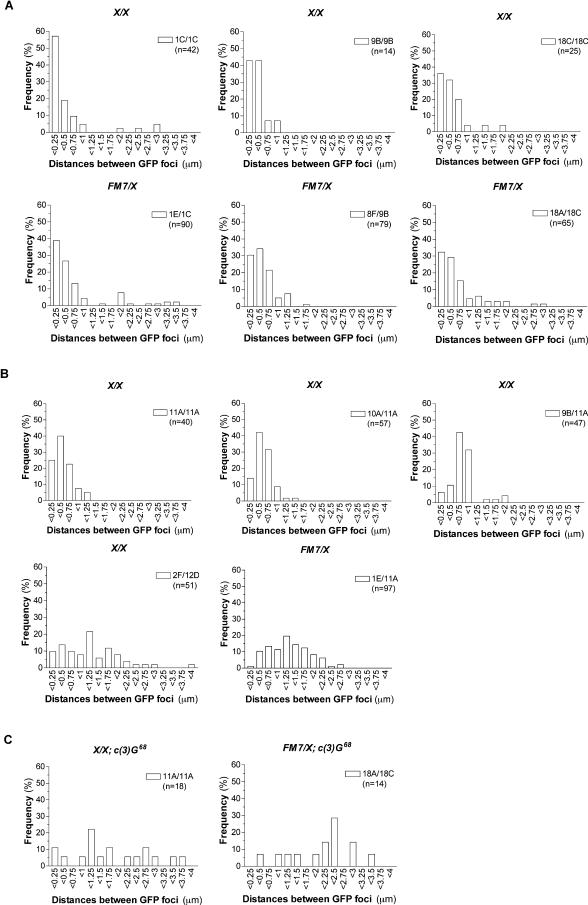
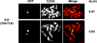
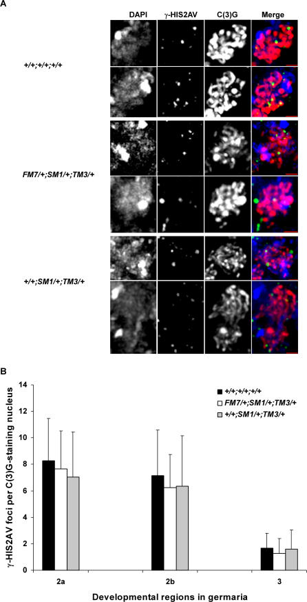
The multiply inverted X chromosome balancer FM7 strongly suppresses, or eliminates, the occurrence of crossing over when heterozygous with a normal sequence homolog. We have utilized the LacI-GFP: lacO system to visualize the effects of FM7 on meiotic pairing, synapsis, and double-strand break formation in Drosophila oocytes. Surprisingly, the analysis of meiotic pairing and synapsis for three lacO reporter couplets in FM7/X heterozygotes revealed they are paired and synapsed during zygotene/pachytene in 70%-80% of oocytes. Moreover, the regions defined by these lacO couplets undergo double-strand break formation at normal frequency. Thus, even complex aberration heterozygotes usually allow high frequencies of meiotic pairing, synapsis, and double-strand break formation in Drosophila oocytes. However, the frequencies of failed pairing and synapsis were still 1.5- to 2-fold higher than were observed for corresponding regions in oocytes with two normal sequence X chromosomes, and this effect was greatest near a breakpoint. We propose that heterozygosity for breakpoints creates a local alteration in synaptonemal complex structure that is propagated across long regions of the bivalent in a fashion analogous to chiasma interference, which also acts to suppress crossing over.


Conflict of interest statement

Competing interests. The authors have declared that no competing interests exist.

Figures

Figure 1
Figure 1. The Multiply Inverted Balancer Chromosome FM7
(A) Schematic diagram of the generation of chromosomal inversions from X to FM7. Color represents the regions that are involved in inversions, and arrows are used to indicate the orientation of each region with respect to the centromere. Circles represent the centromeres. The numbers below each structure represent positions of the standard polytene map. (B) A hypothetical structure displaying the pairing relations of the two homologs in an FM7/X heterozygote. (C) A picture of DAPI-stained chromosome at meiotic metaphase I in FM7/X oocytes (courtesy of W. Gilliland). The two X and 4th chromosomes are positioned between the two autosomal bivalents and the poles, with the dot-like 4th chromosomes located closer to the poles. The FM7 chromosome (denoted by the brighter DAPI staining at its tip) is located between the autosomes and the upper pole, while the normal sequence X chromosome lies in the lower half of the spindle. Note that the X and FM7 chromosomes are not connected by chiasmata.
Figure 2
Figure 2. Synapsed lacO Foci Flanking Stretches of SC in FM7/X or X/X Oocytes
In both X/X and FM7/X oocytes, paired lacO foci flank regions of SC. The positions of paired/synapsed lacO sites studied in X/X and FM7/X oocytes in zygotene/pachytene are shown at the left-most of each row. One to three optical sections show two partially overlapping or adjacent GFP foci (green) associated with a segment of SC (red). Distances between those GFP foci are shown at the right-most in each row. Due to the difficulty of accurately measuring the distance between overlapping foci, “<0.25” is used. Bars = 1 μm.
Figure 3
Figure 3. Distributions of Distances between GFP Foci in Both X/X and FM7/X Oocytes
These data report, in histogram form, the distribution of distances between GFP foci in those oocytes with two distinct GFP foci (including those oocytes with overlapping foci). For those oocytes with overlapping foci, the distance is measured as < 0.25 μm. (A) The distribution of distances in oocytes containing two lacO insertions at allelic sites in X/X females (upper panel) and nearby lacO couplets in FM7/X oocytes (lower panel). (B) The distribution of distances for non-allelic lacO insertion sites in X/X and FM7/X females. The distribution for an allelic pair of lacO insertions at 11A is provided as a control. (C) The distribution of distances in oocytes containing two lacO insertions at an allelic site (11A) in X/X; c(3)G females (left) and a nearby lacO couplet (18A/18C) in FM7/X ; c(3)G oocytes (right panel).
Figure 4
Figure 4. Synapsed and Unsynapsed lacO Sites in X/X (10A/11A) Oocytes
Note that in the lower (unsynapsed) case, the two GFP foci are displaced along the length of the same stretch of SC. In these images, which consist of one to two optical sections, two GFP foci (green) are associated with a segment of SC (red). Distances between those GFP foci are shown at the right-most in each row. Bars = 1 μm.
Figure 5
Figure 5. Unpaired lacO Sites in c(3)G Mutant Oocytes for Both X/X and FM7/X Oocytes
Cytoplasmic protein marker ORB was used to identify the 16-cell cysts. It is present from region 2a, where it is evenly distributed in the 16 cells. At region 2b and region 3, ORB concentrates in the pro-oocytes and the oocyte. The GFP foci (green) in merge images in conjunction with DNA (blue, DAPI staining) and ORB (red). Two optical sections are shown in wildtype, while six to seven optical sections are shown in c(3)G mutants. Bars = 1 μm.
Figure 6
Figure 6. DSB Formation on the Same Stretch of SC As Paired lacO Couplets in FM7/X and X/X Oocytes
The representatives from FM7/X (18A/18C) and X/X (18C/18C) oocytes are shown in this figure. Single C(3)G-positive (red) meiotic cells are shown in each row. DSBs are indicated by γ-HIS2AV staining (blue, arrows). GFP foci (green, arrows) represent the pairing FM7 and X or X and X chromosomes. Two to three optical sections of images were projected. Bars = 1 μm.
Figure 7
Figure 7. DSBs in Oocytes Heterozygous for Three Balancer Chromosomes
(A) Representatives from the oocytes with normal or heterozygous balancer chromosomes. Maximum intensity projection of image stacks of nuclei showing C(3)G (red) in conjunction with DNA (blue, DAPI staining) and DSB (green, γ-HIS2AV staining). Bars = 1 μm. (B) Distribution graph representing the number of γ-HIS2AV foci per C(3)G staining nucleus at different developmental regions in germaria. The sample size of oocytes in wildtype is 64, 34, and 9, representing region 2a, 2b, and 3; in FM7/+;SM1/+;TM3/+, 48, 43, and 11; in +/+;SM1/+;TM3/+, 66, 38, and 10, respectively. Data are presented as means ± SD.

References

    1. Sherizen D, Jang JK, Bhagat R, Kato N, McKim KS. Meiotic recombination in Drosophila females depends on chromosome continuity between genetically defined boundaries. Genetics. 2005;169:767–781. - PMC - PubMed
    1. Vazquez J, Belmont AS, Sedat JW. The dynamics of homologous chromosome pairing during male Drosophila meiosis. Curr Biol. 2002;12:1473–1483. - PubMed
    1. Hiraoka Y, Dernburg AF, Parmelee SJ, Rykowski MC, Agard DA, et al. The onset of homologous chromosome pairing during Drosophila melanogaster embryogenesis. J Cell Biol. 1993;120:591–600. - PMC - PubMed
    1. Roeder GS. Meiotic chromosomes: It takes two to tango. Genes Dev. 1997;11:2600–2621. - PubMed
    1. Weiner BM, Kleckner N. Chromosome pairing via multiple interstitial interactions before and during meiosis in yeast. Cell. 1997;77:977–991. - PubMed

Publication types

Substances