. 2006 Jan;4(1):e6.
doi: 10.1371/journal.pbio.0040006.

Functional amyloid formation within mammalian tissue

Affiliations

Functional amyloid formation within mammalian tissue

Douglas M Fowler et al. PLoS Biol. 2006 Jan.

Abstract

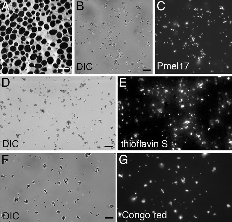
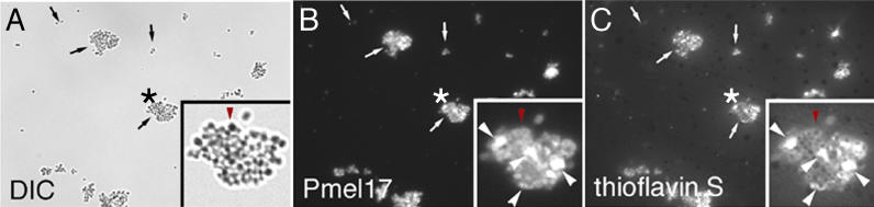
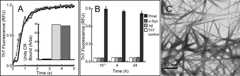
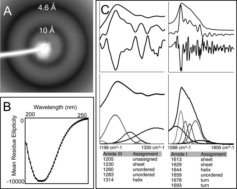
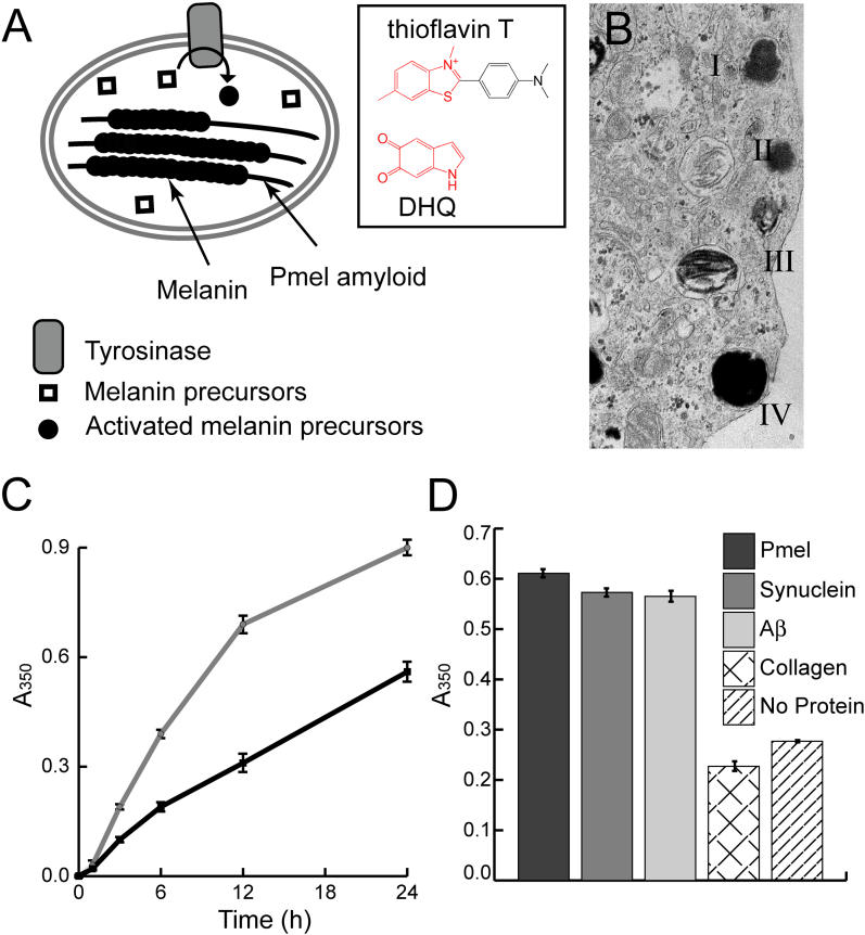
Amyloid is a generally insoluble, fibrous cross-beta sheet protein aggregate. The process of amyloidogenesis is associated with a variety of neurodegenerative diseases including Alzheimer, Parkinson, and Huntington disease. We report the discovery of an unprecedented functional mammalian amyloid structure generated by the protein Pmel17. This discovery demonstrates that amyloid is a fundamental nonpathological protein fold utilized by organisms from bacteria to humans. We have found that Pmel17 amyloid templates and accelerates the covalent polymerization of reactive small molecules into melanin-a critically important biopolymer that protects against a broad range of cytotoxic insults including UV and oxidative damage. Pmel17 amyloid also appears to play a role in mitigating the toxicity associated with melanin formation by sequestering and minimizing diffusion of highly reactive, toxic melanin precursors out of the melanosome. Intracellular Pmel17 amyloidogenesis is carefully orchestrated by the secretory pathway, utilizing membrane sequestration and proteolytic steps to protect the cell from amyloid and amyloidogenic intermediates that can be toxic. While functional and pathological amyloid share similar structural features, critical differences in packaging and kinetics of assembly enable the usage of Pmel17 amyloid for normal function. The discovery of native Pmel17 amyloid in mammals provides key insight into the molecular basis of both melanin formation and amyloid pathology, and demonstrates that native amyloid (amyloidin) may be an ancient, evolutionarily conserved protein quaternary structure underpinning diverse pathways contributing to normal cell and tissue physiology.


Figures

Figure 1
Figure 1. Purified Melanosomes Stain with Amyloidophilic Dyes
Melanosomes were isolated from bovine RPE and choroid and visualized using transmission electron microscopy (A; scale bar = 1 μm), differential interference contrast microscopy (DIC) (B, D, and F; scale bars = 10 μm), indirect immunofluorescence using a Pmel17-specific antibody (C), or the thioflavin S (E) or Congo red (G) amyloidophilic fluorophores. Images (B) and (C), (D) and (E), and (F) and (G) are paired.
Figure 2
Figure 2. Pmel17 and Thioflavin S Fluorescence Overlap in the Detergent-Insoluble Melanosome Fraction
A 1% Triton-X 100 detergent-insoluble fraction was prepared from purified melanosomes and visualized using differential interference contrast microscopy (DIC) (A), indirect immunofluorescence using a Pmel17-specific antibody (B), or thioflavin S fluorescence (C). Arrows denote Pmel17-containing insoluble clusters of variable size. Asterisks indicate the enlarged cluster shown in the lower righthand corner of each panel. In the insets, the large white arrowheads denote Pmel17-positive structures (shown in [B]) that directly overlap with thioflavin S staining (shown in [C]); the red arrowhead in (A) denotes a residual dense melanin-containing granule lacking Pmel17 (shown in [B]) that does not stain with thioflavin S (shown in [C]).
Figure 3
Figure 3. rMα Rapidly Forms Thioflavin T– and Congo Red–Positive Fibers under Nondenaturing Conditions
(A) rMα samples (in 8 M GdmCl to preserve an unfolded, nonaggregated state) were diluted by manual mixing to start an amyloid fiber formation time course (monitored by thioflavin T fluorescence) at varying pHs: pH 7.4 (black line, triangles), pH 6.0 (dark grey line, circles), and pH 4.85 (light grey line, squares); control (thioflavin T buffer) (black line, white diamonds ). The inset bar graph reflects endpoint Congo red binding of equimolar amounts of deposits of Mα formed at pH 7.4 (dark grey), Aβ 1–40 fibers associated with Alzheimer disease (light grey), and control (Congo red buffer) (black). (B) rMα (Pmel) forms thioflavin T (ThT)–positive aggregates at least four orders of magnitude faster than either α-synuclein (α-Syn) or Aβ when all three polypeptides are diluted from 8 M GdmCl into physiological buffer (error bars represent the standard deviation of triplicate samples). (C) Transmission electron micrograph of typical rMα amyloid fibers with an average diameter of 10 nm, formed under nondenaturing conditions.
Figure 4
Figure 4. rMα Fibers Have a Cross-β Sheet Structure
(A) X-ray powder diffraction of lyophilized rMα fibers formed in vitro exhibit a very strong reflection at 4.6 Å and a strong reflection at 10 Å, which is expected of an amyloid cross-β sheet structure. (B) The far-UV CD spectra of soluble Mα aggregates formed at low concentrations to avoid precipitation support a predominantly β-sheet structure. Mα aggregates are approximately 11% α-helix, 32% β-sheet, 23% β-turn, and 33% disordered, based on curve fitting with a basis set of 43 soluble proteins. Since β-sheet content is estimated using a set of proteins not composed of cross-β sheet structures, the potential error in the estimate cannot be determined. (C) The attenuated total reflectance FT-IR spectrum of aggregated rMα in the solid state supports a β-sheet-rich structure. Peaks in the amide III (top left, upper curve) and I (top right, upper curve) regions were identified using Fourier self-deconvolution (top left and right, middle curve) and confirmed by second derivative analysis (top left and right, bottom curve). Peak assignments are listed, and were used to fit the original spectrum using fixed Gaussian peaks at the assigned positions (bottom). Peaks assigned to β-sheet regions of the spectrum accounted for a large percentage of the total area in the amide I and III regions.
Figure 5
Figure 5. Amyloid, Including rMα, Specifically Accelerates Melanin Synthesis
(A) In melanosomes, assembly of activated melanin precursors, generated by tyrosinase, occurs along Pmel17 fibers. The boxed portion of (A) illustrates the amyloid-binding dye thioflavin T and the activated melanin precursor DHQ, which possess similar core structures. This suggests an explanation for the ability of Pmel17 to concentrate and organize melanin precursors, thereby enabling melanogenesis. (B) In vivo, melanosome maturation is a four-step process (I–IV) in which initial formation of the Pmel17 fibrillar matrix (II) enables subsequent melanin polymerization along the Pmel17 fibers (III) (Adapted with permission from [16].) (C) A time course of melanin synthesis in vitro shows that insoluble rMα amyloid increases the amount of insoluble melanin formed per unit time (grey line) versus a control reaction lacking rMα (black line). (D) Melanin synthesis after 20 h was also evaluated in the presence of insoluble rMα amyloid, α-synuclein amyloid, Aβ amyloid, and collagen IV α-helical fibers. The melanin precursor D,L-DOPA was incubated in the presence of the enzyme tyrosinase and the amyloid of interest at room temperature. Melanin content of each reaction condition was measured by pelleting insoluble melanin, dissolving it in 1 M NaOH, and measuring the absorbance at 350 nm. Supernatant melanin content was equal for all samples. In (C) and (D) error bars represent the standard deviation between triplicate samples.

References

    1. Petkova AT, Leapman RD, Guo Z, Yau WM, Mattson MP, et al. Self-propagating, molecular-level polymorphism in Alzheimer's beta-amyloid fibrils. Science. 2005;307:262–265. - PubMed
    1. Chen S, Berthelier V, Hamilton JB, O'Nuallain B, Wetzel R. Amyloid-like features of polyglutamine aggregates and their assembly kinetics. Biochemistry. 2002;41:7391–7399. - PubMed
    1. Sekijima Y, Wiseman RL, Matteson J, Hammarstrom P, Miller SR, et al. The biological and chemical basis for tissue-selective amyloid disease. Cell. 2005;121:73–85. - PubMed
    1. Tanaka M, Chien P, Yonekura K, Weissman JS. Mechanism of cross-species prion transmission: An infectious conformation compatible with two highly divergent yeast prion proteins. Cell. 2005;121:49–62. - PubMed
    1. Dobson CM. Protein chemistry: In the footsteps of alchemists. Science. 2004;304:1259–1262. 1261. - PubMed

Publication types

MeSH terms