Comparative Study
. 2005 Nov 29;102(48):17495-500.
doi: 10.1073/pnas.0508950102. Epub 2005 Nov 21.

Crosstalk in G protein-coupled receptors: changes at the transmembrane homodimer interface determine activation

Affiliations
Comparative Study

Crosstalk in G protein-coupled receptors: changes at the transmembrane homodimer interface determine activation

Wen Guo et al. Proc Natl Acad Sci U S A. .

Abstract

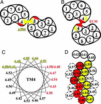
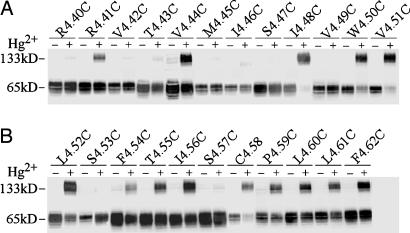
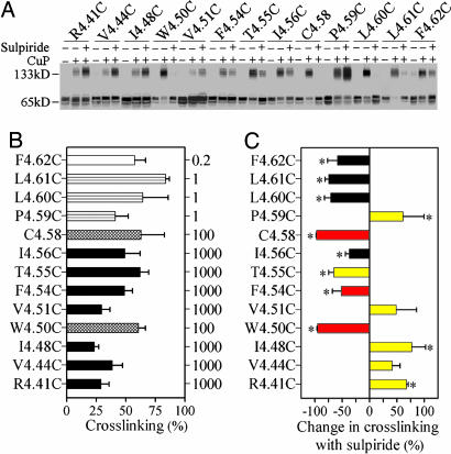
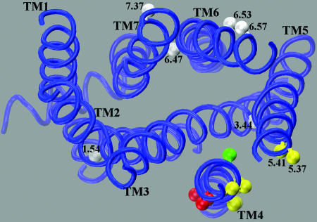
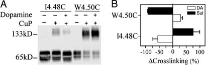
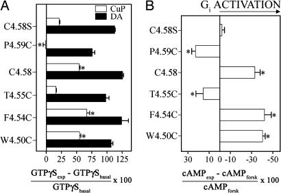
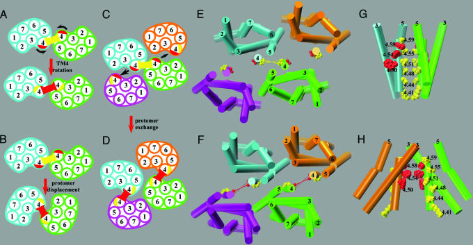
Functional crosstalk between G protein-coupled receptors in a homo- or heterodimeric assembly likely involves conformational changes at the dimer interface, but the nature of this interface is not yet established, and the dynamic changes have not yet been identified. We have mapped the homodimer interface in the dopamine D2 receptor over the entire length of the fourth transmembrane segment (TM4) by crosslinking of substituted cysteines. Their susceptibilities to crosslinking are differentially altered by the presence of agonists and inverse agonists. The TM4 dimer interface in the inverse agonist-bound conformation is consistent with the dimer of the inactive form of rhodopsin modeled with constraints from atomic force microscopy. Crosslinking of a different set of cysteines in TM4 was slowed by inverse agonists and accelerated in the presence of agonists; crosslinking of the latter set locks the receptor in an active state. Thus, a conformational change at the TM4 dimer interface is part of the receptor activation mechanism.


Figures

Fig. 1.
Fig. 1.
Models of the rhodopsin dimer interface. Schematic arrangements illustrating the symmetric TM4-TM5 interface (yellow) of mouse rhodopsin in disk membranes in the dark as modeled by Liang et al. (21) based on AFM (A) and the symmetric TM4 interface (red) of squid rhodopsin deduced from ECM of 2D crystalline arrays in reconstituted membrane (22) (B). (C) Helical wheel representation of TM4 showing the indexed positions of the residues. Trp-4.50 (see Methods for indexing) is conserved in 96% of opsins and 99% of amine receptors and allows us to unambiguously align the D2R sequence with the rhodopsin sequence and structure. Residues at the AFM interface are shown in yellow, and those at the ECM interface are shown in red. The positions more extracellular than 4.59 are not colored due to their location in the last distorted turn of helix in bovine rhodopsin and the ambiguity as to the exact end of TM4 in D2R. (D) Helical net of TM4 colored as in C. The extracellular end of TM4 is at the top of the figure. Positions for which the corresponding cysteine mutants in D2R were crosslinked by CuP (see Fig. 3) are shown as solid circles, and those that were not crosslinked are shown as dashed circles.
Fig. 2.
Fig. 2.
Crosslinking of D2R TM4 cysteine mutants with mercuric chloride. Stably expressed cysteine mutants in TM4 from R1504.40C to F1724.62C [denoted by using the indexing system described in Methods (24)] were crosslinked by 20 μM HgCl2 at 25°C for 10 min, stopped with N-ethylmaleimide and analyzed by immunoblotting. All experiments were repeated at least three times, and a representative experiment is shown.
Fig. 3.
Fig. 3.
Effects of sulpiride on crosslinking of D2R TM4 cysteine mutants with copper phenanthroline. (A) Cysteine mutants were crosslinked by copper sulfate and 1,10-phenanthroline in a 1:2 molar ratio at the Cu2+ concentrations indicated in B at 25°C for 10 min in the absence or presence of 10 μM sulpiride. All experiments were repeated at least three times, and a representative experiment is shown. (B) Cysteine mutants were crosslinked by CuP at the concentrations in μM shown on the right axis at 25°C for 10 min, and the fraction of crosslinking is shown as mean ± SEM (n = 3-8). (C) The sulpiride-induced change in crosslinking fraction for each mutant is shown as mean ± SEM (n = 3-5). Crosslinking in the presence or absence of sulpiride was compared by one-way ANOVA and Bonferroni post hoc test with statistical significance at the P < 0.05 level indicated by *. Bars are shown in yellow or red for those positions predicted to be at the dimer interface in the AFM or ECM models, respectively (see Fig. 1).
Fig. 4.
Fig. 4.
Homology model of the D2R based on the structure of bovine rhodopsin (52). The Cβ positions that were crosslinked by CuP are shown in yellow or red depending on their predicted location at the AFM or ECM interfaces (see Fig. 1). Position 4.56, which is predicted to face inward is shown in green. For simplicity, we omit the segment 4.60-4.62, which is the most extracellular part of TM4 and may not be in an α-helical conformation. Endogenous cysteines that did not crosslink, including 1.54, 3.44, and 6.47, are shown in gray, as are the positions of substituted cysteines 6.53, 6.57, and 7.37 in TM6 and TM7 that did not crosslink despite their predicted positions facing outward in the rhodopsin monomer.
Fig. 5.
Fig. 5.
Effects of dopamine on crosslinking of selected D2R TM4 cysteine mutants with copper phenanthroline. (A) I1584.48C and W1604.50C were crosslinked by the concentrations of CuP indicated in Fig. 3B in the absence or presence of 10 μM dopamine and detected by immunoblotting. (B) The sulpiride- and dopamine-induced changes in crosslinking fraction for each mutant are shown as mean ± SEM (n = 3-5) for dopamine (open bars) or sulpiride (filled bars). DA, dopamine.
Fig. 6.
Fig. 6.
Activation of unliganded D2R results from crosslinking selected TM4 cysteine mutants. (A) GTPγS binding to membranes prepared after treating cells stably expressing the appropriate TM4 cysteine mutants with 100 μM CuP (open bars) or by untreated membranes exposed to 10 μM dopamine for 15 min at 30°C (filled bars). The dopamine- or CuP-induced change in GTPγS binding (GTPγSexp) compared with untreated membranes exposed to vehicle alone (GTPγSbasal) is shown as mean ± SEM (n = 3-5). The effects were analyzed by one-way ANOVA and Dunnett's post hoc test, and statistical significance, comparing the effect of CuP treatment on each cysteine mutant with that on uncrosslinked C1684.58S treated with the same concentration of CuP, at the P < 0.05 level is indicated by *. (B) The change in [cAMP] in the presence of forskolin (100 μM) after treatment with CuP (100 μM) in cells stably expressing the appropriate cysteine mutants is shown as the mean ± SEM (n = 3-6). One-way ANOVA and Dunnett's post hoc test were performed as in A. The x axis is plotted so that activation of the receptor, which causes inhibition of adenylyl cylase through Gi, is shown to the right of the y intercept. In C1684.58S, the maximal change of cAMP induced by dopamine was -74% (23). DA, dopamine.
Fig. 7.
Fig. 7.
Illustration of potential dimer rearrangements and molecular model representations of the proposed dimer interfaces. (A) Rotation of TM4 that could bring the red interface closer together in the active state. (B) A protomer displacement in which the same two protomers move so that they now interact at the red interface. (C and D) A protomer exchange in which crosslinking is between different protomer pairs in the inactive state (C) and in the active state (D). (E and G) Extracellular and side views, respectively, of a symmetric TM4-TM5 interface (yellow) as proposed by Liang et al. (21) incorporated into a tetrameric arrangement. (F and H) Extracellular and side views of a symmetric TM4 interface (red) deduced from the squid rhodopsin 2D electron density map (22) incorporated into an alternative tetrameric arrangement. These two models are proposed to correspond to the inactive and active states, respectively. The backbone of the transmembrane segments is rendered as cylinders. E-H were prepared by using VMD (53) for a homology model of the D2R based on the structure of bovine rhodopsin (52). For simplicity, we omit the segment 4.60-4.62, which is the most extracellular part of TM4 and may not be in an α-helical conformation.

References

    1. Gether, U. (2000) Endocr. Rev. 21, 90-113. - PubMed
    1. Terrillon, S. & Bouvier, M. (2004) EMBO Rep. 5, 30-34. - PMC - PubMed
    1. Milligan, G. (2004) Mol. Pharmacol. 66, 1-7. - PubMed
    1. Angers, S., Salahpour, A. & Bouvier, M. (2002) Annu. Rev. Pharmacol. Toxicol. 42, 409-435. - PubMed
    1. Park, P. S., Filipek, S., Wells, J. W. & Palczewski, K. (2004) Biochemistry 43, 15643-15656. - PMC - PubMed

Publication types

MeSH terms