. 2006 Jan;45(1):1-9.
doi: 10.1002/mc.20160.

Effects of MAP kinase inhibitors on epidermal growth factor-induced neoplastic transformation of human keratinocytes

Affiliations

Effects of MAP kinase inhibitors on epidermal growth factor-induced neoplastic transformation of human keratinocytes

Hideya Mizuno et al. Mol Carcinog. 2006 Jan.

Abstract

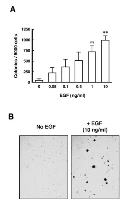
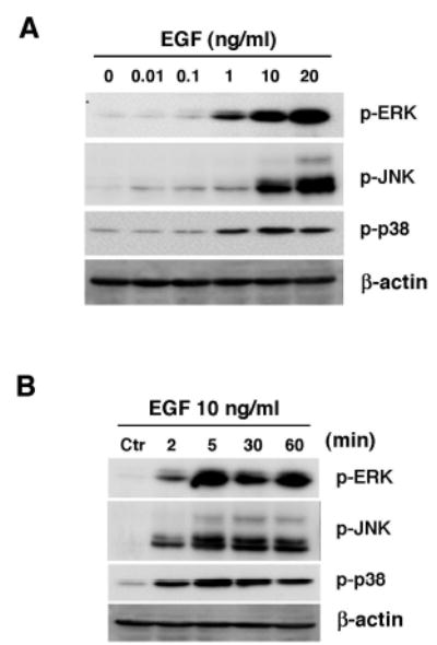
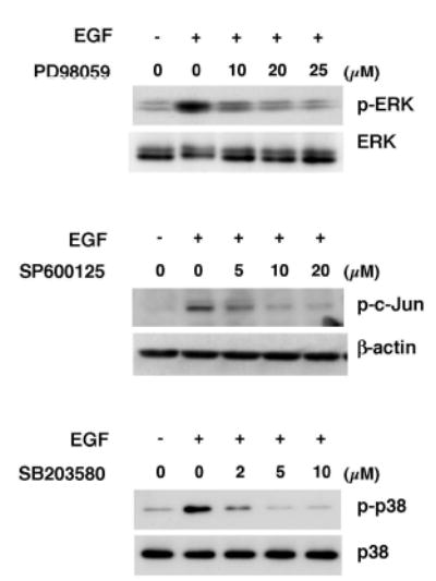
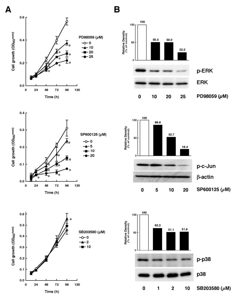
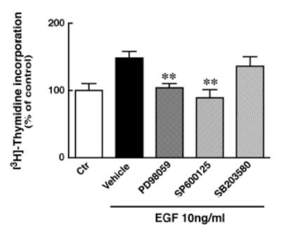
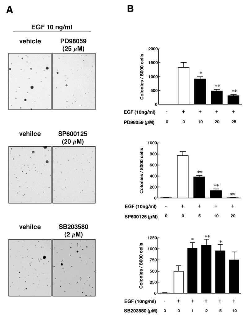
We previously reported data regarding the mechanism of neoplastic transformation in JB6 Cl41 mouse skin epidermal cells. However, experimental in vitro models for studying neoplastic transformation of human cells could provide further insight into the mechanisms of human cancer development. In this study, we have established a neoplastic transformation model with HaCaT cells, a human keratinocyte cell line, and showed the usefulness of this cell line for studying the mechanisms of neoplastic transformation. Epidermal growth factor (EGF) treatment induced a dose-dependent anchorage-independent cell transformation in HaCaT cells. Furthermore, PD98059, a mitogen-activated protein (MAP) kinase/ERK kinase (MEK) inhibitor, or SP600125, c-Jun N-terminal kinase (JNK) inhibitor, decreased cell growth, EGF-induced DNA synthesis and transformation. Unlike observations in the JB6 mouse epidermal cell model, SB203580, a stress-activated protein kinase-2/p38 alpha and beta (p38) inhibitor, increased EGF-induced transformation in HaCaT cells. These results suggest that extracellular-signal regulated kinase (ERK), JNK, or p38 are implicated in EGF-induced neoplastic transformation of human cells.


Figures

Figure 1
Figure 1
Effect of EGF on neoplastic transformation in HaCaT cells. HaCaT cells (8000 cells) were exposed to EGF at various concentration (0 – 10 ng/ml). Cell colonies were scored after 14 days of incubation at 37 °C in a 5% CO2 incubator. (A) Quantitation of total colonies from triplicate samples. Data indicate the mean ± S.D. ** = p < 0.01 compared to untreated control. (B) EGF (10 ng/ml) induces transformation in HaCaT cells.
Figure 2
Figure 2
EGF-induced phosphorylation of ERK, JNK and p38 kinase in HaCaT cells. (A) After starving for 36 h in serum-free DMEM, HaCaT cells were treated with different concentrations of EGF (0 – 20 ng/ml) for 15 min. (B) After starving for 36 h in serum-free DMEM, HaCaT cells were incubated with EGF at 10 ng/ml for the indicated time. Proteins in total cell lysates were separated by SDS-PAGE and immunoblotted with anti-phospho ERK, anti-phospho JNK, anti-phospho p38 or anti-β-actin, as indicated.
Figure 3
Figure 3
Effects of MAP kinase inhibitors on EGF-induced signal transduction. After starving for 36 h in serum-free DMEM, HaCaT cells were incubated with PD98059, a MEK inhibitor, SP600125, a JNK inhibitor, or SB203580, a p38 inhibitor, at different concentrations or with their vehicle, 0.1% DMSO as a control, for 1h. Then, HaCaT cells were treated with EGF at 10 ng/ml for 15 min. Proteins in total cell lysates were separated by SDS-PAGE and immunoblotted with anti-phospho ERK, anti-ERK, anti-phospho c-Jun, anti-β-actin, anti-phospho p38 or anti-p38, as indicated.
Figure 4
Figure 4
Effects of MAP kinase inhibitors on growth of HaCaT cells. (A) HaCaT cells (1000 cells) were seeded in 96-well plates. After culturing for 24 h, medium was replaced with fresh DMEM containing 10% FBS. After culturing for 2–3 h, HaCaT cells were incubated with different concentrations of inhibitors as for Figure 3. After additional culturing for the indicated time, cell viability was measured by the MTS assay (Promega; Madison, WI). Data indicate mean ± S.D. (n = 4). * = p < 0.05 compared to control. (B) After culturing for 24 h in DMEM containing 10% FBS, HaCaT cells were incubated with inhibitor as for Figure 3 for 48 h. After adjusting protein amount, proteins in total cell lysates were separated by SDS-PAGE and immunoblotted with anti-phospho ERK, anti-ERK, anti-phospho c-Jun, anti-β-actin, anti-phospho p38 or anti-p38, as indicated.
Figure 5
Figure 5
Effects of MAP kinase inhibitors on EGF-dependent DNA synthesis in HaCaT cells. HaCaT cells (8000 cells) were seeded in 96-well plates. After culturing for 24 h, media were replaced with DMEM containing 0.05% FBS and cells were incubated for 24 h. HaCaT cells were incubated with inhibitors as for Figure 3 for 1 h. Then, HaCaT cells were treated with 10 ng/ml EGF. After culturing for 18 h, [3H]-Thymidine (0.5 μCi/well) was added to each well, and then cells were incubated for another 6 h. After harvesting of cells, the incorporation of the [3H]-thymidine was measured by liquid scintillation counting. Data indicate mean ± S.D. (n =5). ** = p < 0.01 compared to vehicle.
Figure 6
Figure 6
Effects of MAP kinase inhibitors on EGF-induced transformation in HaCaT cells. HaCaT cells (8000 cells) were exposed simultaneously to 10 ng/ml EGF and different concentrations of PD98059 (MEK inhibitor), SP600125 (JNK inhibitor), or SB203580 (p38 inhibitor) or their vehicle, 0.1% DMSO, as a control, in 0.33% agar BME containing 10% FBS over 0.5% agar BME containing 10% FBS. Cell colonies were scored after 14 days of incubation at 37 °C in a 5% CO2 incubator. (A) PD98059 and SP600125, but not SB203580, inhibit EGF-induced transformation. (B) Quantitation of total colonies from triplicate samples. Data indicate the mean ± S.D. * = p < 0.05; ** = p < 0.01 compared to EGF alone.

Similar articles

Cited by

References

    1. Lage A, Crombet T, Gonzalez G. Targeting epidermal growth factor receptor signaling: early results and future trends in oncology. Ann Med. 2003;35(5):327–336. - PubMed
    1. Stanton P, Richards S, Reeves J, et al. Epidermal growth factor receptor expression by human squamous cell carcinomas of the head and neck, cell lines and xenografts. Br J Cancer. 1994;70(3):427–433. - PMC - PubMed
    1. Huang C, Ma WY, Dong Z. Requirement for phosphatidylinositol 3-kinase in epidermal growth factor-induced AP-1 transactivation and transformation in JB6 P+ cells. Mol Cell Biol. 1996;16(11):6427–6435. - PMC - PubMed
    1. Huang C, Ma WY, Young MR, Colburn N, Dong Z. Shortage of mitogen-activated protein kinase is responsible for resistance to AP-1 transactivation and transformation in mouse JB6 cells. Proc Natl Acad Sci U S A. 1998;95(1):156–161. - PMC - PubMed
    1. Dong Z, Huang C, Ma WY. PI-3 kinase in signal transduction, cell transformation, and as a target for chemoprevention of cancer. Anticancer Res. 1999;19(5A):3743–3747. - PubMed

Publication types

MeSH terms

Substances