. 2006 Mar 15;107(6):2453-60.
doi: 10.1182/blood-2005-07-2831. Epub 2005 Nov 22.

Keratinocyte growth factor (KGF) is required for postnatal thymic regeneration

Affiliations

Keratinocyte growth factor (KGF) is required for postnatal thymic regeneration

Onder Alpdogan et al. Blood. .

Abstract

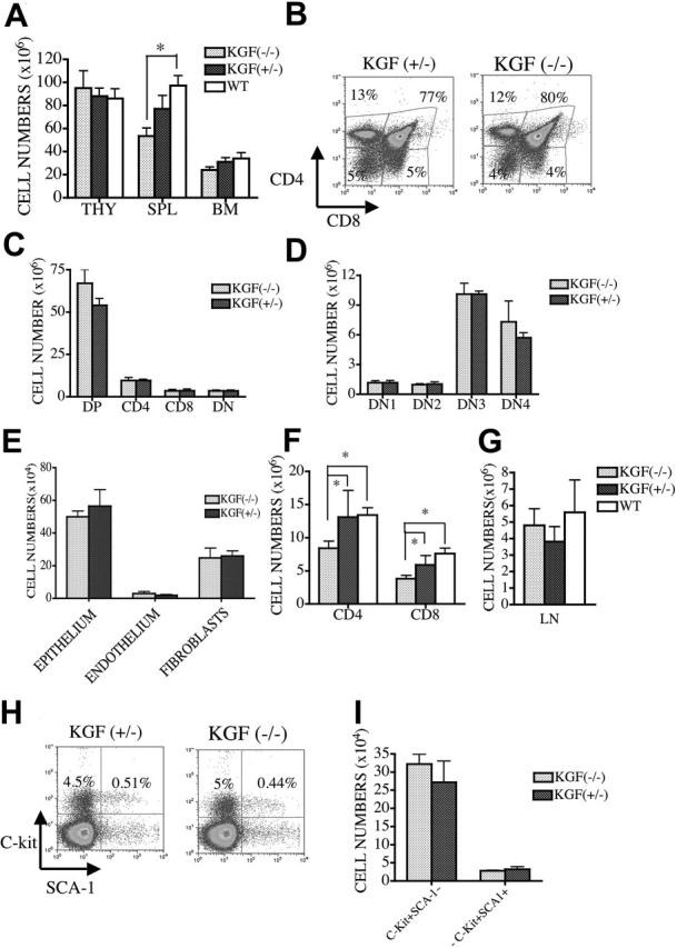
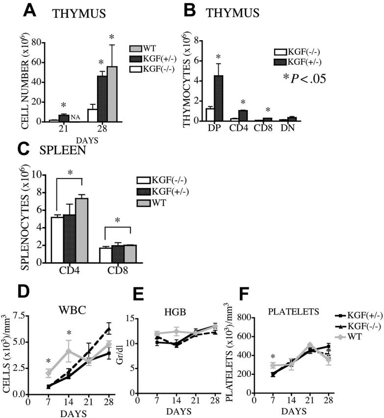
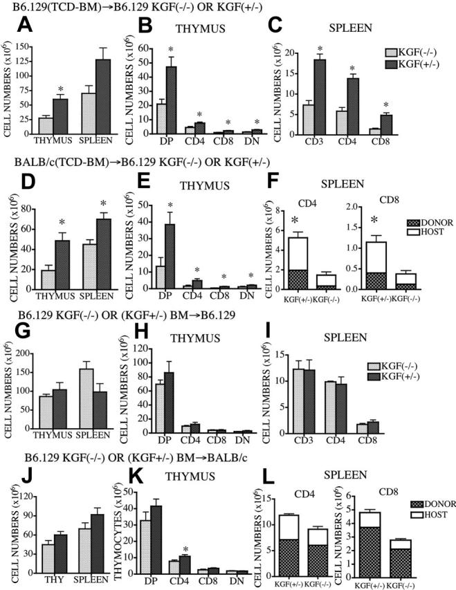
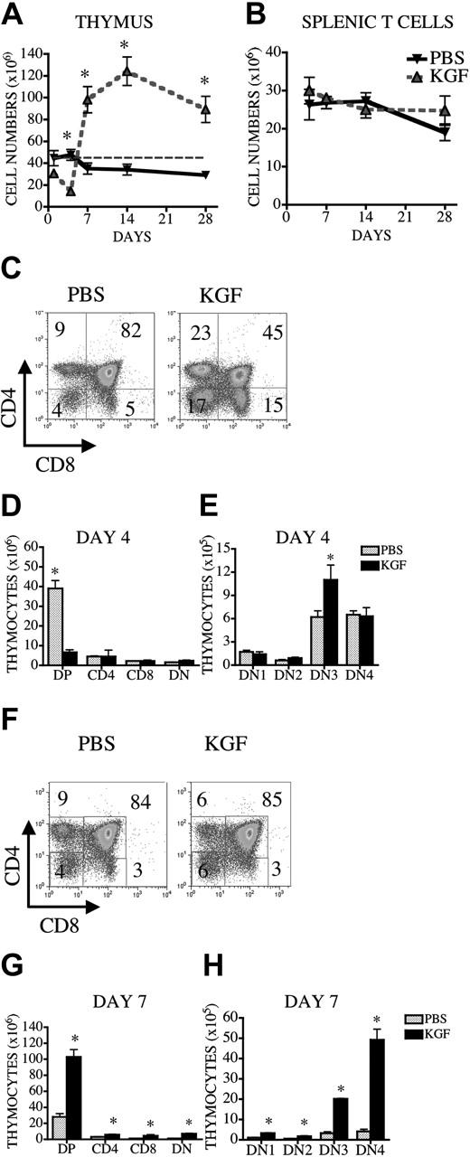
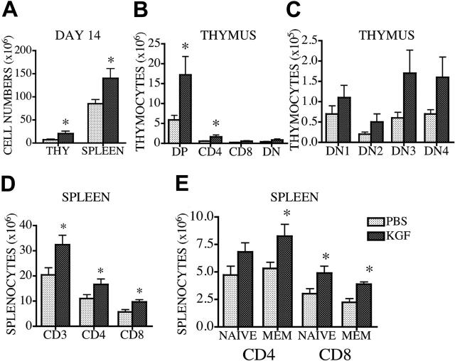
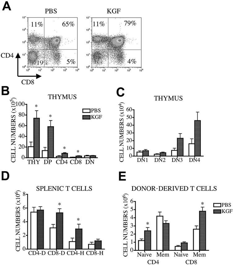
Keratinocyte growth factor (KGF) is a member of the fibroblast growth factor family that mediates epithelial cell proliferation and differentiation in a variety of tissues, including the thymus. We studied the role of KGF in T-cell development with KGF-/- mice and demonstrated that thymic cellularity and the distribution of thymocyte subsets among KGF-/-, wildtype (WT), and KGF+/- mice were similar. However, KGF-/- mice are more vulnerable to sublethal irradiation (450 cGy), and a significant decrease was found in thymic cellularity after irradiation. Defective thymopoiesis and peripheral T-cell reconstitution were found in KGF-/- recipients of syngeneic or allogeneic bone marrow transplant, but using KGF-/- mice as a donor did not affect T-cell development after transplantation. Despite causing an early developmental block in the thymus, administration of KGF to young and old mice enhanced thymopoiesis. Exogenous KGF also accelerated thymic recovery after irradiation, cyclophosphamide, and dexamethasone treatment. Finally, we found that administering KGF before bone marrow transplantation (BMT) resulted in enhanced thymopoiesis and peripheral T-cell numbers in middle-aged recipients of an allogeneic BM transplant. We conclude that KGF plays a critical role in postnatal thymic regeneration and may be useful in treating immune deficiency conditions.


Figures

Figure 1.
Figure 1.
KGF-/- mice have intact T-cell development in the thymus. (A) Nine- to 12-week-old KGF-/-, KGF+/-, and WT controls (B6.129) were killed, and thymic, splenic, and BM cellularities were determined. (B-D) Ten- to 12-week-old KGF-/- and KGF+/- mice were killed and stained with anti-CD4, -CD8, -CD3, -CD44, and -CD25 antibodies, and thymic subpopulations were determined (DP, CD4+CD8+; DN, CD4-CD8-; DN1, CD44+CD25-CD4-CD8-CD3-; DN2, CD44+CD25+CD4-CD8-CD3-; DN3, CD44-CD25+CD4-CD8-CD3-; DN4, CD44-CD25-CD4-CD8-CD3-). (E) Thymic stromal cell populations were determined in KGF-/- and KGF+/- mice (according to the protocol described in “Materials and methods”). (F) Splenocytes from KGF-/- and KGF+/- mice were stained with CD4- and CD8- antibodies, and total CD4+ and CD8+ T-cell numbers were determined by flow cytometric analyses and splenic counts. (G) Peripheral lymph node (axillary and inguinal) cellularities of KGF-/-, KGF+/-, and WT mice were determined. (H-I) Bone marrow cells from KGF-/- and KGF+/- mice were stained with lineage-specific antibodies (anti-CD3, -CD11b, -Gr-1, -B220, and -NK1.1), anti-C-kit, and anti-SCA-1 antibodies. Stem cell populations were determined by multicolor flow cytometric analysis of lineage-negative cells, and the BM cell count from one mouse leg. Values represent mean (± SEM) and n = 6 to 12 mice per group. *P < .05.
Figure 2.
Figure 2.
KGF is required for thymic reconstitution after sublethal irradiation. KGF-/-, KGF+/-, and WT mice were sublethally irradiated (450 cGy), and mice were harvested at day 21 and at day 28 after irradiation. (A-B) Thymic cellularity was determined, and DP, DN, CD4+, and CD8+ thymocyte numbers were calculated by flow cytometric analysis and thymic counts. (C) Splenic cellularity was determined, and splenic CD3+CD4+, and CD3+CD8+ T-cell numbers were calculated by flow cytometric analysis and splenic counts. (D-F) Peripheral blood was obtained from these mice at different time points, and white blood cell (WBC) counts, platelet counts, and hemoglobin levels were determined by an automated cell counter. Values represent mean (± SEM) and n = 3-10 per group. *P < .05.
Figure 3.
Figure 3.
Host-derived KGF is required for thymic reconstitution after syngeneic and allogeneic BMT. Lethally irradiated (1100 cGy) KGF-/- or KGF+/- mice were transplanted with B6.129-WT bone marrow (A-C) or BALB/c BM (D-F). Lethally irradiated (1100 cGy) B6.129 (G-H) or (900 cGy) BALB/c mice (J-L) were transplanted with KGF-/- or KGF+/- BM and were killed at day 28. Thymic and splenic cellularity was determined, and splenic CD3+, CD4+, and CD8+ T-cell numbers were calculated by flow cytometric analysis. Anti-H2d antibody was used to determine the chimerism in allogeneic experiments. Values represent mean (± SEM) and n = 5-11 per group. *P < .05.
Figure 4.
Figure 4.
KGF administration enhances thymopoiesis in young and middleaged normal mice. Normal 3-month-old CBA mice received 5 mg/kg KGF or PBS subcutaneously on 3 consecutive days. Mice were harvested at different time points after KGF administration, and thymic and splenic cellularity were determined. (A) Thymic cellularity at days 1, 4, 7, 14, and 28 after KGF administration in young mice. Dotted gray line represents normal number of thymocytes (3-month-old CBA). (B) Splenic CD3+ T-cell numbers at days 4, 7, 14, and 28 after KGF administration in young mice. (C-H) Thymic cellularity and the number of thymocyte subsets were determined at days 4 and 7 after the final administration of KGF. Thymocytes were stained, and subpopulation numbers were calculated as described in Figure 1. Values represent mean (± SEM) and n = 4-8 per group. *P < .05.
Figure 5.
Figure 5.
KGF administration enhances thymopoiesis and peripheral T-cell development in 18-month-old mice. Eighteen-month-old CBA mice received 5 mg/kg KGF or PBS subcutaneously on 3 consecutive days. Mice were harvested 14 days after KGF administration, and thymic and splenic cellularity were determined. (A) Thymic and splenic cellularity are depicted. (B-C) Thymic subpopulations were calculated as described in Figure 1. (D) Splenic T-cell numbers were determined as described in Table 1. (E) Splenocytes were stained with anti-CD4, -CD8, and -CD44 antibodies, and the percentages of T cells (naive CD4, CD4+CD44-; memory CD4, CD4+CD44+; naive CD8, CD8+CD44-; and memory CD8, CD8+CD44+) were determined by multicolor flow cytometry. Values represent mean (± SEM) and n = 4-8 per group. *P < .05.
Figure 6.
Figure 6.
Pretransplantation administration of KGF in middle-aged recipients of allogeneic BM transplant increases thymopoiesis. Lethally irradiated (1300 cGy) 12-month-old CBA mice were transplanted with T-cell-depleted B10.BR BM (10 × 106). Recipients received either KGF (5 mg/kg/d) or PBS subcutaneously from days -6 to -4. Mice were harvested at day 56, and thymic and splenic cellularities were determined. (A-C) Thymic subpopulations were calculated as described in Figure 1. (D) The number of donor- or host-derived splenic CD4+ and CD8+ T cells from recipients of allogeneic BM transplant are shown. (E) Splenocytes stained with anti-CD4, -CD8, -CD44, and -Ly9.1 antibodies and donor-derived naive and memory T-cell numbers were calculated as described in Figure 5. Values represent mean (± SEM) and n = 6 mice per group. *P < .05.

Similar articles

Cited by

References

    1. Danilenko DM. Preclinical and early clinical development of keratinocyte growth factor, an epithelial-specific tissue growth factor. Toxicol Pathol. 1999;27: 64-71. - PubMed
    1. Danilenko DM, Ring BD, Trebasky BD, et al. Keratinocyte growth factor alters thymocyte development via stimulation of thymic epithelial cells [abstract]. FASEB J. 1996;10: A1148.
    1. Rubin JS, Osada H, Finch PW, Taylor WG, Rudikoff S, Aaronson SA. Purification and characterization of a newly identified growth factor specific for epithelial cells. Proc Natl Acad Sci U S A. 1989;86: 802-806. - PMC - PubMed
    1. Finch PW, Cunha GR, Rubin JS, Wong J, Ron D. Pattern of keratinocyte growth factor and keratinocyte growth factor receptor expression during mouse fetal development suggests a role in mediating morphogenetic mesenchymal-epithelial interactions. Dev Dyn. 1995;203: 223-240. - PubMed
    1. Finch PW, Rubin JS, Miki T, Ron D, Aaronson SA. Human KGF is FGF-related with properties of a paracrine effector of epithelial cell growth. Science. 1989;245: 752-755. - PubMed

Publication types

Substances