

The muscular dystrophies: from genes to therapies
- PMID: 16305275
- PMCID: PMC4496952
The muscular dystrophies: from genes to therapies
Abstract
The genetic basis of many muscular disorders, including many of the more common muscular dystrophies, is now known. Clinically, the recent genetic advances have improved diagnostic capabilities, but they have not yet provided clues about treatment or management. Thanks to better management strategies and therapeutic interventions, however, many patients with a muscular dystrophy are more active and are living longer. Physical therapists, therefore, are more likely to see a patient with a muscular dystrophy, so understanding these muscle disorders and their management is essential. Physical therapy offers the most promise in caring for the majority of patients with these conditions, because it is unlikely that advances in gene therapy will significantly alter their clinical treatment in the near future. This perspective covers some of the basic molecular biological advances together with the clinical manifestations of the muscular dystrophies and the latest approaches to their management.
Figures
References
-
- Dalkilic I, Kunkel LM. Muscular dystrophies: genes to pathogenesis. Curr Opin Genet Dev. 2003;13:231–238. - PubMed
-
- Murray JM, Davies KE, Harper PS, et al. Linkage relationship of a cloned DNA sequence on the short arm of the X chromosome to Duchenne muscular dystrophy. Nature. 1982;300:69–71. - PubMed
-
- Hoffman EP, Brown RH, Jr, Kunkel LM. Dystrophin: the protein product of the Duchenne muscular dystrophy locus. Cell. 1987;51:919–928. - PubMed
-
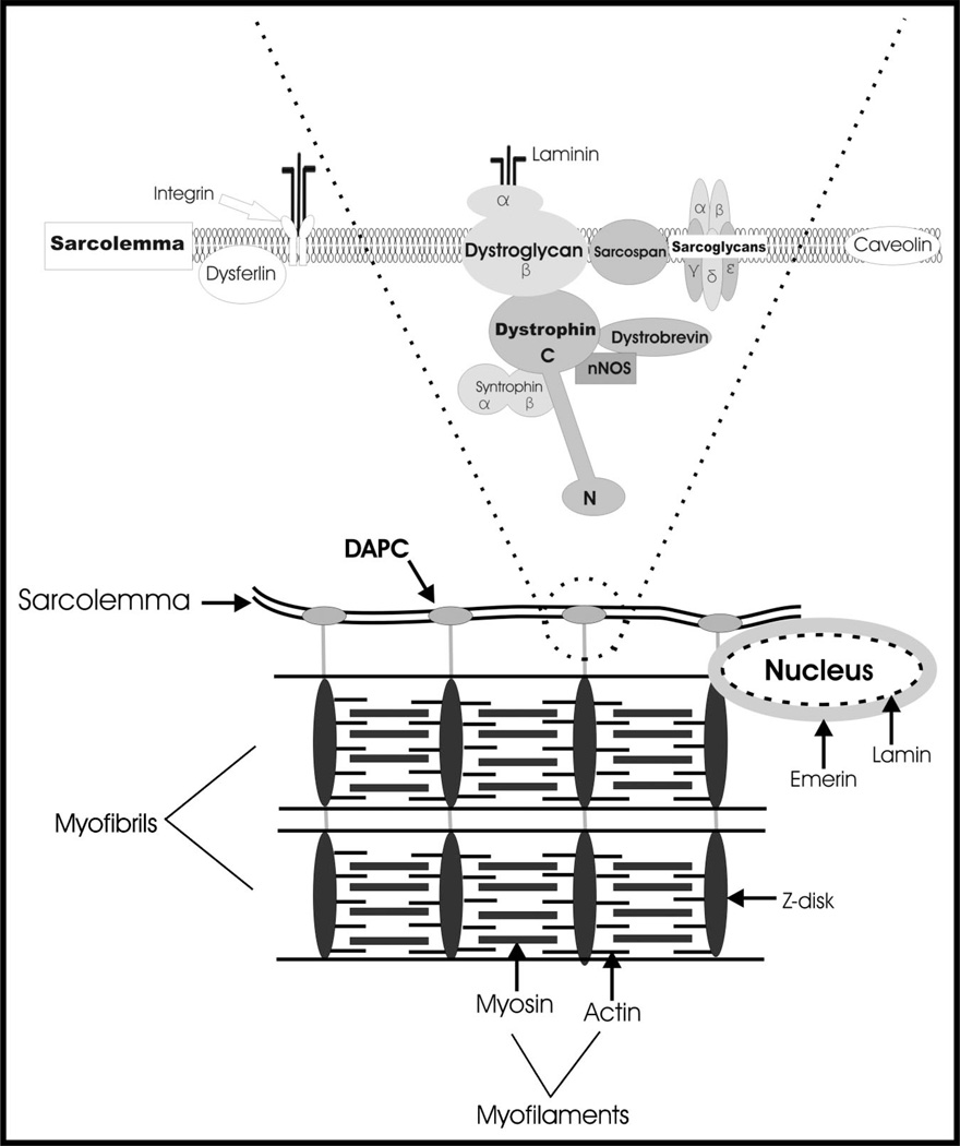
- Ehmsen J, Poon E, Davies K. The dystrophin-associated protein complex. J Cell Sci. 2002;115:2801–2803. - PubMed
Publication types
MeSH terms
Substances
Grants and funding
LinkOut - more resources
Full Text Sources
Other Literature Sources
Medical