. 2006 Jan 27;281(4):1943-55.
doi: 10.1074/jbc.M510504200. Epub 2005 Nov 22.

Sequence, distance, and accessibility are determinants of 5'-end-directed cleavages by retroviral RNases H

Affiliations

Sequence, distance, and accessibility are determinants of 5'-end-directed cleavages by retroviral RNases H

Sharon J Schultz et al. J Biol Chem. .

Abstract

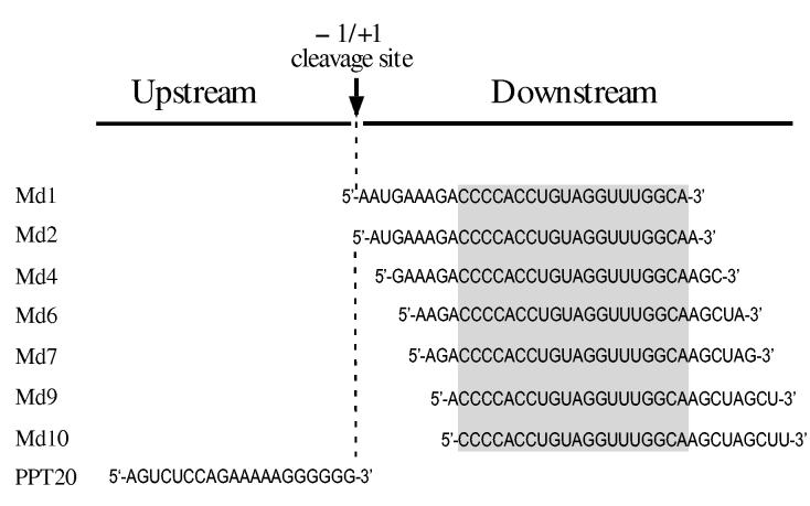
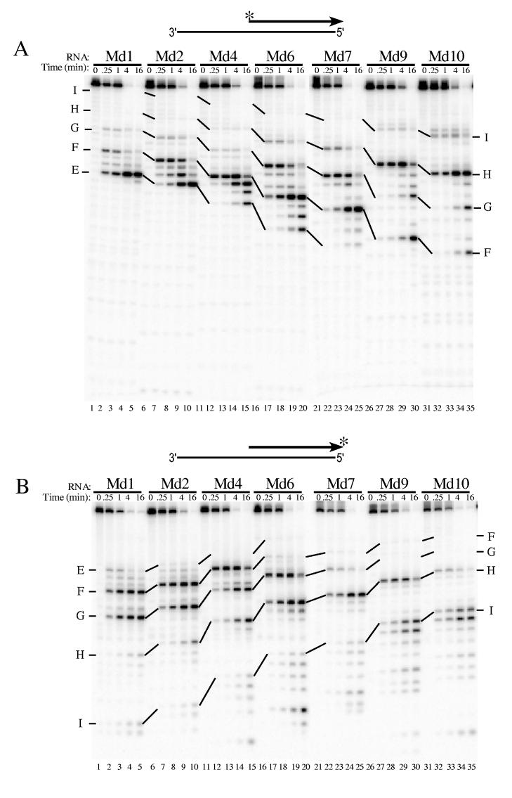
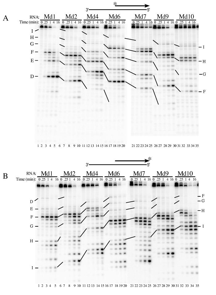
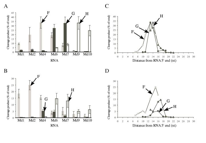
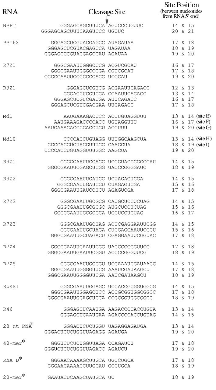
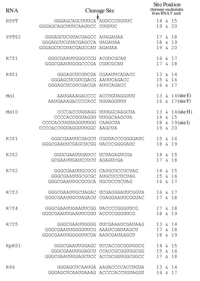
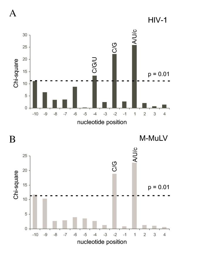
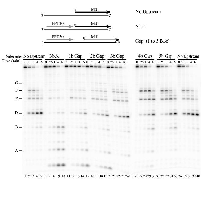
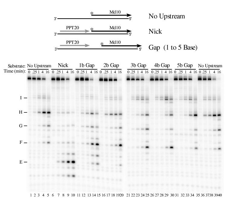
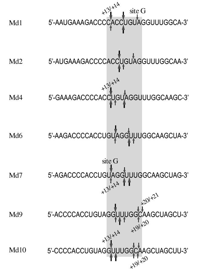
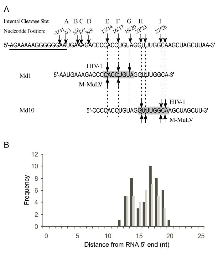
The RNase H activity of reverse transcriptase is essential for retroviral replication. RNA 5'-end-directed cleavages represent a form of RNase H activity that is carried out on RNA/DNA hybrids that contain a recessed RNA 5'-end. Previously, the distance from the RNA 5'-end has been considered the primary determinant for the location of these cleavages. Employing model hybrid substrates and the HIV-1 and Moloney murine leukemia virus reverse transcriptases, we demonstrate that cleavage sites correlate with specific sequences and that the distance from the RNA 5'-end determines the extent of cleavage. An alignment of sequences flanking multiple RNA 5'-end-directed cleavage sites reveals that both enzymes strongly prefer A or U at the +1 position and C or G at the -2 position, and additionally for HIV-1, A is disfavored at the -4 position. For both enzymes, 5'-end-directed cleavages occurred when sites were positioned between the 13th and 20th nucleotides from the RNA 5'-end, a distance termed the cleavage window. In examining the importance of accessibility to the RNA 5'-end, it was found that the extent of 5'-end-directed cleavages observed in substrates containing a free recessed RNA 5'-end was most comparable to substrates with a gap of two or three bases between the upstream and downstream RNAs. Together these finding demonstrate that the selection of 5'-end-directed cleavage sites by retroviral RNases H results from a combination of nucleotide sequence, permissible distance, and accessibility to the RNA 5'-end.


Figures

Fig. 1.
Fig. 1.
RNA oligonucleotides used in RNase H cleavage assays. All RNAs contain M-MuLV sequences from the PPT region, which contains the -1/+1 site where cleavage occurs to generate the plus-strand primer and which corresponds to nucleotides 7815 and 7816 in the M-MuLV sequence (55),. PPT20 is upstream of this site and spans nucleotides -20 to -1. The seven downstream RNAs are named as Moloney downstream (Md) followed by the nucleotide position of the 5′ terminus relative to the -1/+1 site. The sequence shared by all of these RNAs is indicated by a shaded box.
Fig. 2.
Fig. 2.
Analysis of 5′ end-directed RNase H cleavages in RNAs Md1 through Md10 using HIV-1 reverse transcriptase. The hybrid substrate (thick line RNA) shown schematically at the top of each panel contained the indicated 5′ end-labeled (A) or 3′ end-labeled (B) RNA. Substrates were treated with HIV-1 reverse transcriptase, aliquots were removed at the indicated times, and samples were analyzed in denaturing 20% polyacrylamide gels. Results were visualized using a Phosphorimager. Products resulting from cleavage at specific sites in Md1 are labeled as sites E — I, and solid lines track cleavage products corresponding to products from cleavage of the same sites in the other substrates. In Md1 (see Fig. 1), the cleavage sites are found between the following nucleotides from the RNA 5′ end: site E is +13/+14, site F is +16/+17, site G is +19/+20, site H is +22/+23, and site I is +27/+28. The cleavage sites identified in Md1 that give rise to the cleavage products shown for Md10 are indicated at the right.
Fig. 3.
Fig. 3.
Analysis of 5′ end-directed RNase H cleavages in RNAs Md1 through Md10 using M-MuLV reverse transcriptase. The hybrid substrate shown schematically at the top of each panel contained the indicated 5′ end-labeled (A) or 3′ end-labeled (B) RNA. Substrates were treated with M-MuLV reverse transcriptase and analyzed as described in Fig. 2. In Md1, the cleavage sites are found between the following nucleotides from the RNA 5′ end: site D is +8/+9, site E is +13/+14, site F is +16/+17, site G is +19/+20, site H is +22/+23, and site I is +27/+28.
Fig. 4.
Fig. 4.
Extent of cleavage and optimal distances for cleavage at sites F, G, and H in RNAs Md1 through Md10 by HIV-1 and M-MuLV reverse transcriptases. The amount of product generated by cleavage (% of total) at sites F, G, and H in the indicated substrates was determined for HIV-1 (A) or M-MuLV (B) reverse transcriptase. Data from the 1 min time points in three (A) or four (B) independent experiments with 5′ end-labeled RNAs were used to determine the amount of product that resulted from the cleavages at site F (gray bars), site G (black bars), or site H (white bars) (± S.D.). These same data were also used to analyze the optimal distance for cleavage of each site relative to the 5′ RNA ends for HIV-1 (C) or M-MuLV (D) reverse transcriptase. The amount of product generated by cleavage (% of total) for sites F (gray squares), G (black circles), or H (open triangles) is plotted as a function of the cleavage site distance in nucleotides from the 5′ end of each substrate.
Fig. 5.
Fig. 5.
Alignment of sequences flanking RNA 5′ end-directed cleavage sites recognized by HIV-1 RNase H. The left column lists the names of the RNAs used in this analysis, and asterisks denote those presented in prior studies. Cleavage sites mapped using RNAs in this study are described in the Materials and Methods. The 28 nt RNA is from (40). The 40-mer RNA from (25) was used as a blunt-end hybrid, but a later study has shown that while the extent of cleavage is diminished, the locations of 5′ end-directed cleavage sites are not affected by a blunt end (40). RNA D is from (29,32,42). The 20-mer RNA is from (45). In the center column, the sequence surrounding each cleavage site is given, with the location of the cleavage site represented as a gap. The right column gives the position of each cleavage site counting from the 5′ end of the RNA.
Fig. 6.
Fig. 6.
Alignment of sequences flanking RNA 5′ end-directed cleavage sites recognized by M-MuLV RNase H. The sequences are presented as described in the legend to Fig. 5. Cleavage sites mapped using RNAs in this study are described in the Materials and Methods.
Fig. 7.
Fig. 7.
Base preferences at nucleotide positions flanking strong and medium RNA 5′ end-directed cleavages sites. The chi-square values of the nucleotide distribution from positions -10 to +4 were calculated by comparison to overall base frequencies and plotted as a function of nucleotide position, with the cleavage site located between positions -1 and +1. (A) Analysis of the 46 sites for HIV-1 shown in Fig. 5. (B) Analysis of the 35 sites for M-MuLV shown in Fig. 6. The dashed line indicates the chi-square value of 11.34, which has a p value of 0.01. Nucleotide positions with scores exceeding 11.34 are considered significant. The preferred nucleotides for each of the significant positions are indicated above the corresponding bar in upper case (strongly preferred) or lower case (acceptable) letters.
Fig. 8.
Fig. 8.
Influence of gap size on 5′ end-directed cleavage of Md1 by M-MuLV reverse transcriptase. 5′ end-labeled Md1 was used as a substrate without upstream RNA (No Upstream), as a substrate with PPT20 annealed immediately upstream (Nick), or as substrates with gaps of 1 to 5 bases between the 3′ terminus of upstream PPT20 and the 5′ end of Md1 (1 to 5 b Gap). Substrates were incubated with M-MuLV reverse transcriptase, and analyzed as described in Fig. 2. In Md1, cleavage site A is +2/+3, site B is +5/+6, and the other sites are as described in Fig. 3.
Fig. 9.
Fig. 9.
Influence of gap size on 5′ end-directed cleavage of Md10 by HIV-1 reverse transcriptase. 5′ end-labeled Md10 was used as a substrate without upstream RNA (No Upstream), in substrate with PPT20 annealed immediately upstream (Nick), or in substrates with gaps of 1 to 5 bases between the 3′ terminus of upstream PPT20 and the 5′ end of Md10 (1 to 5 b Gap). Substrates were incubated with HIV-1 reverse transcriptase, and analyzed as described in Fig. 2. Products resulting from cleavage at specific positions in Md10 are indicated at left, as described in Figs. 2 and 3. The smallest cleavage product (most distinct in lanes 7 - 10 below site E) corresponds to cleavage between the first and second nucleotides of Md10.
Fig. 10.
Fig. 10.
Comparison of HIV-1 and M-MuLV RNase H 5′ end-directed cleavages in the sequences of RNAs Md1 - Md10. The sequences of the 29-mer RNAs Md1 through Md10 are aligned by the RNA 5′ ends to compare the positions of 5′ end-directed cleavage sites. In each sequence, the extent of cleavage at a site is indicated as strong (large arrows) or medium (small arrows) for HIV-1 reverse transcriptase (above) or M-MuLV reverse transcriptase (below). As described in the Discussion, the range of the closest and furthest independent 5′ end-directed cleavage sites is indicated by the positions of the bordering nucleotides from the RNA 5′ end, the position of site G in substrates Md1 and Md7 is indicated, and the grey box highlights nucleotide positions +13 and +20 that include the range of distances where the 5′ end-directed cleavages occur.
Fig. 11.
Fig. 11.
“Cleavage Window” model for sequence and distance as determinants of 5′ end-directed cleavage. (A) The sequence of the M-MuLV region surrounding the -1/+1 site is shown, beginning at -20 and ending at +40, with the PPT underlined. In this sequence, the locations of the most prominent internal cleavage sites recognized by M-MuLV or HIV-1 reverse transcriptase (23) are indicated by arrows. Above each arrow is the 5′ and 3′ nucleotide positions bordering the cleavage site and the letter designation given to that site. Site C is an internal cleavage site recognized by HIV-1 RNase H that is not observed in 5′ end-directed cleavages, possibly because it is too close to the 5′ end. The sequences of Md1 and Md10 are aligned below with vertical dashed lines indicating the positions of 5′ end-directed cleavage sites. The “cleavage window” that defines eligible sites for 5′ end-directed cleavage is indicated by the shaded box. (B) A frequency distribution plot relative to specific positions that are used for 5′ end-directed cleavages for HIV-1 (black bars) or M-MuLV (grey bars), based upon the 46 sequences shown for HIV-1 in Fig. 5 and the 35 sequences shown for M-MuLV shown in Fig. 6.

Similar articles

Cited by

References

    1. Arts EJ, LeGrice SFJ. Prog. Nucleic Acid Res. Mol. Biol. 1998;58:339–393. - PubMed
    1. Coffin JM, Hughes SH, Varmus HE. Retroviruses. Cold Spring Harbor Laboratory Press; Plainview, N. Y.: 1997. - PubMed
    1. di Marzo Veronese F, Copeland TD, DeVico AL, Rahman R, Oroszlan S, Gallo RC, Sarngadharan MG. Science. 1986;231:1289–1291. - PubMed
    1. Telesnitsky A, Goff SP. Proc. Natl. Acad. Sci. U.S.A. 1993;90:1276–1280. - PMC - PubMed
    1. Misra HS, Pandey PK, Pandey VN. J. Biol. Chem. 1998;273:9785–9789. - PubMed

Publication types