. 2006 Jan 20;281(3):1840-52.
doi: 10.1074/jbc.M509866200. Epub 2005 Nov 23.

Opposite roles of FAP-1 and dynamin in the regulation of Fas (CD95) translocation to the cell surface and susceptibility to Fas ligand-mediated apoptosis

Affiliations

Opposite roles of FAP-1 and dynamin in the regulation of Fas (CD95) translocation to the cell surface and susceptibility to Fas ligand-mediated apoptosis

Vladimir N Ivanov et al. J Biol Chem. .

Abstract

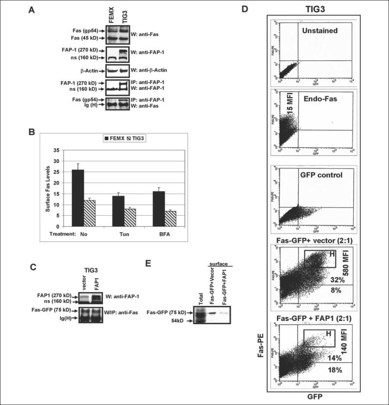
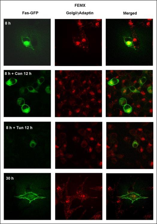
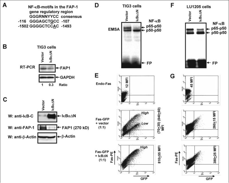
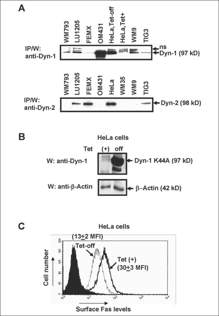
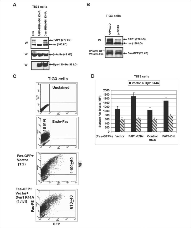
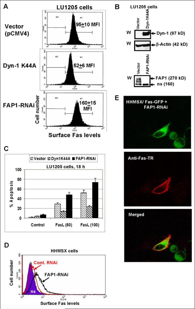
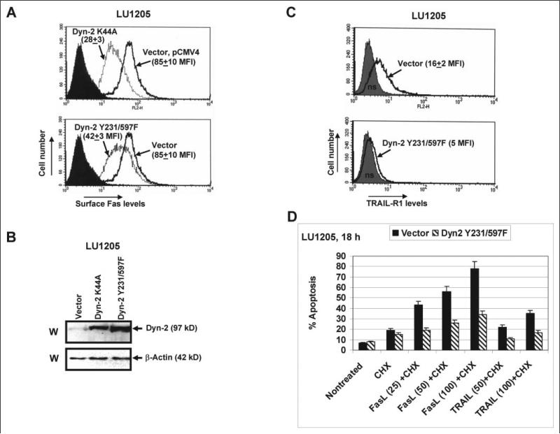
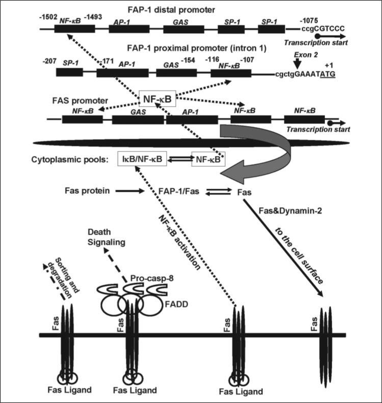
Human melanoma is the most aggressive form of skin cancer and is extremely resistant to radiation and chemotherapy. One of the critical parameters of this resistance is down-regulation of Fas (CD95) cell-surface expression. Using TIG3 normal human fibroblasts and human melanoma cell lines, we investigated transcriptional regulation of FAP-1, a regulator of Fas translocation in the cell. Protein-tyrosine phosphatase FAP-1 (PTPN13, PTP-BAS) interacts with human Fas protein and prevents its export from the cytoplasm to the cell surface. In contrast, dynamin-2 facilitates Fas protein translocation from the Golgi apparatus via the trans-Golgi network to the cell surface. Suppression of dynamin functions by dominant negative dynamin K44A blocks Fas export, whereas the down-regulation of FAP-1 expression by specific RNA interference restores Fas export (a phenomenon that could still be down-regulated in the presence of dominant-negative dynamin). Based on the FAP-1- and dynamin-dependent regulation of Fas translocation, we have created human melanoma lines with different levels of surface expression of Fas. Treatment of these melanoma lines with soluble Fas ligand resulted in programmed cell death that was proportional to the pre-existing levels of surface Fas. Taking into consideration the well known observations that FAP-1 expression is often up-regulated in metastatic tumors, we have established a causal connection between high basal NF-kappaB transcription factor activity (which is a hallmark of many types of metastatic tumors) and NF-kappaB-dependent transcriptional regulation of FAP-1 gene expression that finally restricts Fas protein trafficking, thereby, facilitating the survival of cancer cells.


Figures

FIGURE 1
FIGURE 1. Fas translocation to the cell surface: the negative effects of tunicamycin, BFA, and FAP-1 overexpression
A, Western blot analysis of protein levels of Fas (a mature glycoprotein of 54 kDa and partially glycosylated protein of 45 kDa) in FEMX human melanoma cells and in TIG3 human embryonic lung fibroblasts; β-actin was used as a protein downloading control; immunoprecipitation with anti-FAP-1 Ab and the subsequent Western analysis with anti-Fas mAb or anti-FAP-1 Ab demonstrate FAP-1/Fas protein association in TIG3 cells: FAP-1 (270 kDa), Fas (gp54); ns, nonspecific band. B, FEMX or TIG3 cells were non-treated or treated with tunicamycin (Tun) or Brefeldin A (BFA) for 16 h; surface Fas levels were determined using cell staining with anti-Fas-PE mAb and subsequent flow cytometry. Error bars represent mean ± S.D. from three independent experiments. C, direct Western analysis was performed for determination of FAP-1 levels after transfection of TIG3 cells with FAP-1 expression construct; immunoprecipitation with the subsequent Western analysis was performed to determine Fas-GFP levels in TIG3 cells, which were transiently transfected with pEF-Fas-GFP wt in the presence or in the absence of FAP-1 expression vector. D, TIG3 cells were transfected with pEF-Fas-GFP and the empty vector pCMV4 (1:1) or with pEF-Fas-GFP and FAP-1 expression constructs (1:1); 24 h after transfection, cells were stained with anti-Fas-PE mAb and analyzed for surface Fas expression by flow cytometry; MFI of surface Fas levels and percentage of the Fas+GFP+ and FasGFP+ cells are indicated; the brightest Fas+ cells were gated as the ‘Hrectangular. Gating of unstained cells, single-stained with anti-Fas-PE mAb cells and GFP-positive cells is indicated. E, surface Fas expression was detected in transfected TIG3 cells by biotinylation of cell-surface proteins with subsequent precipitation of biotinylated proteins by Streptavidin-agarose beads and identification of surface proteins using immunoblotting and anti-Fas Ab. Fas-GFP fused protein (75 kDa) was detected on the cell surface after transfection of Fas-GFP; cotransfection with FAP-1 expression construct down-regulated Fas-GFP surface levels.
FIGURE 2
FIGURE 2. Fas-GFP-fused protein translocation to the cell surface in FEMX: the negative effects of tunicamycin
FEMX melanoma cells were transiently transfected with a pEF-Fas-GFP expression construct and analyzed 8, 20, and 30 h after transfection; some cell cultures were treated with tunicamycin for 12 h. Confocal analysis of Fas-GFP (green) and a Golgi marker γ-adaptin (red; secondary Ab labeled with Texas Red) was performed for the determination of subcellular localization.
FIGURE 3
FIGURE 3. Super-stable inhibitor IκBαΔN down-regulates FAP-1 expression and up-regulates surface Fas-GFP expression
A, putative NF-κB-binding elements in the FAP-1 gene regulatory region are indicated. B, TIG3 cells were transfected by the empty vector pCMV4 or by expression construct encoding super-stable IκBαΔN; corresponding changes in the endogenous FAP-1 mRNA levels following transfection of IκBαΔN were determined with RT-PCR; normalization was based on glyceraldehyde-3-phosphate dehydrogenase (GAPDH) levels. C, Western blot analysis of overexpressed IκBαΔN (detected with Ab against C-terminal IκB) and of endogenous FAP-1 levels has been performed 24 h after transfection; actin was used as a protein loading control. D, NF-κB p65-p50 DNA-binding activity (determined by electrophoretic mobility shift assay) was substantially suppressed in the presence of overexpressed IκBαΔN (FP, free oligonucleotide probe). E, cotransfection of Fas-GFP expression construct with IκBαΔN expression construct (which blocked FAP-1 expression) resulted in the notable increase of Fas-GFP translocation to the surface of TIG3 cells that was determined by staining TIG3 cells with anti-Fas-PE mAb and the subsequent flow cytometry; ‘High’ and ‘Low’ indicate positions of the cell subpopulations with high and low levels of surface expression of Fas receptor; MFI values (±S.D. of parallel repeats) are indicated. Results of typical experiment from three independent experiments are presented. F, NF-κB p65-p50 DNA-binding activity (determined by electrophoretic mobility shift assay) was partially suppressed in the presence of overexpressed IκBαΔN in LU1205 cells (FP, free oligonucleotide probe). G, cotransfection of Fas-GFP expression construct with IκBαΔN expression construct resulted in the notable increase of Fas-GFP translocation to the surface of LU1205 cells that was determined by staining LU1205 cells with anti-Fas-PE mAb and subsequent flow cytometry. Data of one typical experiment (from three independent experiments) are presented.
FIGURE 4
FIGURE 4. NF-κB activation by IKKβ or MEKK1Δ up-regulates FAP-1 expression
A, transfection and overexpression of IKKβ S178E/S181E or MEKK1Δ in TIG3 cells up-regulated the NF-κB DNA-binding activity and increased endogenous FAP-1 levels; non-inducible NF-Y DNA-binding activity served as loading control. B, regulation of pFAP1-Luc reporter activity by inhibitor and activators of the NF-κB activity; the FAP-1 gene intron 1 contains several putative binding sites for transcription factors, including SP-1, NF-κB, AP-1, and STATs; a 0.5-kb fragment of this region was cloned in the pGL3-basic reporter; a standard luciferase reporter assay was performed for wild-type or NF-κB-mutated reporter constructs in the presence of indicated regulators of NF-κB activity. C, NF-κB-dependent down-regulation of Fas-GFP translocation has been determined by FACS analysis; the percentages of gated cells in the regions with high and low Fas surface expression and their MFI are indicated. Error bars represent mean ± S.D. from three independent experiments. D, overexpression of IκBαΔN did not affect exogenous FAP1-dependent down-regulation of Fas-GFP translocation. TIG3 cells were transfected with pEF-Fas-GFP together with four different combinations of expression constructs: pCMV4 (vector), pCMV4-IκBΔN plus pCMV4, pCMV4-FAP1 plus pCMV4, and pCMV4-FAP1 plus pCMV4-IκBΔN. Surface Fas expression has been determined by FACS analysis using anti-Fas-PE mAb. Error bars represent mean ± S.D. from three independent experiments.
FIGURE 5
FIGURE 5. Dominant-negative dynamin-1 K44A down-regulates Fas surface expression
A, levels of endogenous dynamin-1 (Dyn-1) or dynamin-2 (Dyn-2) in the indicated cell lines were determined using immunoprecipitation with anti-Dyn-1 or anti-Dyn-2 mAbs followed by Western blot analysis with the same mAbs; human melanoma lines, TIG3 fibroblasts and HeLa cells with Tet-off control of Dyn-1 K44A expression were used. B and C, HeLa Tet-off system for controlled expression of Dyn-1 K44A has been used; overexpression of Dyn-1 K44A (24 h after tetracycline withdrawn) has resulted in down-regulation of endogenous surface Fas receptor expression, which was determined by FACS analysis. Results of typical experiment from four independent experiments are presented.
FIGURE 6
FIGURE 6. Functional interaction of FAP-1 and dynamin regulates Fas-GFP surface levels in TIG3 cells
A, TIG3 cells were transfected by empty vector pRS, combinations of pRS-FAP-1 RNAi with Dyn-1 K44A or control RNAi with Dyn-1 K44A; Western blot analysis was performed for detection FAP-1 and Dyn-1 K44A levels 48 h after transfection. B, TIG3 cells were transiently transfected with the empty vector pcDNA3 or with the expression construct encoding dominant-negative FAP-1ΔCD in the presence of Fas-GFP expression vector; levels of Fas-GFP were determined by IP/W, whereas levels of (FAP-1 plus FAP1ΔCD) were determined by direct Western analysis 48 h after transfection. C and D, TIG3 cells were transfected with Fas-GFP in the indicated combinations of the empty vector, FAP-1 RNAi, control RNAi, Dyn-1 K44A, and FAP1-DN (FAP-1ΔCD). FACS analysis of TIG3 cells was performed 48 h after transfection, and cells were stained with anti-Fas-PE mAb and analyzed by the flow cytometry. Results of one typical experiment (C) and of three independent experiments (D) are presented. Error bars represent mean ± S.D. from three independent experiments.
FIGURE 7
FIGURE 7. Regulation of surface Fas expression and susceptibility to the Fas-mediated apoptosis by dynamin and FAP1 in melanoma cells
A, LU1205 melanoma cells were stably transfected with either Dyn-1 K44A or FAP1 RNAi expression constructs; surface Fas expression in transfected cell lines was determined by FACS analysis; MFI is indicated. B, Western blot analysis of Dyn-1 and FAP1 levels in transfected LU1205 cells. C, apoptosis was induced by soluble FasL (50–100 ng/ml) and cycloheximide (1 μg/ml) treatment for 18 h; apoptosis levels were determined using propidium iodide staining of DNA and the flow cytometry. Error bars represent mean ± S.D. from three independent experiments. D and E, Fas-GFP-fused protein translocation to the cell surface in HHMSX cells. HHMSX melanoma cells were transiently transfected with a pEF-Fas-GFP and FAP-1 RNAi or pEF-Fas-GFP and control RNAi constructs and analyzed 40 h after transfection. Surface Fas expression was determined by using anti-Fas-PE mAb and FACS analysis (D). Confocal analysis of Fas-GFP-fused protein (green) and surface Fas expression (red; using primary anti-Fas mAb and secondary Ab labeled with Texas Red) was performed for the determination of subcellular localization (E).
FIGURE 8
FIGURE 8. Regulation of surface Fas expression and susceptibility to the Fas- and TRAIL-R1-mediated apoptosis by dynamin-2 in melanoma cells
A, LU1205 melanoma cells were stably transfected with either the empty vector pCMV4 or Dyn-2 K44A or Dyn-2 Y231F/Y597F expression constructs; surface Fas expression in transfected cell lines was determined by FACS analysis; MFI is indicated. B, Western blot analysis of Dyn-2 levels in indicated transfected cell lines using anti-Dyn-2 mAb. C, surface TRAIL-R1 expression in transfected cell lines was determined by FACS analysis; MFI is indicated. D, apoptosis levels were determined 18 h after treatments with recombinant FasL (25–100 ng/ml) and recombinant TRAIL (50–100 ng/ml) together with cycloheximide (CHX, 1 μg/ml) using propidium iodide-staining DNA and flow cytometry. Error bars represent mean ± S.D. from three independent experiments.
FIGURE 9
FIGURE 9. The Fas cycle in the cell: the role of NF-κB in the regulation of the total and surface Fas expression
NF-κB plays a dual role in the regulation of the surface Fas expression by controlling transcription of both genes: Fas and a suppressor of Fas protein export, FAP-1. Both genes contain the NF-κB-binding sites in their promoters. Dynamin-2 facilitates Fas translocation from the Golgi and TGN to cell surface. Finally, FasL/Fas interaction, in addition to the canonical induction of death signaling, may induce the NF-κB signaling pathway, up-regulating FAP-1 gene expression with subsequent restriction of the Fas export to the cell surface. (See an additional description of this figure under “Discussion.”)

Similar articles

Cited by

References

    1. Nagata S. Annu. Rev. Genet. 1999;33:29–55. - PubMed
    1. Debatin KM, Krammer PH. Oncogene. 2004;23:2950–2966. - PubMed
    1. Zheng Y, Ouaaz F, Bruzzo P, Singh V, Gerondakis S, Beg AA. J. Immunol. 2001;166:4949–4957. - PubMed
    1. Ivanov VN, Ronai Z. Oncogene. 2000;19:3003–3012. - PubMed
    1. Chan H, Bartos DP, Owen-Schaub LB. Mol. Cell. Biol. 1999;19:2098–2108. - PMC - PubMed

Publication types

MeSH terms