. 2005 Dec 6;102(49):17658-63.
doi: 10.1073/pnas.0507222102. Epub 2005 Nov 23.

Bacterial DNA segregation by dynamic SopA polymers

Affiliations

Bacterial DNA segregation by dynamic SopA polymers

Grace E Lim et al. Proc Natl Acad Sci U S A. .

Abstract

Many bacterial plasmids and chromosomes rely on ParA ATPases for proper positioning within the cell and for efficient segregation to daughter cells. Here we demonstrate that the F-plasmid-partitioning protein SopA polymerizes into filaments in an ATP-dependent manner in vitro, and that the filaments elongate at a rate that is similar to that of plasmid separation in vivo. We show that SopA is a dynamic protein within the cell, undergoing cycles of polymerization and depolymerization, and shuttling back and forth between nucleoprotein complexes that are composed of the SopB protein bound to sopC-containing plasmids (SopB/sopC). The dynamic behavior of SopA is critical for Sop-mediated plasmid DNA segregation; mutations that lock SopA into a static polymer in the cell inhibit plasmid segregation. We show that SopA colocalizes with SopB/sopC in the cell and that SopB/sopC nucleates the assembly of SopA and is required for its dynamic behavior. When SopA is polymerized in vitro in the presence of SopB and sopC-containing DNA, SopA filaments emanate from the plasmid DNA in radial asters. We propose a mechanism in which plasmid separation is driven by the polymerization of SopA, and we speculate that the radial assembly of SopA polymers is responsible for positioning plasmids both before and after segregation.


Figures
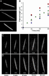
Fig. 1.
Fig. 1.
Filament formation by SopA in vitro.(A) Filaments were grown in the presence of ATP and stained with Nile red as described in Materials and Methods. (Scale bar, 1 μm.) (B) The distance between the ends of a growing filament were measured and the lengths were plotted vs. time. The average elongation rate for the six filaments is 0.18 μm/min. (C and D) Time-lapse microscopy of filament formation. The elongation of a single filament is followed in each row. Numbers at the bottom of each micrograph indicate filament length in μm.
Fig. 2.
Fig. 2.
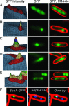
Localization of SopA-GFP and SopB-CFP. (AE) SopA-GFP (green) was expressed from plasmid pGEL10 by induction with 2 μM IPTG, as described in Materials and Methods. Cell membranes were stained with FM 4-64 (red). (A) Strain JP5053, which contains no other Sop system components. (BE) Strain JP5036, which contains an intact Sop system. Foci (B) were observed in 60% of the cells, and hazes (C and D) were observed in 35% of the cells. In the fluorescence intensity plots, colors indicate pixel intensity on a linear scale in arbitrary units: blue, 0–200; red, 200–400; yellow, 400–600; green, 600–800; purple, 800–1,000; and orange, 1,000–1,200. (F) Colocalization of SopA-GFP foci with SopB-CFP foci in strain JP5067. SopA-GFP (green) was expressed from plasmid pGEL10 by induction with 2 μM IPTG, and SopB-CFP (blue) was expressed from plasmid pGEL26 by induction with arabinose as described in Materials and Methods. (Scale bars, 1 μm.)
Fig. 3.
Fig. 3.
Time-lapse microscopy of SopA-GFP. SopA-GFP (green) was expressed from plasmid pGEL10 in strain JP5036 by induction with 2 μM IPTG, as described in Materials and Methods. (Upper) Cell membranes were stained with FM 4-64 (red). Time in minutes is shown in white text in the top right corner of each panel. (Scale bar, 1 μm.) (Lower) Intensity plot of GFP fluorescence. Colors indicate pixel intensity, as in Fig. 2. The oscillation was observed in 98% of the cells in the field (n = 196).
Fig. 4.
Fig. 4.
Recruitment and nucleation of SopA by SopB/sopC.(AC) Radial aster formation by SopA in vitro when the other Sop components are present. Assembly reactions with SopA, SopB, plasmid pGEL30, and ATP were carried out as described in Materials and Methods. (A and B) Staining was with Nile red. (C) SopB was covalently labeled with Alexa 488 (green), and pGEL30 DNA was stained with DAPI (blue). Nile red stains both SopA and SopB. No asters are observed if any of the components are omitted from the assembly reactions (data not shown). (D and E) SopB recruits SopA. In strain JP5081, SopA-GFP (green) was expressed from plasmid pGEL10 by induction with IPTG, and SopB-CFP (blue) was expressed from plasmid pGEL26 by induction with arabinose, as described in Materials and Methods and Results.(D) Before induction of SopB-CFP, SopA-GFP gives rise to diffuse fluorescence. (E) After induction of SopB-CFP, SopA-GFP colocalizes with SopB-CFP at the plasmid. Cell membranes were stained with FM 4-64 (red). (Scale bars, 1 μm.) In the absence of SopA-GFP, SopB-CFP fluorescence tracks with the chromosome, owing to its ability to bind to DNA nonspecifically in the absence of sopC DNA (Fig. 7).
Fig. 5.
Fig. 5.
Properties of the SopA1 mutant. (A) SopA1-GFP (green) was expressed from plasmid pGEL11 in strain JP5034 by induction with 100 μM IPTG as described in Materials and Methods. Cell membranes were stained with FM 4-64 (red). (B) FRAP analysis of a SopA1-GFP (green) filament in strain JP5034. No fluorescence recovery is apparent at 5 min after bleaching. Cell membranes were stained with FM 4-64 (red). (C and D) Strains JP5036 (C) and JP5037 (D) were plated on LB agar plates containing 40 μg/ml X-Gal and incubated overnight at 30°C. The blue (Lac+) colonies of JP5036 indicate that F′lac is stably inherited. The white colonies and sectored blue colonies of JP5037 indicate that F′lac is unstable. (EH) SopA-GFP was expressed from plasmid pGEL10 by induction with 100 μM IPTG, and SopA1 was expressed from plasmid pGEL39 by induction with 0.2% arabinose for 30 min. (E and F) Strain JP5119. (G and H) Strain JP5121. Before induction of SopA1, SopA-GFP tracks the chromosome (E) or forms foci and hazes (G); after induction, SopA-GFP is polymerized into filaments that link the cells in chains (F and H). (Top) Cell membranes stained with FM 4-64 (red). (Middle) SopA-GFP (green). (Bottom) Overlay. (Scale bars, 1 μm.)
Fig. 6.
Fig. 6.
Proposed mechanism for plasmid segregation as mediated by the Sop system. (A) Before plasmid replication, a radial array of SopA polymers (red) assembled on a nucleoprotein complex of SopB (green) bound to sopC-containing plasmid (blue) centers the plasmid at midcell through a force-balancing mechanism. (B) After replication, polymerization of SopA between the SopB/sopC complexes separates the daughter plasmids. (C) The radial arrays of SopA polymers emanating from the two SopB/sopC complexes center the plasmids with respect to each other and with respect to the cell boundaries by a force-balancing mechanism.

References

    1. Gadde, S. & Heald, R. (2004) Curr. Biol. 14, R797–R805. - PubMed
    1. Kline-Smith, S. L. & Walczak, C. E. (2004) Mol. Cell 15, 317–327. - PubMed
    1. Gitai, Z., Thanbichler, M. & Shapiro, L. (2005) Trends Microbiol. 13, 221–228. - PubMed
    1. Gerdes, K., Møller-Jensen, J., Ebersbach, G., Kruse, T. & Nordström, K. (2004) Cell 116, 359–366. - PubMed
    1. Thanbichler, M., Viollier, P. H. & Shapiro, L. (2005) Curr. Opin. Genet. Dev. 15, 153–162. - PubMed

Publication types

MeSH terms