. 2005 Nov 23;25(47):10815-21.
doi: 10.1523/JNEUROSCI.3249-05.2005.

The adult mouse hippocampal progenitor is neurogenic but not a stem cell

Affiliations

The adult mouse hippocampal progenitor is neurogenic but not a stem cell

Natalie D Bull et al. J Neurosci. .

Abstract

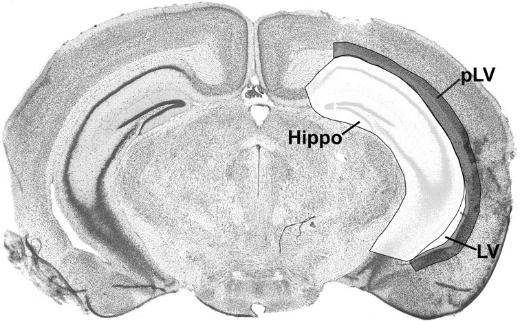
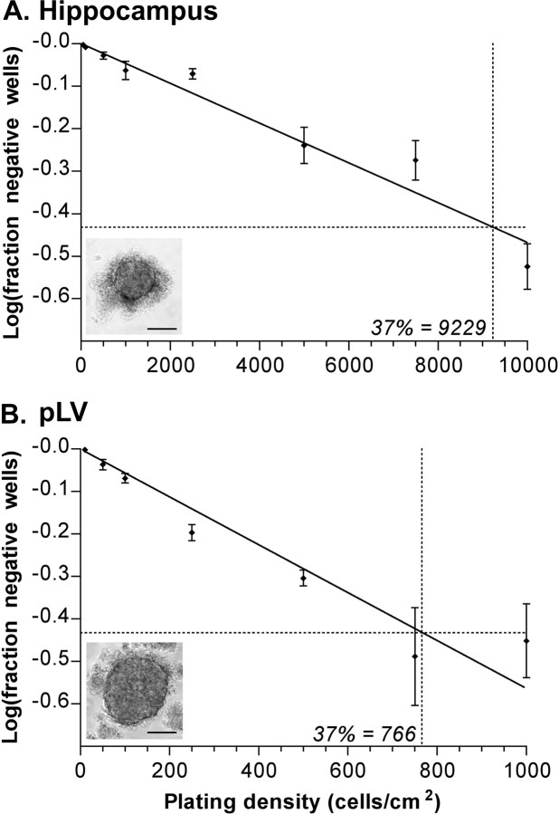
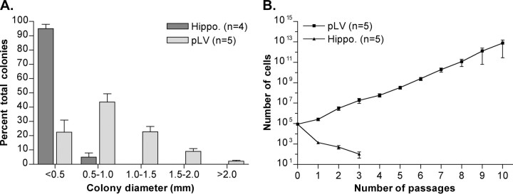
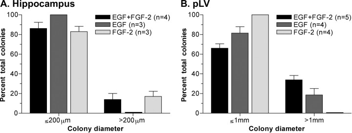
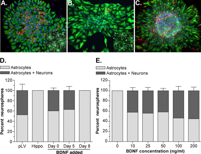
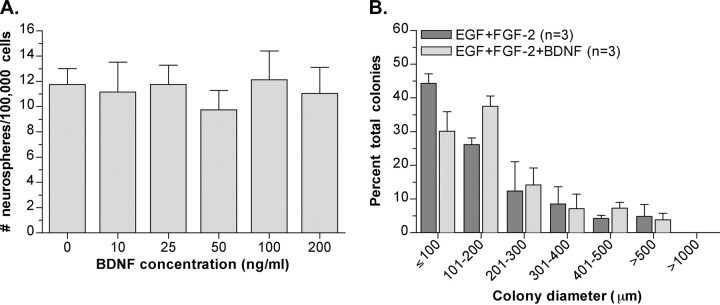
The aim of this investigation was to characterize the proliferative precursor cells in the adult mouse hippocampal region. Given that a very large number of new hippocampal cells are generated over the lifetime of an animal, it is predicted that a neural stem cell is ultimately responsible for maintaining this genesis. Although it is generally accepted that a proliferative precursor resides within the hippocampus, contradictory reports exist regarding the classification of this cell. Is it a true stem cell or a more limited progenitor? Using a strict functional definition of a neural stem cell and a number of in vitro assays, we report that the resident hippocampal precursor is a progenitor capable of proliferation and multipotential differentiation but is unable to self-renew and thus proliferate indefinitely. Furthermore, the mitogen FGF-2 stimulates proliferation of these cells to a greater extent than epidermal growth factor (EGF). In addition, we found that BDNF was essential for the production of neurons from the hippocampal progenitor cells, being required during proliferation to trigger neuronal fate. In contrast, a bona fide neural stem cell was identified in the lateral wall of the lateral ventricle surrounding the hippocampus. Interestingly, EGF proved to be the stronger mitogenic factor for this cell, which was clearly a different precursor from the resident hippocampal progenitor. These results suggest that the stem cell ultimately responsible for adult hippocampal neurogenesis resides outside the hippocampus, producing progenitor cells that migrate into the neurogenic zones and proliferate to produce new neurons and glia.


Figures

Figure 1.
Figure 1.
Diagram of primary tissue dissected for in vitro assay of stem-cell activity. A2-mm-thick coronal slice was cut between -1.22 and -2.70 mm, relative to bregma, as determined from an adult mouse brain atlas (Paxinos and Franklin, 2001). The slice was placed caudal side up and the tissue dissected using two pairs of curved forceps. First, the midbrain was removed, and then the hippocampus, as a whole, was separated gently from the corpus callosum. Finally, the pLV, including the corpus callosum, was pinched away from the cortex. Hippo, Hippocampus; LV, lateral ventricle. This image is from Paxinos and Franklin (2001) with permission.
Figure 2.
Figure 2.
Proliferative cell frequency when primary tissue was cultured with both EGF and FGF-2. A, Primary hippocampal neurosphere limiting dilution assay (n = 9; mean ± SEM). The intercept of log(37% negative wells) gave a neurosphere-forming frequency of one neurosphere for every 9229 cells plated. Inset, Representative photograph of a primary hippocampal neurosphere. Scale bar, 100 μm. B, Primary pLV neurosphere limiting dilution assay (n = 6; mean ± SEM). Neurosphere-forming frequency was calculated as one neurosphere for every 766 cells plated. Inset, Representative photograph of a primary pLV neurosphere. Scale bar, 100 μm.
Figure 3.
Figure 3.
Potential for long-term proliferation in vitro in the presence of both EGF and FGF-2. A, Colony diameters (in millimeters) when primary hippocampal and pLV cells were cultured in the neural colony-forming cell assay (mean ± SEM). B, Expansion of cell numbers when primary hippocampal and pLV cells were cultured in the neurosphere assay through multiple passages (mean ± SEM). Hippo., Hippocampus.
Figure 4.
Figure 4.
Differential effects of EGF and FGF-2 on proliferation. A, Colony diameters (in micrometers) produced by primary hippocampal cells when cultured in the presence of EGF plus FGF-2 or each growth factor separately in the neural colony-forming cell assay (mean ± SEM). B, Colony diameters (in millimeters) when primary pLV cells were cultured in the presence of EGF plus FGF-2 or each growth factor alone in the neural colony-forming cell assay (mean ± SEM).
Figure 5.
Figure 5.
Differentiation of primary neurospheres. A-C, Representative differentiated neurospheres immunolabeled for GFAP (green) and βIII tubulin (red) and counterstained with DAPI (blue). Primary pLV neurosphere (A), primary hippocampal neurosphere grown in the presence of EGF and FGF-2 (B), and a primary hippocampal neurosphere cultured in the presence of EGF, FGF-2, and BDNF (100 ng/ml) (C) are shown. A, B, Insets, Immunolabeling for the oligodendrocyte marker O4 (green) in representative differentiated pLV (A) and hippocampal (B) neurospheres. D, Percentage of differentiated neurospheres containing cells immunopositive for either GFAP alone (astrocytes) or GFAP plus βIII tubulin (neurons) after growth in the presence of EGF and FGF-2 or with BDNF (100 ng/ml) added to hippocampal cultures at different time points during neurosphere proliferation (n = 3; mean ± SEM). E, Percentage of differentiated neurospheres containing astrocytes alone or astrocytes plus neurons when primary hippocampal cells were cultured in increasing concentrations of BDNF (n = 3; mean ± SEM). Hippo., Hippocampus.
Figure 6.
Figure 6.
Effect of BDNF on hippocampal neurosphere frequency and proliferative potential. A, Primary hippocampal neurosphere frequency when cells were cultured in the presence of EGF and FGF-2 plus BDNF at increasing concentrations (n = 4; mean ± SEM). B, Colony diameters (in micrometers) when primary hippocampal cells were cultured in the neural colony-forming cell assay in the presence of EGF and FGF-2 or EGF and FGF-2 plus BDNF (100 ng/ml). Values are mean ± SEM.

References

    1. Adams DJ, Reynolds BA, Louis SA, Rietze RL (2004) The neurosphere assay overestimates the frequency of adult neural stem cells. Soc Neurosci Abstr 30: 383.6.
    1. Altman J, Das GD (1965) Autoradiographic and histological evidence of postnatal hippocampal neurogenesis in rats. J Comp Neurol 124: 319-335. - PubMed
    1. Becq H, Jorquera I, Ben-Ari Y, Weiss S, Represa A (2005) Differential properties of dentate gyrus and CA1 neural precursors. J Neurobiol 62: 243-261. - PubMed
    1. Cameron HA, Woolley CS, McEwen BS, Gould E (1993) Differentiation of newly born neurons and glia in the dentate gyrus of the adult rat. Neuroscience 56: 337-344. - PubMed
    1. Cheng Y, Black IB, DiCicco-Bloom E (2002) Hippocampal granule neuron production and population size are regulated by levels of bFGF. Eur J Neurosci 15: 3-12. - PubMed

Publication types

Substances