. 2005 Nov 23;25(47):10884-93.
doi: 10.1523/JNEUROSCI.2909-05.2005.

Choroid plexus megalin is involved in neuroprotection by serum insulin-like growth factor I

Affiliations

Choroid plexus megalin is involved in neuroprotection by serum insulin-like growth factor I

Eva Carro et al. J Neurosci. .

Abstract

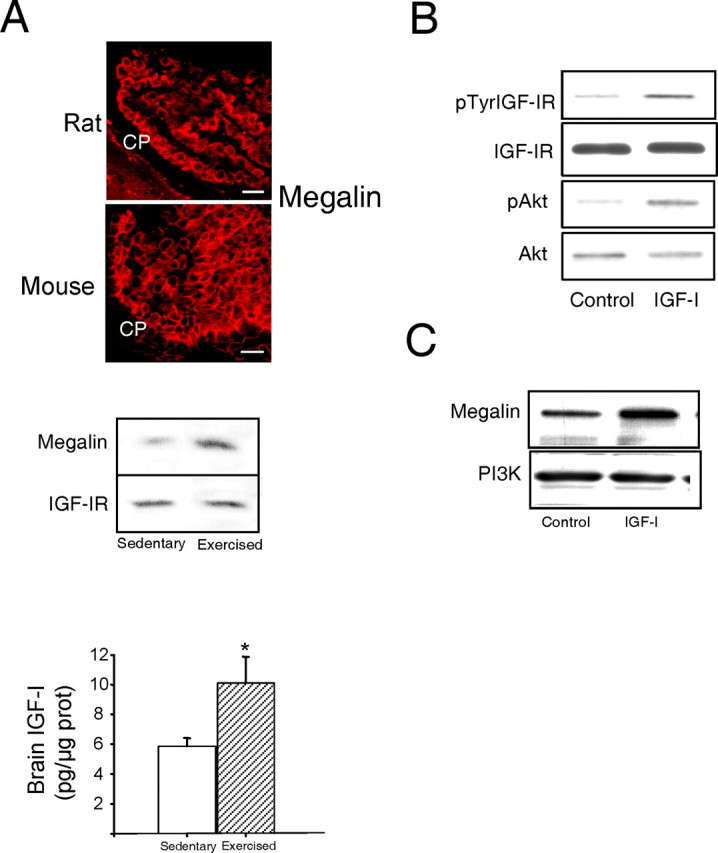
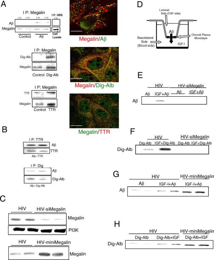
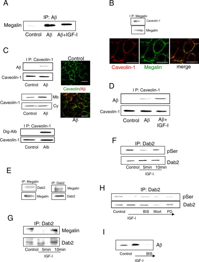
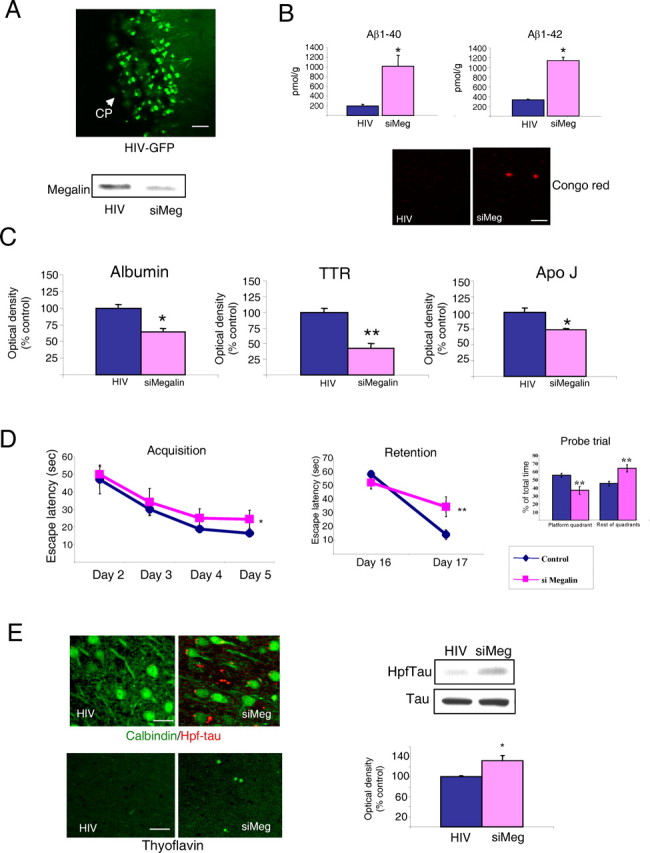
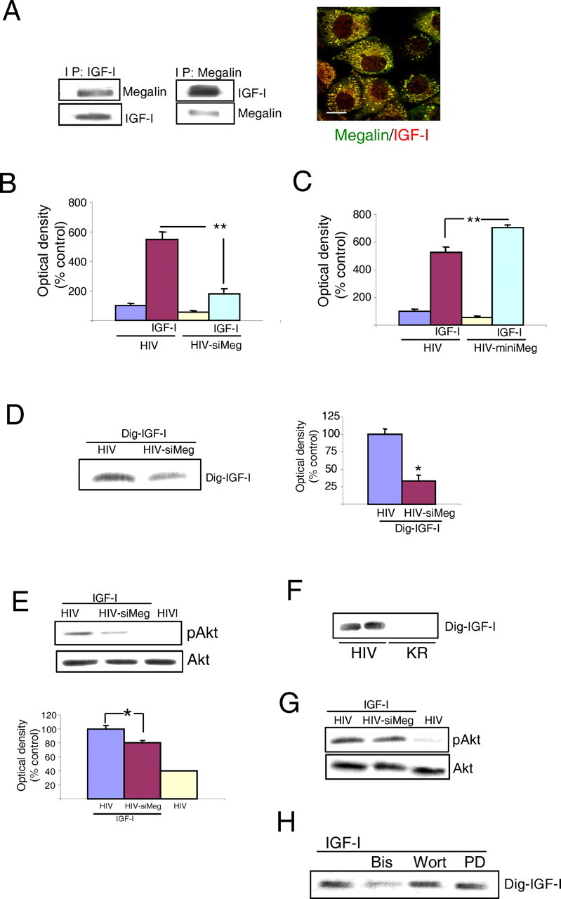
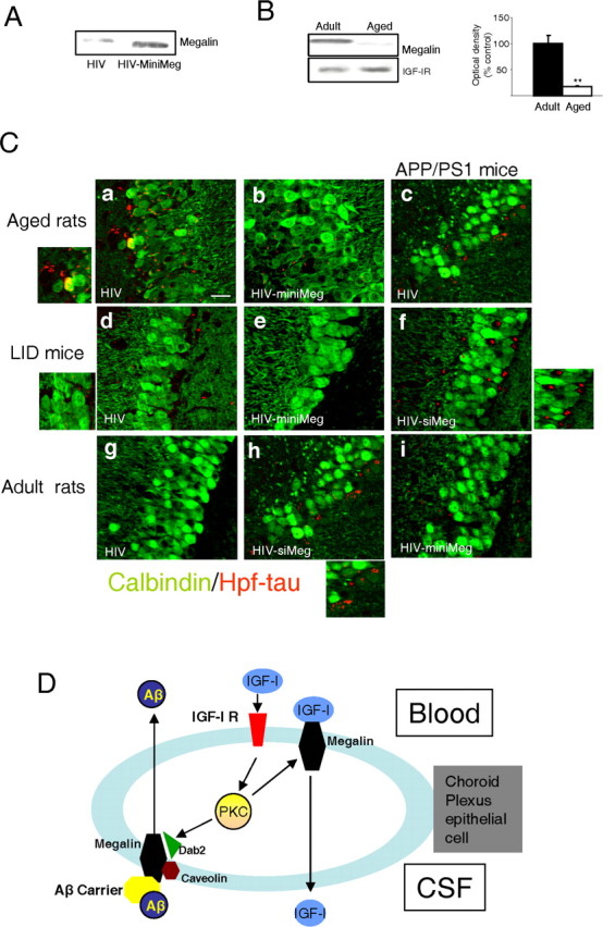
The involvement of circulating insulin-like growth factor I (IGF-I) in the beneficial effects of physical exercise on the brain makes this abundant serum growth factor a physiologically relevant neuroprotective signal. However, the mechanisms underlying neuroprotection by serum IGF-I remain primarily unknown. Among many other neuroprotective actions, IGF-I enhances clearance of brain amyloid beta (Abeta) by modulating transport/production of Abeta carriers at the blood-brain interface in the choroid plexus. We found that physical exercise increases the levels of the choroid plexus endocytic receptor megalin/low-density lipoprotein receptor-related protein-2 (LRP2), a multicargo transporter known to participate in brain uptake of Abeta carriers. By manipulating choroid plexus megalin levels through viral-directed overexpression and RNA interference, we observed that megalin mediates IGF-I-induced clearance of Abeta and is involved in IGF-I transport into the brain. Through this dual role, megalin participates in the neuroprotective actions of IGF-I including prevention of tau hyperphosphorylation and maintenance of cognitive function in a variety of animal models of cognitive loss. Because we found that in normal aged animals, choroid plexus megalin/LRP2 is decreased, an attenuated IGF-I/megalin input may contribute to increased risk of neurodegeneration, including late-onset Alzheimer's disease.


Figures

Figure 1.
Figure 1.
Modulation of choroid plexus megalin. A, Photomicrograph, Megalin immunoreactivity (red) is highly abundant in rat and mouse choroid plexus (CP). Histograms, IGF-Ilevelsin cerebellum of C57BL/6 mice were increased after exercise (n = 3-4; *p < 0.05 vs sedentary mice by Student's t test). Error bars represent SEM. Blot, Exercised mice show significantly increased megalin levels in choroid plexus, whereas levels of IGF-IRs in choroid plexus remained unchanged. A representative blot is shown. Quantitative densitometry showed a more than twofold increase in megalin levels (n = 5; p < 0.05 vs sedentary mice by Student's t test). Scale bars, 20 μm. B, Injection of IGF-I (10 μg) into the carotid of adult Wistar rats stimulates auto-phosphorylation of IGF-IRs in choroid plexus as well as phosphorylation of Akt, a canonical downstream signal of IGF-I. Levels of IGF-IRs and Akt remain unchanged (n = 4). p, Phosphorylated. C, Exposure of rat choroid plexus epithelial cells to IGF-I (100 nm) resulted in significantly increased levels of megalin 24 h later. A representative blot is shown. Levels of megalin were normalized to PI3K levels in choroid plexus. Quantitative densitometry showed a twofold increase (n = 5; p < 0.05 vs control).
Figure 2.
Figure 2.
Megalin is involved in IGF-I-induced transport of Aβ through choroid plexus. A, Blots, Immunoprecipitation with anti-megalin of rat choroid plexus cell extracts followed by blotting with respective antibodies revealed association of megalin with exogenously added Aβ, digoxigenin-labeled albumin (Dig-Alb), and TTR. No bands are seen in control cultures treated with vehicle. Immunoprecipitation with nonspecific serum [normal rabbit serum (NRS)] showed no unspecific association of Aβ. Reciprocal coimmunoprecipitation confirmed these interactions (data not shown, but see Fig. 3A). Representative blots are shown (n = 4). Micrographs, Megalin colocalizes with exogenously added Aβ and the Aβ carriers albumin (Dig-Alb) and TTR in choroid plexus cultures. Confocal analysis shows merging of immunostaining signals. Scale bars, 10 μm. IP, Immunoprecipitation. B, Albumin and TTR bind to Aβ in choroid plexus cultures. Choroid plexus cultures were treated with either TTR plus Aβ or Dig-Alb plus Aβ and cell lysates immunoprecipitated with either anti-TTR (top blots) or anti-digoxigenin and blotted with anti-Aβ. Duplicate treatments are shown (n = 4). C, Levels of choroid plexus megalin in rats are modified by viral expression of megalin siRNA (top blot) or miniMegalin (bottom blot). Note that levels of the unrelated protein PI3Kp85 remain undisturbed after expression of siMegalin, suggesting specificity of RNA interference. Two animals per group are shown (n = 6). D, In vitro double-chamber well used to mimic the blood (lower chamber)-CSF (upper chamber) interface. Choroid plexus epithelial cells were grown in the floor of the upper compartment on top of a porous membrane. Cells were infected with either empty virus (HIV) or HIV vector expressing either siRNA megalin or miniMegalin. IGF-I was added to the lower chamber, whereas Aβ and/or its carriers were added in the upper chamber. E, In HIV-infected rat cells, IGF-I promotes translocation of Aβ from the upper to the lower compartment. This effect is blocked in cells infected with megalin siRNA (2 replicates are shown in siMegalin-treated cultures; n = 4). F, siMegalin also blocks translocation of albumin induced by IGF-I (n=4). G, H, Increased levels of megalin after infection of cells with HIV-miniMegalin enhances IGF-I-induced transport of Aβ (G) and albumin (H) across rat choroid plexus epithelial cells. Representative blots are shown in duplicate wells. At least three independent experiments were run for each condition.
Figure 3.
Figure 3.
Mechanism of IGF-I-induced transport of Aβ1-40 through choroid plexus cells. A, IGF-I does not modulate the association of Aβ with megalin. The amount of Aβ complexed with megalin did not change after exposure of rat choroid plexus cultures to IGF-I. Cells were immunoprecipitated with anti-Aβ and blotted with anti-megalin. B, Megalin in rat choroid plexus cells is constitutively associated with caveolin-1. Megalin coimmunoprecipitated and colocalized by immunocytochemical confocal analysis with caveolin-1. Scale bars, 10 μm. C, Megalin-mediated transport of Aβ across rat choroid plexus cells involves association of the Aβ/carrier complex with caveolin. Top blots, Caveolin-1 coimmunoprecipitates with exogenous Aβ in choroid plexus cultures. No association is seen in untreated cultures (Control). Middle blots, The amount of caveolin-1 present in the membrane (Mb) of choroid plexus cells is increased after addition of Aβ, whereas the cytoplasmic fraction (Cy) is not modified. Bottom blots, In cultured choroid plexus cells, caveolin-1 associates with albumin, an Aβ carrier (Carro et al., 2002), as determined by coimmunoprecipitation. Dig-Alb, Digoxigenin-labeled albumin. Right micrographs, Peri-membrane localization of caveolin-1 staining is apparent in control rat cultures. After adding Aβ, caveolin-1 colocalizes with it. Scalebars, 10 μm. D, IGF-I increases the amount of Aβ associated with caveolin-1. E, Megalin coimmunoprecipitates with Dab2 in choroid plexus epithelial cell cultures. Reciprocal coimmunoprecipation confirmed association of Dab2 with megalin. F, IGF-I induces transient Ser-dephosphorylation of Dab2 in rat choroid plexus cells. Total Dab2 levels remained unchanged. p, Phosphorylated. G, IGF-I induces transient decoupling of Dab2 and megalin. Note that the time course coincides with dephosphorylation of Dab2 by IGF-I. Total Dab2 levels remain constant. H, IGF-I-induced dephosphorylation of Dab2 is blocked by bisindolylamine (BIS), a PKC inhibitor, whereas wortmannin (Wort; which inhibits PI3K) or PD98059 (PD; which blocks MAPK activation) did not affect IGF-I action. I, Blockade of PKC activity with bisindolylamine (BIS) abrogates IGF-I-induced translocation of Aβ across the epithelial monolayer. Representative blots are shown throughout. A minimum of four experiments were performed for each condition shown in this figure. IP, Immunoprecipitation.
Figure 4.
Figure 4.
Blockade of megalin in choroid plexus of adult rats. A, Photomicrograph, GFP expression in rat choroid plexus cells 3 months after a single intracerebroventricular injection of HIV-GFP (n=6). Blot, Intraventricular HIV-si Megalin (siMeg) administration resulted in marked decrease in choroid plexus megalin levels 6 months later compared with HIV-injected controls. CP, Choroid plexus. Scale bar, 30 μm. B, Levels of Aβ1-40 and Aβ1-42 in rat brain cortex are markedly increased 6 months after blockade of choroid plexus megalin by intracerebroventricular injection of HIV-siMeg. Scarce and small Congo red+ deposits were found only in brain cortex of siMeg rats. Scale bar, 20μm. C, Brain cortex levels of Aβ carriers albumin, TTR, and apolipoprotein j (Apo J) were significantly decreased in siMeg rats 6 months after injection of the viral vector. D, Performance in the water maze was also impaired by siMeg administration. Both learning (acquisition phase) and memory (retention) in the water maze were significantly deteriorated 6 months later in siMeg-injected rats (n= 6). Probe trials indicate that whereas control (HIV) rats spent significantly longer time in the platform quadrant than in the rest of quadrants, siMeg animals were swimming in each quadrant the same time (no significant preference for the platform quadrant). Consequently, we also found significant differences between control and siMeg animals in platform quadrant preferences (n=4-6 per group). *p < 0.05; **p < 0.01 versus HIV-injected controls, by ANOVA followed by post hoc Student's t test. E, Brain Hpf-tau levels significantly increased after blockade of choroid plexus megalin (representative blot and quantitation histograms), with Hpf-tau-positive deposits present only in HIV-siMeg treated animals (top photomicrographs). Thyoflavin staining of Hpf-tau deposits were present only in megalin-blocked rats (bottom). Scale bars, 20 μm. *p < 0.05; **p < 0.01 versus HIV-injected controls, by Student's t test. n = 4-6 in all experiments. Error bars represent SEM.
Figure 5.
Figure 5.
Transport of IGF-I across the choroid plexus. A, Megalin in rat choroid plexus cells interacts with IGF-I. Blots, Reciprocal coimmunoprecipitation of megalin with IGF-I after adding the latter to choroid plexus cultures. Photomicrographs, Megalin (green) and IGF-I (red) colocalize in cultured choroid plexus epithelial cells. Scale bar, 10 μm. B, Rat choroid plexus cells expressing siMegalin translocate significantly less IGF-I from the lower to the upper culture chamber (see Fig. 1 E). **p < 0.01 versus HIV-infected cells by Student's t test. C, When rat choroid plexus cells express miniMegalin, the amount of translocated IGF-I is significantly increased. **p < 0.01 by Student's t test. D, Rats infected with HIV-siMeg in the choroid plexus translocate significantly less Dig-IGF-I from the blood into the CSF. Representative blot and quantitative histograms are shown. *p < 0.05 versus HIV-infected rats by Student's t test (n = 4). E, In rats expressing siMeg in the choroid plexus, intracarotid injection of IGF-I (10 μg per rat) results in significantly lower stimulation of Akt in brain parenchyma (hippocampus) 1 h later. Representative blots and quantitation histograms are shown. Total Akt remained unchanged. F, Rats expressing a KR IGF-IR in choroid plexus (HIV-KR rats) did not translocate Dig-IGF-I from the blood into the CSF. The blot shows Dig-IGF-I in CSF of HIV (control) and HIV-KR rats. Two rats per treatment are shown (n = 5). G, Megalin is not required for IGF-I-induced activation of Akt in rat choroid plexus. After infection of choroid plexus with HIV-siMeg, intracarotid IGF-I is still able to stimulate phosphorylation of Akt in choroid plexus. H, Inhibition of PKC with bisindolylamine (Bis), but not of PI3K/Akt with wortmannin (Wort) or MAPK with PD98059 (PD), abrogates passage of Dig-IGF-I across a rat choroid plexus epithelial monolayer. Representative blots are shown. At least three independent invitro experiments were run for each condition. IP, Immunoprecipitation; siMeg, siMegalin; miniMeg, miniMegalin; p, phosphorylated.
Figure 6.
Figure 6.
Role of megalin in choroid plexus. A, HIV-directed expression of miniMegalin (miniMeg) in choroid plexus of old rats results in increased megalin levels 6 months after injection, compared with HIV-injected controls. Representative blot is shown (n=4). B, Old rats have decreased megalin content in choroid plexus compared with adult animals. Representative blot and quantitation histograms. **p< 0.01 versus adult rats by Student's ttest (n=4). Error bars represent SEM. C, Hpf-tau deposits (red) within the pyramidal cell layer of the hippocampus in different rodent models of cognitive deterioration after modulation of mega lin levels in choroid plexus. Neurons are stained with calbindin (green). Ca,Cb, Hpf-tau staining, readily detectable in aged rats (30 months of age), is less prominent in aged rats expressing miniMeg in choroid plexus for 6 months. The left inset shows magnification of deposits in Ca. Cc, Cf, Ci, Scattered Hpf-tau deposits in APP/PS1 mice become more apparent after reduction of megalin levels with siMeg (Cf), whereas they are not substantially modified after miniMeg expression (Ci). The right inset shows magnification of deposits inCf. Cd, Ce, LID mice have low levels of Hpf-tau deposits that disappear after miniMeg expression for 3 months. The right inset shows magnification of small deposits in Cd. Cg, Ch, Injection of HIV-siMeg to adult rats resulted in the appearance of scattered Hpf-tau deposits in the hippocampus. The inset in Ch shows magnification of the deposits. Representative hippocampal sections are shown. Changes in Hpf-tau levels were quantified by Western blot (see Results). Control animals received void HIV vector intracerebroventricular injections in all cases. Scale bar, 20 μm. min Meg, miniMegalin; siMeg, siMegalin. D, Functional interactions between serum IGF-I and choroid plexus megalin. Serum IGF-I stimulates its receptor at epithelial choroid plexus cells to induce its own transport across the cell via megalin. Serum IGF-I also enhances transport by megalin of Aβ complexed with its carriers from the CSF to the blood through a caveolin-dependent endocytic pathway involving IGF-I-induced decoupling of Dab2 to megalin. IGF-I requires PKC activity, both to favor transport of Aβ/carrier complexes as well as to be transported itself by megalin. Whether megalin binds IGF-I bound to the IGF-I receptor or free IGF-I requires additional analysis.

References

    1. Aberg MA, Aberg ND, Hedbacker H, Oscarsson J, Eriksson PS (2000) Peripheral infusion of IGF-I selectively induces neurogenesis in the adult rat hippocampus. J Neurosci 20: 2896-2903. - PMC - PubMed
    1. Adlard PA, Perreau VM, Pop V, Cotman CW (2005) Voluntary exercise decreases amyloid load in a transgenic model of Alzheimer's disease. J Neurosci 25: 4217-4221. - PMC - PubMed
    1. Avila J, Lucas JJ, Perez M, Hernandez F (2004) Role of tau protein in both physiological and pathological conditions. Physiol Rev 84: 361-384. - PubMed
    1. Barth JL, Argraves WS (2001) Cubilin and megalin: partners in lipoprotein and vitamin metabolism. Trends Cardiovasc Med 11: 26-31. - PubMed
    1. Biere AL, Ostaszewski B, Stimson ER, Hyman BT, Maggio JE, Selkoe DJ (1996) Amyloid beta-peptide is transported on lipoproteins and albumin in human plasma. J Biol Chem 271: 32916-32922. - PubMed

Publication types

Substances

LinkOut - more resources