. 2005 Nov 23;25(47):10913-21.
doi: 10.1523/JNEUROSCI.2922-05.2005.

Neural activity controls the synaptic accumulation of alpha-synuclein

Affiliations

Neural activity controls the synaptic accumulation of alpha-synuclein

Doris L Fortin et al. J Neurosci. .

Abstract

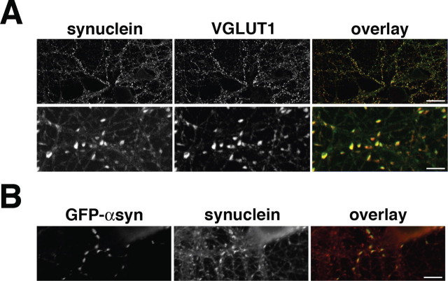
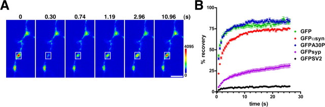
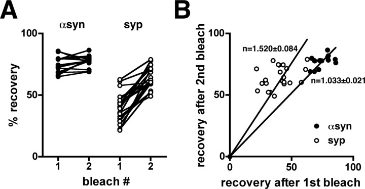
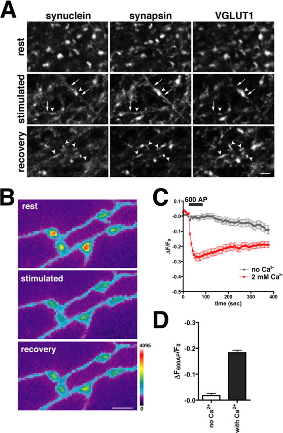
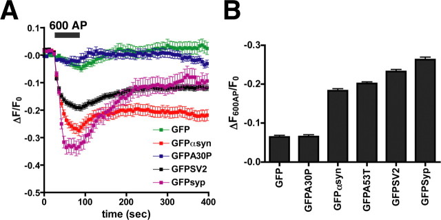
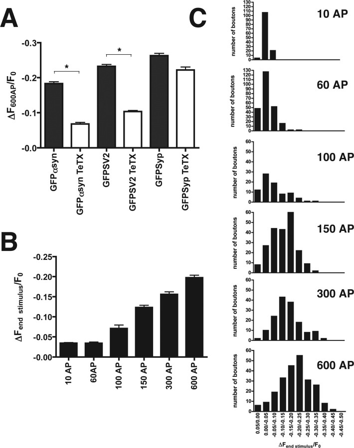
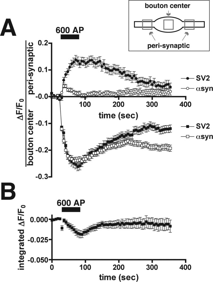
The presynaptic protein alpha-synuclein has a central role in Parkinson's disease (PD). However, the mechanism by which the protein contributes to neurodegeneration and its normal function remain unknown. Alpha-synuclein localizes to the nerve terminal and interacts with artificial membranes in vitro but binds weakly to native brain membranes. To characterize the membrane association of alpha-synuclein in living neurons, we used fluorescence recovery after photobleaching. Despite its enrichment at the synapse, alpha-synuclein is highly mobile, with rapid exchange between adjacent synapses. In addition, we find that alpha-synuclein disperses from the nerve terminal in response to neural activity. Dispersion depends on exocytosis, but unlike other synaptic vesicle proteins, alpha-synuclein dissociates from the synaptic vesicle membrane after fusion. Furthermore, the dispersion of alpha-synuclein is graded with respect to stimulus intensity. Neural activity thus controls the normal function of alpha-synuclein at the nerve terminal and may influence its role in PD.


Figures

Figure 1.
Figure 1.
Steady-state enrichment of α-synuclein at the synapse. A, Endogenous α-synuclein colocalizes with VGLUT1 at the synapses of cultured hippocampal neurons. In the overlay images, α-synuclein and VGLUT1 are pseudocolored green and red, respectively. Colocalized α-synuclein and VGLUT1 appear yellow in the overlay. Scale bars: top, 20 μm; bottom, 4 μm. B, GFP-α-synuclein (GFP-αsyn) localizes to the synapses of transfected rat hippocampal neurons. Transfection leads to a modest ∼50% increase in the expression of total α-synuclein (synuclein) detected using an antibody that recognizes both rat and human proteins. GFP-α-synuclein is pseudocolored green in the overlay, whereas total α-synuclein (transfected and endogenous) is red. Scale bar, 6 μm.
Figure 2.
Figure 2.
α-Synuclein interacts transiently with synaptic components. A, A single, isolated hippocampal bouton expressing GFP-α-synuclein was photobleached at high laser power, and recovery was monitored every 400 ms thereafter. GFP-α-synuclein recovers rapidly after photobleaching. The two synapses outside the bleached box show a reduction in fluorescence during recovery of the bleached region. The color scale is shown to the right. Scale bar, 2 μm. B, The intensity of fluorescence at the bleached synapse was quantified and expressed as a percentage of initial fluorescence, with fluorescence after the bleach defined as zero. GFP (green) and GFP-A30P (blue) recover with similar, rapid kinetics after photobleaching, consistent with their behavior as soluble proteins. In contrast, GFP-α-synuclein (red) recovers more slowly, indicating a distinct but rapidly reversible interaction with synaptic components. GFP-synapsin (GFPsyp; purple) recovers more slowly than α-synuclein, consistent with a higher affinity for synaptic vesicles. GFP-SV2 (black) does not recover after photobleaching, indicating that synaptic vesicles do not exchange between synapses, at least during this time scale. Data shown are averages ± SEM of 30-40 boutons per construct.
Figure 3.
Figure 3.
GFP-α-synuclein has no immobile fraction. A, To assess a possible immobile fraction of α-synuclein, individual boutons were bleached consecutively, and the extent of recovery normalized to the fluorescence observed at the start of each bleach. The extent of fluorescence recovery for GFP-α-synuclein (αsyn) remains the same after the second bleach, excluding an immobile fraction. In contrast, the recovery of synapsin (syp) increases after the second bleach, consistent with an immobile fraction. B, The extent of recovery for GFP-α-synuclein after bleach 1 and 2 (filled) falls on a line with slope ∼1, confirming a similar extent of recovery after both bleaches and no immobile fraction. In the case of synapsin (open), however, the ratio of recovery exceeds 1, indicating a larger recovery after the second bleach and hence an immobile fraction.
Figure 4.
Figure 4.
α-Synuclein disperses in response to depolarization. A, Neurons were fixed (rest), depolarized with 45 mm KCl and immediately fixed (stimulated), or depolarized followed by a 10 min recovery (recovery) before fixation. Synaptic boutons were identified by VGLUT1 staining and are indicated with arrowheads. Similar to synapsin, endogenous α-synuclein disperses from boutons after depolarization. Unlike synapsin, however,α-synuclein does not accumulate in the axon (arrows) after stimulation. Ten minutes after recovery, synapsin has reclustered in the synaptic terminal and colocalizes with VGLUT1. α-Synuclein does not reaccumulate at the synapse in this time frame. Scalebar, 2 μm. B, At rest, GFP-α-synuclein is enriched at synaptic boutons and disperses during stimulation with 600 action potentials (AP) delivered at 10Hz. Minimal reclustering of the protein has occurred by 5 min (recovery). The color scale is shown to the right. Scalebar, 2 μm. C, No dispersion of α-synuclein occurs during stimulation with 600 AP in calcium-free medium (gray). After stimulation in the absence of calcium, the cells were washed in calcium-containing medium for 10 min and were then restimulated (red). The dispersion of α-synuclein thus depends on calcium entry. The traces are the average ± SEM dispersion at 30 synapses from one representative cell. D, Average dispersion of GFP-α-synuclein during sequential stimulation in calcium-free and calcium-containing medium. Medium was exchanged during a 10 min rest separating the two stimulation rounds. Error bars indicate SEM. p<0.0001, Student's ttest; n = 138 synapses from three cells.
Figure 5.
Figure 5.
Synaptic vesicle proteins disperse with different kinetics after stimulation. A, GFP-α-synuclein (GFP αsyn) and GFP-A53T-α-synuclein (data not shown) disperse more slowly than GFP-synapsin (GFP-syp) after 600 action potentials (AP) delivered at 10 Hz. In contrast, GFP and GFP-A30P-α-synuclein (GFPA30P) do not disperse after 600 AP, consistent with their behavior as soluble proteins. The kinetics of α-synuclein dispersion most closely resembles that of GFP-SV2. The traces shown are averages ± SEM of 22-54 boutons from one representative cell for each construct. B, Average maximal dispersion of GFP-tagged proteins immediately after 600 AP. Error bars indicate SEM (n > 300 boutons from 6-9 cells for each construct).
Figure 6.
Figure 6.
The dispersion of α-synuclein depends on exocytosis and shows a graded response to increasing numbers of action potentials (AP). A, The average maximal dispersion after 600 AP in cells expressing GFP-α-synuclein (GFP αsyn), GFPSV2, or GFP-synapsin (GFPsyp) either with or without tetanus toxin pretreatment. Tetanus toxin greatly reduces the activity-dependent dispersion of α-synuclein and GFP-SV2. In contrast, tetanus toxin does not affect the dispersion of synapsin, consistent with its dissociation before synaptic vesicle exocytosis. Error bars indicate SEM. *p < 0.0001, Student's t test; n > 140 boutons from four to nine cells for each construct and treatment. B, Average maximal dispersion of α-synuclein increases with the number of action potentials, all delivered at 10 Hz. Error bars indicate SEM (n > 80 boutons from 3-5 cells for each of the different stimulus durations). C, Frequency histograms of GFP-α-synuclein dispersion. The number of boutons exhibiting different levels of dispersion at the end of the stimulus is plotted as a function of initial fluorescence binned in 5% increments. Increasing AP number produces greater dispersion, and the distributions show a graded rather than all-or-none, bimodal response.
Figure 7.
Figure 7.
α-Synuclein dissociates from synaptic vesicles during exocytosis. A, The activity-dependent dispersion of GFP-SV2 (SV2; filled squares) from synaptic boutons is accompanied by an increase in fluorescence in the perisynaptic area (filled circles). After endocytosis and the reclustering of synaptic vesicles, the fluorescence of GFP-SV2 returns to the center of the bouton. In contrast, GFP-α-synuclein (αsyn) does not accumulate perisynaptically (open circles) during dispersion from the bouton (open squares), consistent with its dissociation from the membrane after synaptic vesicle exocytosis. A small, brief increase in perisynaptic fluorescence follows the onset of stimulation and may reflect the rapid diffusion of GFP-α-synuclein away from the bouton. The inset indicates the regions used for quantification of fluorescence at the center of the bouton and in the perisynaptic area. The traces are averages ± SEM from one representative cell for each construct (n = 30 boutons for SV2 and 75 boutons for α-synuclein). B, Average integrated fluorescence intensity of GFP-α-synuclein over the entire field declines by 1.8 ± 0.4% after 600 action potentials (AP). Please note the difference in scale from A. The trace shows average ± SEM fluorescence (n = 5 fields).

Similar articles

Cited by

References

    1. Abeliovich A, Schmitz Y, Farinas I, Choi-Lundberg D, Ho WH, Castillo PE, Shinsky N, Verdugo JM, Armanini M, Ryan A, Hynes M, Phillips H, Sulzer D, Rosenthal A (2000) Mice lacking alpha-synuclein display functional deficits in the nigrostriatal dopamine system. Neuron 25: 239-252. - PubMed
    1. Cabin DE, Shimazu K, Murphy D, Cole NB, Gottschalk W, McIlwain KL, Orrison B, Chen A, Ellis CE, Paylor R, Lu B, Nussbaum RL (2002) Synaptic vesicle depletion correlates with attenuated synaptic responses to prolonged repetitive stimulation in mice lacking α-synuclein. J Neurosci 22: 8797-8807. - PMC - PubMed
    1. Chandra S, Chen X, Rizo J, Jahn R, Sudhof TC (2003) A broken alpha-helix in folded alpha-synuclein. J Biol Chem 278: 15313-15318. - PubMed
    1. Chandra S, Fornai F, Kwon HB, Yazdani U, Atasoy D, Liu X, Hammer RE, Battaglia G, German DC, Castillo PE, Sudhof TC (2004) Double-knockout mice for alpha- and beta-synucleins: effect on synaptic functions. Proc Natl Acad Sci USA 101: 14966-14971. - PMC - PubMed
    1. Chi P, Greengard P, Ryan TA (2001) Synapsin dispersion and reclustering during synaptic activity. Nat Neurosci 4: 1187-1193. - PubMed

Publication types

LinkOut - more resources