. 2006 Jan 6;98(1):141-8.
doi: 10.1161/01.RES.0000197783.70106.4a. Epub 2005 Nov 23.

Cardiomyocyte cell cycle activation ameliorates fibrosis in the atrium

Affiliations

Cardiomyocyte cell cycle activation ameliorates fibrosis in the atrium

Hidehiro Nakajima et al. Circ Res. .

Abstract

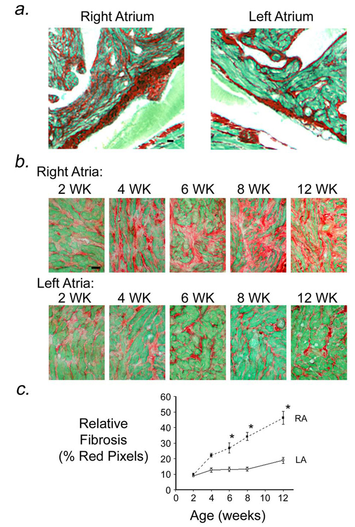
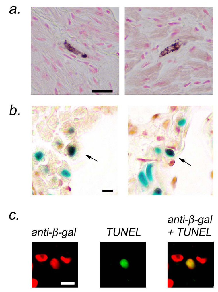
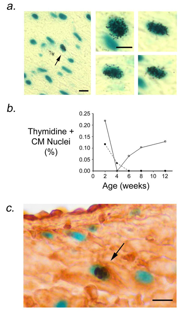
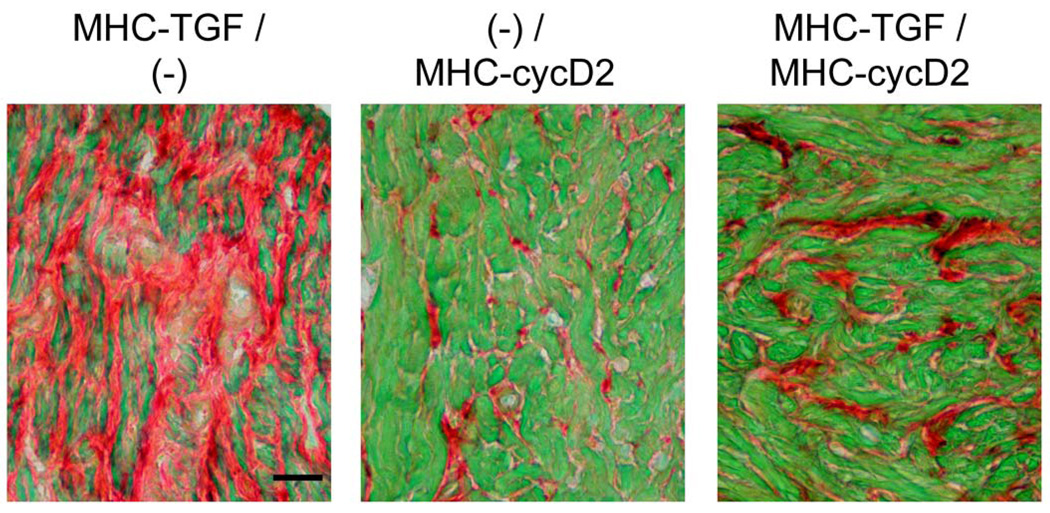
MHC-TGFcys33ser transgenic mice have elevated levels of active transforming growth factor (TGF)-beta1 in the myocardium. Previous studies have shown that these animals develop atrial, but not ventricular, fibrosis. Here we show that atrial fibrosis was accompanied with cardiomyocyte apoptosis. Although similar levels of cardiomyocyte apoptosis were present in the right and left atria of MHC-TGFcys33ser hearts, the extent of fibrosis was more pronounced in the right atrium. Thus, additional factors influence the degree of atrial fibrosis in this model. Tritiated thymidine incorporation studies revealed cardiomyocyte cell cycle activity in left atrial cardiomyocytes, but not in right atrial cardiomyocytes. These observations suggested that cardiomyocyte cell cycle activation ameliorated the severity of atrial fibrosis. To directly test this hypothesis, MHC-TGFcys33ser mice were crossed with MHC-cycD2 mice (which have constitutive cardiomyocyte cell cycle activity in the right atrium). Mice inheriting both transgenes exhibited right atrial cardiomyocyte cell cycle activity and a concomitant reduction in the severity of right atrial fibrosis, despite the presence of a similar level of cardiomyocyte apoptosis as was observed in mice inheriting the MHC-TGFcys33ser transgene alone. These data support the notion that cardiomyocyte cell cycle induction can antagonize fibrosis in the myocardium.


Figures

Figure 1
Figure 1
Atrial fibrosis in MHC-TGFcys33 ser transgenic hearts. (a) Sections from the right and left atria of an 18 month old MHC-TGFcys33 ser transgenic mouse, stained with Sirius Red and Fast Green. Bar = 20 microns. (b) Sections of right and left atria from individual MHC-TGFcys33 ser / MHC-nLAC transgenic hearts at 2, 4, 6, 8 and 12 weeks of age, stained with Sirius Red and Fast Green. Bar = 20 microns. (c) Quantitation of collagen content in the right (RA, filled squares) and left (LA, open squares) atria of MHC-TGFcys33 ser / MHC-nLAC mice at the indicated ages. Data is from 5 mice per time point. *: p<0.05, right atria vs. left atria.
Figure 2
Figure 2
Atrial cardiomyocyte apoptosis in MHC-TGFcys33 ser / MHC-nLAC double transgenic mice. (a) Activated caspase 3 immune assay for cardiomyocyte apoptosis. Cardiomyocytes at early stages of apoptosis were identified by the presence of activated caspase 3 immune reactivity (cytoplasmic brown signal in rod-shaped cells). Bar = 20 microns. (b) ISEL assay for cardiomyocyte apoptosis. Sections were stained with X-GAL to identify cardiomyocyte nuclei (blue signal) and for ISEL activity to identify apoptotic cells (HRP-conjugated reaction, dark brown signal). Apoptotic cardiomyocytes have brown and blue signals over the nucleus (arrows). Bar = 10 microns. (c) TUNEL assay for cardiomyocyte apoptosis. Sections were stained for beta-galactosidase immune reactivity to identify cardiomyocyte nuclei (rhodamine conjugated secondary antibody, red signal, left panel) and for TUNEL activity to identify apoptotic cells (FITC-conjugated secondary antibody, green signal, middle panel). Apoptotic cardiomyocytes are identified in the merged image (yellow signal, right panel). Bar = 10 microns.
Figure 3
Figure 3
Cardiomyocyte DNA synthesis in left atria from MHC-TGFcys33 ser / MHC-nLAC double transgenic mice. (a) Low (left panel) and high (right panels) views of cardiomyocytes synthesizing DNA. Sections were stained with X-GAL to identify cardiomyocyte nuclei (blue signal) and processed for autoradiography to identify DNA synthesis (nuclear silver grains). Arrow in left panel indicates a cardiomyocyte nucleus which was synthesizing DNA. Bar = 10 microns. (b) Quantitation of cardiomyocyte DNA synthesis in right (filled squares) and left (open squares) atria of MHC-TGFcys33 ser at the indicated ages. Data is from 5 mice per time point. *: p<0.05, left atria vs. right atria. (c) Phosphorylated histone H3 immune reactivity (dark brown signal, horseradish peroxidase-conjugated secondary antibody) in cardiomyocyte nuclei (blue signal, X-GAL staining) of left atrial cardiomyocytes of MHC-TGFcys33 ser / MHC-nLAC double transgenic mice at 12 weeks of age. Bar = 10 microns.
Figure 4
Figure 4
Reduced right atrial fibrosis in mice carrying both the MHC-TGFcys33 ser and the MHC-cycD2 transgenes. Right atria from MHC-TGFcys33 ser / (−) (left panel), (−) / MHC-cycD2 (middle panel) or MHC-TGFcys33 ser / MHC-cycD2 (right panel) mice, stained with Sirius Red and Fast Green. Bar = 20 microns.

References

    1. Swynghedauw B. Molecular mechanisms of myocardial remodeling. Physiol Rev. 1999;79:215–262. - PubMed
    1. Kostin S, Pool L, Elsasser A, Hein S, Drexler HC, Arnon E, Hayakawa Y, Zimmermann R, Bauer E, Klovekorn WP, Schaper J. Myocytes die by multiple mechanisms in failing human hearts. Circ Res. 2003;92:715–724. - PubMed
    1. Butt RP, Laurent GJ, Bishop JE. Collagen production and replication by cardiac fibroblasts is enhanced in response to diverse classes of growth factors. Eur J Cell Biol. 1995;68:330–335. - PubMed
    1. Boluyt MO, O'Neill L, Meredith AL, Bing OH, Brooks WW, Conrad CH, Crow MT, Lakatta EG. Alterations in cardiac gene expression during the transition from stable hypertrophy to heart failure. Marked upregulation of genes encoding extracellular matrix components. Circ Res. 1994;75:23–32. - PubMed
    1. Butt RP, Laurent GJ, Bishop JE. Mechanical load and polypeptide growth factors stimulate cardiac fibroblast activity. Ann N Y Acad Sci. 1995;752:387–393. - PubMed

Publication types