. 2005 Dec;79(24):15016-26.
doi: 10.1128/JVI.79.24.15016-15026.2005.

A point mutation within the replicase gene differentially affects coronavirus genome versus minigenome replication

Affiliations

A point mutation within the replicase gene differentially affects coronavirus genome versus minigenome replication

Carmen Galán et al. J Virol. 2005 Dec.

Abstract

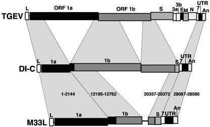
During the construction of the transmissible gastroenteritis virus (TGEV) full-length cDNA clone, a point mutation at position 637 that was present in the defective minigenome DI-C was maintained as a genetic marker. Sequence analysis of the recovered viruses showed a reversion at this position to the original virus sequence. The effect of point mutations at nucleotide 637 was analyzed by reverse genetics using a TGEV full-length cDNA clone and cDNAs from TGEV-derived minigenomes. The replacement of nucleotide 637 of TGEV genome by a T, as in the DI-C sequence, or an A severely affected virus recovery from the cDNA, yielding mutant viruses with low titers and small plaques compared to those of the wild type. In contrast, T or A at position 637 was required for minigenome rescue in trans by the helper virus. No relationship between these observations and RNA secondary-structure predictions was found, indicating that mutations at nucleotide 637 most likely had an effect at the protein level. Nucleotide 637 occupies the second codon position at amino acid 108 of the pp1a polyprotein. This position is predicted to map in the N-terminal polyprotein papain-like proteinase (PLP-1) cleavage site at the p9/p87 junction. Replacement of G-637 by A, which causes a drastic amino acid change (Gly to Asp) at position 108, affected PLP-1-mediated cleavage in vitro. A correlation was found between predicted cleaving and noncleaving mutations and efficient virus rescue from cDNA and minigenome amplification, respectively.


Figures

FIG. 1.
FIG. 1.
Genetic structure of the TGEV genome and defective RNAs. The bar on top represents the TGEV virus genome, in which the different genes are depicted as boxes. The sequence relationship between the parental genome and minigenomes DI-C and M33L is indicated by shadowed polygons. The nucleotide positions of each discontinuous region in the TGEV genome are indicated for the minigenome M33L. L, leader; ORF1a and ORF1b, replicase genes; S, spike gene; 3a and 3b, nonstructural 3a and 3b genes; E, envelope protein gene; M, membrane protein gene; N, nucleoprotein gene; 7, nonstructural gene 7; UTR, untranslated region; An, poly(A).
FIG. 2.
FIG. 2.
Analysis of genetic markers of rTGEV viruses recovered from cDNA. The genetic structure of the TGEV cDNA clone and the positions of the genetic markers (indicated by arrows) are illustrated. Abbreviations are as in Fig. 1. The nucleotide sequence of the genetic markers is indicated for the parental virus (PUR-MAD), the rescued recombinant virus (rTGEV), the cDNA clone (pBAC-TGEVFL), and the minigenome from which it was engineered (DI-C).
FIG. 3.
FIG. 3.
Phenotype of recombinant viruses with point mutations at position 637. (A) Schematic diagram showing the genome organization of the TGEV cDNA constructs with the four nucleotide substitutions at position 637. Nucleotide substitutions and the resulting amino acid changes are indicated. The names of the recombinant viruses are indicated on the right side of the panel. (B) Virus rescue. TGEV cDNAs with the desired mutations at position 637 were transfected into BHK-pAPN cells as described in Materials and Methods and virus titers were determined at the indicated times posttransfection by plaque assay on ST cells. (C) Plaque morphology of the recombinant viruses. The phenotypes of rTGEV-637C and rTGEV-637A were similar to those of rTGEV-637G and rTGEV-637T, respectively.
FIG. 4.
FIG. 4.
Rescue of M33L mutants. (A) Scheme of the experimental procedure used to evaluate minigenome amplification. The bar on top represents the M33L cDNA, in which nucleotide 637 and the mutations introduced at this position are indicated. The discontinuous viral regions used for the specific design of the forward primer for Q-RT-PCR quantification are amplified under the M33L scheme. The specific oligonucleotides are indicated by arrows. T7-driven transcripts were transfected to helper TGEV-infected ST cells and amplified by five serial virus passages in confluent ST cell monolayers (P0 to P5). Total RNA was extracted from each virus passage to analyze minigenome amplification by Q-RT-PCR. Abbreviations are as in Fig. 1. T7, T7 promoter; Rz, ribozyme of hepatitis delta virus; T7t, T7 transcription termination signal. (B) Q-RT-PCR analysis. The RNA accumulation of M33L minigenome mutants at the successive viral passages, expressed in relative units, was determined by Q-RT-PCR. M33L-637 mutants are indicated at the right of each curve, named by the letter M followed by the corresponding mutation. Q-RT-PCR analyses of a noninfected (MOCK), a nontransfected (TGEV), and a nontemplated (NTC) control are also represented. RNA input indicates the quantification of a duplicate sample of each RNA transfection extracted at 4 h posttransfection. The standard deviation of replicate quantifications (when significant) is indicated by error bars.
FIG. 5.
FIG. 5.
Rescue of DI-C mutants. (A) Scheme of DI-C cDNA in which nucleotide 637 and the specific oligonucleotides designed to amplify the first discontinuous genomic region are indicated by arrows. Abbreviations are as in Fig. 4. (B) RNA quantification. The RNA accumulation of DI-C mutants at the successive viral passages, represented in relative units, was analyzed by Q-RT-PCR. Analyses of noninfected (MOCK) and nontransfected (TGEV) controls are also represented. RNA input indicates the quantification of a duplicate sample of each RNA transfection extracted at 4 h posttransfection. The standard deviation of replicate quantifications (when significant) is indicated by error bars.
FIG. 6.
FIG. 6.
In silico analysis of the RNA secondary structure of the TGEV domain containing nucleotide 637. (A) Secondary structure of the RNA sequence derived from the TGEV mutants. The secondary structure was predicted for each mutant using the Mfold 3.1 software. Predicted minimum energy structures were common for sequences containing G or A at position 637 (folding A) or those containing T or C (folding B). Only the RNA motif containing nucleotide 637 is shown, with an arrow indicating the nucleotide at this position. (B) Relationship between structure predictions of the RNA domain containing each substitution for nucleotide 637 and the observed phenotypes of the viruses and minigenomes with the corresponding mutations.
FIG. 7.
FIG. 7.
Point mutations at nucleotide 637 affected N-terminal replicase processing in vitro. (A) Schematic representation of the amino-terminal region of ORF1a for HCoV-229E and TGEV. Arrows represent PLP-1 cleavage sites that have been experimentally shown (continuous lines) or inferred by sequence comparison (dotted lines). Replicase sequence containing the codon affected by mutations at position 637 is depicted. The two possible cleavage sites (black arrowheads) at the N terminus of the TGEV replicase were predicted by sequence alignment with HCoV-229E. Amino acids flanking the two potential PLP-1 cleavage sites are designated according to the nomenclature introduced by Schechter and Berger (38) as NH2-P3-P2-P1↓P1′-P2′-P3′-COOH. (B) trans-Cleavage assay. The PLP-1 domain and the pp1a N-terminal 610 amino acids with the wild-type sequence G at position 637 or an A at the same position, leading to the least conservative amino acid change (Gly108Asp), were generated as described in Materials and Methods. After translation, 1 volume of [35S]methionine-labeled substrate was incubated for 14 h at 30°C in the absence or presence of 2.8, 5, or 10 volumes of enzyme reaction mixture. The cleavage reaction products were separated by sodium dodecyl sulfate-10% polyacrylamide gel electrophoresis and analyzed by fluorography. Molecular mass markers are given on the left in kDa, and the uncleaved substrate and C-terminal cleavage product are indicated by arrows on the right.

Similar articles

Cited by

References

    1. Almazán, F., J. M. González, Z. Pénzes, A. Izeta, E. Calvo, J. Plana-Durán, and L. Enjuanes. 2000. Engineering the largest RNA virus genome as an infectious bacterial artificial chromosome. Proc. Natl. Acad. Sci. USA 97:5516-5521. - PMC - PubMed
    1. Baker, S. C., C. K. Shieh, L. H. Soe, M. F. Chang, D. M. Vannier, and M. M. C. Lai. 1989. Identification of a domain required for autoproteolytic cleavage of murine coronavirus gene A polyprotein. J. Virol. 63:3693-3699. - PMC - PubMed
    1. Bonilla, P. J., S. A. Hughes, and S. R. Weiss. 1997. Characterization of a second cleavage site and demonstration of activity in trans by the papain-like proteinase of the murine coronavirus mouse hepatitis virus strain A59. J. Virol. 71:900-909. - PMC - PubMed
    1. Brian, D. A., and R. S. Baric. 2005. Coronavirus genome structure and replication. Curr. Top. Microbiol. Immunol. 287:1-30. - PMC - PubMed
    1. Brierley, I., P. Digard, and S. C. Inglis. 1989. Characterization of an efficient coronavirus ribosomal frameshifting signal: requirement for an RNA pseudoknot. Cell 57:537-547. - PMC - PubMed

Publication types

MeSH terms

Substances