. 2005 Dec;79(24):15064-73.
doi: 10.1128/JVI.79.24.15064-15073.2005.

Expression of hemagglutinin esterase protein from recombinant mouse hepatitis virus enhances neurovirulence

Affiliations

Expression of hemagglutinin esterase protein from recombinant mouse hepatitis virus enhances neurovirulence

Lubna Kazi et al. J Virol. 2005 Dec.

Abstract

Murine hepatitis virus (MHV) infection provides a model system for the study of hepatitis, acute encephalitis, and chronic demyelinating disease. The spike glycoprotein, S, which mediates receptor binding and membrane fusion, plays a critical role in MHV pathogenesis. However, viral proteins other than S also contribute to pathogenicity. The JHM strain of MHV is highly neurovirulent and expresses a second spike glycoprotein, the hemagglutinin esterase (HE), which is not produced by MHV-A59, a hepatotropic but only mildly neurovirulent strain. To investigate a possible role for HE in MHV-induced neurovirulence, isogenic recombinant MHV-A59 viruses were generated that produced either (i) the wild-type protein, (ii) an enzymatically inactive HE protein, or (iii) no HE at all (A. Lissenberg, M. M. Vrolijk, A. L. W. van Vliet, M. A. Langereis, J. D. F. de Groot-Mijnes, P. J. M. Rottier, and R. J. de Groot, J. Virol. 79:15054-15063, 2005 [accompanying paper]). A second, mirror set of recombinant viruses was constructed in which, in addition, the MHV-A59 S gene had been replaced with that from MHV-JHM. The expression of HE in combination with A59 S did not affect the tropism, pathogenicity, or spread of the virus in vivo. However, in combination with JHM S, the expression of HE, regardless of whether it retained esterase activity or not, resulted in increased viral spread within the central nervous system and in increased neurovirulence. Our findings suggest that the properties of S receptor utilization and/or fusogenicity mainly determine organ and host cell tropism but that HE enhances the efficiency of infection and promotes viral dissemination, at least in some tissues, presumably by serving as a second receptor-binding protein.


Figures

FIG. 1.
FIG. 1.
Schematic representation of recombinant viruses differing in HE expression. (A) Schematic representation of MHV genome organization (top). Genes for the replicase, the structural proteins HE, S, E, M, and N, and the nonstructural proteins 2a, 4a and b, and 5a are depicted as open boxes. The bottom panel shows the relevant nucleotide and amino acid sequences of the HE variants. HE+ designates the wild-type HE of MHV strain S; HE0 designates an enzymatically inactive HE in which the active-site serine has been mutated to threonine; and HE designates a truncated HE protein (nucleotides 101 to 104 were deleted from the HE gene, causing a frameshift mutation immediately downstream of codon 33 [23a]). Recombinant viruses were generated that carry either the S gene of MHV-A59 (B; S gene indicated by a dark gray box) or that of MHV-JHM (C; S gene indicated by a light gray box). Within each set, the viruses are isogenic except for the HE gene, which is either wild type (HE+) or encodes an inactive acetylesterase (HE0) or a truncated HE polypeptide (HE). HE+, HE0, and HE are represented by striped bars, white bars, and black bars, respectively.
FIG. 2.
FIG. 2.
Viral titers in brains and livers of mice infected with rMHV-A59S-HE+, rMHV-A59S-HE0, and rMHV-A59S-HE. Four-week-old C57BL/6 mice were inoculated by either the intracranial (A) or intranasal (B) route. Viruses were titrated from brain and liver lysates from mice sacrificed at the indicated times. Each time point represents the mean titer from four or five mice. (A) Viral titers on day 5 p.i. in the brains (left panel) and livers (right panel) of mice inoculated by the intracranial route with 6,000 PFU of rMHV-A59S-HE+, rMHV-A59S-HE0, and rMHV-A59S-HE. (B) Viral titers on days 3, 5, and 7 p.i. in the brains (left panel) and livers (right panel) of mice after intranasal inoculation with 10,000 PFU of rMHV-A59S-HE+ (striped bars), rMHV-A59S-HE0 (open bars), and rMHV-A59S-HE (closed bars).
FIG. 3.
FIG. 3.
Virions of rMHV-A59S-HE+ and rMHV-A59S-HE0 isolated from the livers and brains of infected mice carry HE in their envelopes. Plaque-purified viruses isolated from liver and brain lysates derived from animals infected with rMHV-A59S-HE+ (A) or rMHV-A59S-HE0 (B and C) were used to infect LR7 cells. Virus-infected cells were metabolically labeled with [35S]methionine from 2 to 10 h p.i., after which time tissue culture supernatants were harvested and virus particles were collected by virus immunopurification with anti-MHV-A59 K135 serum as described in Materials and Methods and in reference . Samples were analyzed in 15% sodium dodecyl sulfate-polyacrylamide gels. The first two lanes of each panel contained proteins from cells infected with rMHV-A59S-HE+ (+) or rMHV-HE (−) controls.
FIG. 4.
FIG. 4.
Replication of rMHV-JHMS-HE+, rMHV-JHMS-HE0, and rMHV-JHMS-HE in L2 cell cultures. L2 monolayers were infected in duplicate with the recombinant viruses at a multiplicity of infection of 1 PFU/cell. At the time points indicated, the cells were lysed and viral titers were determined. Each time point represents the mean of duplicate samples. Circles, rMHV-JHMS-HE+; squares, rMHV-JHMS-HE0; triangles, rMHV-JHMS-HE.
FIG. 5.
FIG. 5.
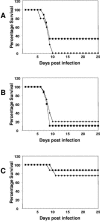
Mortality of mice infected with rMHVs expressing the JHM spike. C57BL/6 mice were infected with 20 PFU/mouse of each virus and observed for 2 weeks. Percentages of survival as a function of time are shown. The data shown represent one of three separate experiments in which two independent recombinants (represented by squares and diamonds) were tested for each virus (n = 10 mice per virus in each experiment). (A) rMHV-JHMS-HE+; (B) rMHV-JHMS-HE0; (C) rMHV-JHMS-HE.
FIG. 6.
FIG. 6.
Replication in C57BL/6 mice of rMHV-JHMS-HE+, rMHV-JHMS-HE0, and rMHV-JHMS-HE. Infectious viruses were titrated from brain lysates derived from mice on day 5 postinfection with 20 PFU of rMHV-JHMS-HE+ (striped bar), rMHV-JHMS-HE0 (open bar), and rMHV-JHMS-HE (filled bar). Each bar represents the average for eight mice. The titers are expressed as log10(PFU/g).
FIG. 7.
FIG. 7.
Viral antigen spread in the brains of mice infected with rMHV-JHMS-HE+, rMHV-JHMS-HE0, and rMHV-JHMS-HE on day 5 p.i. Viral antigen was detected in sagittal brain sections by staining with an antinucleocapsid monoclonal antibody as described in Materials and Methods. The top panels show sections from the midbrain, and the bottom panels show sections from the basal forebrain. From left to right, sections of animals infected with rMHV-JHMS-HE+, rMHV-JHMS-HE0, and rMHV-JHMS-HE are shown. Magnification, ×10.

Similar articles

Cited by

References

    1. Bond, C. W., J. L. Leibowitz, and J. A. Robb. 1979. Pathogenic murine coronaviruses. II. Characterization of virus-specific proteins of murine coronaviruses JHMV and A59V. Virology 94:371-384. - PMC - PubMed
    1. Bosch, B. J., R. van der Zee, C. A. de Haan, and P. J. Rottier. 2003. The coronavirus spike protein is a class I virus fusion protein: structural and functional characterization of the fusion core complex. J. Virol. 77:8801-8811. - PMC - PubMed
    1. Brian, D. A., B. G. Hogue, and T. E. Kienzle. 1995. The coronavirus hemagglutinin esterase glycoprotein, p. 165-179. In S. G. Siddell (ed.), The coronaviridae. Plenum Press, New York, N.Y.
    1. Bridger, J. C., E. O. Caul, and S. I. Egglestone. 1978. Replication of an enteric bovine coronavirus in intestinal organ cultures. Arch. Virol. 57:43-51. - PMC - PubMed
    1. Cavanagh, D. 1995. The coronavirus surface glycoprotein, p. 73-113. In S. G. Siddell (ed.), The coronaviridae. Plenum Press, New York, N.Y.

Publication types

LinkOut - more resources