. 2005 Dec;79(24):15074-83.
doi: 10.1128/JVI.79.24.15074-15083.2005.

Ubiquitination of the prototype foamy virus envelope glycoprotein leader peptide regulates subviral particle release

Affiliations

Ubiquitination of the prototype foamy virus envelope glycoprotein leader peptide regulates subviral particle release

Nicole Stanke et al. J Virol. 2005 Dec.

Abstract

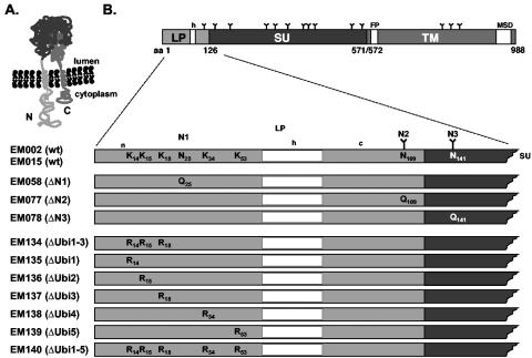
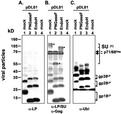
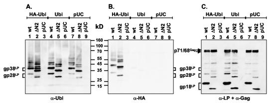
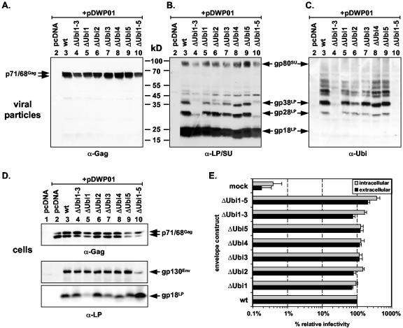
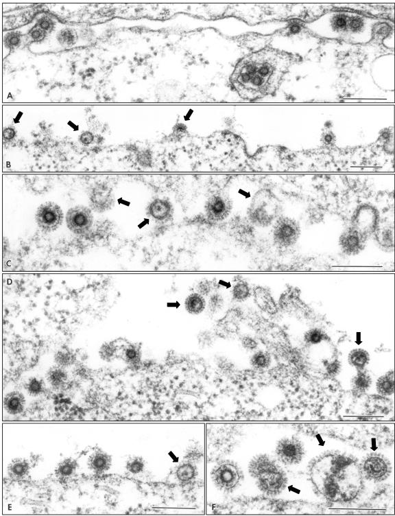
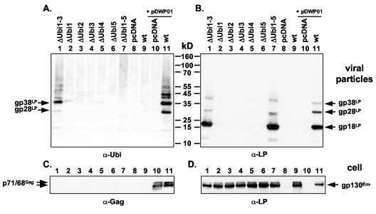
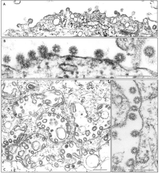
Foamy virus (FV) particle egress is unique among retroviruses because of its essential requirement for Gag and Env coexpression for budding and particle release. The FV glycoprotein undergoes a highly unusual biosynthesis resulting in the generation of three particle-associated, mature subunits, leader peptide (LP), surface (SU), and transmembrane (TM), derived from a precursor protein by posttranslational proteolysis mediated by furin or furinlike proteases. Previously at least three LP products of different molecular weights were detected in purified FV particles. Here we demonstrate that the higher-molecular-weight forms gp28LP and gp38LP are ubiquitinated variants of the major gp18LP cleavage product, which has a type II membrane topology. Furthermore, we show that all five lysine residues located within the N-terminal 60-amino-acid cytoplasmic domain of gp18LP can potentially be ubiquitinated, however, there seems to be a preference for using the first three. Inactivation of ubiquitination sites individually resulted in no obvious phenotype. However, simultaneous inactivation of the first three or all five ubiquitination sites in gp18LP led to a massive increase in subviral particles released by these mutant glycoproteins that were readily detectable by electron microscopy analysis upon expression of the ubiquitination-deficient glycoprotein by itself or in a proviral context. Surprisingly, only the quintuple ubiquitination mutant showed a two- to threefold increase in single-cycle infectivity assays, whereas all other mutants displayed infectivities similar to that of the wild type. Taken together, these data suggest that the balance between viral and subviral particle release of FVs is regulated by ubiquitination of the glycoprotein LP.


Figures

FIG. 1.
FIG. 1.
Schematic illustration of PFV Env membrane topology and expressions constructs. (A) Schematic illustration of the membrane topology of the PFV Env precursor protein as suggested from previous analysis (14, 17). (B) Schematic organization of the PFV Env protein. The N-terminal region spanning the complete leader peptide and partial sequences of the adjacent surface domain is enlarged. Individual lysine residues in the cytoplasmic domain of the LP and the asparagine residues of the first three potential N-glycosylation sites are indicated for the wild-type constructs (wt, EM002 or EM015). N-glycosylation sites used are indicated with a Y. For the individual mutant constructs amino acid sequences deviating at specific positions from the wild-type sequence are indicated. LP: leader peptide; SU: surface subunit; TM: transmembrane subunit; h: hydrophobic region of the LP; FP: fusion peptide; MSD: membrane-spanning domain; N: N terminus; C: C terminus; N1 to N3: potential N-glycosylation sites 1 to 3.
FIG. 2.
FIG. 2.
Glycosylation analysis of PFV particles. PFV particles generated by transfection of 293T cells with the replication-deficient PFV proviral vector pDL01 (lanes 1 to 3) or empty expression vector (lane 4) were purified by ultracentrifugation through 20% sucrose. Subsequently viral particles were digested with N-glycosidase F (PNGaseF) or endoglycosidase H (EndoH) or mock incubated (mock) followed by sodium dodecyl sulfate (SDS)-polyacrylamide gel electrophoresis (PAGE) and Western blot analysis using polyclonal rabbit sera specific for (A) PFV Env LP (α-LP), (B) PFV Env LP/SU (α-LP/SU) and PFV Gag (α-Gag), or (C) ubiquitin (α-Ubi). Viral proteins are indicated on the right. The different glycosylated and deglycosylated forms of the PFV SU subunit are marked by asterisks. The band of about 25 kDa in all lanes in panel C represents a staining artifact in this specific experiment that was present even in lanes loaded only with sample buffer.
FIG. 3.
FIG. 3.
Labeling of particle-associated PFV Env with HA-tagged ubiquitin. Mutant PFV particles were generated by transient cotransfection of 293T cells with the Gag/Pol-expressing PFV vector pDWP01 and the PFV Env expression construct or empty vector as indicated. In addition, HA-tagged ubiquitin (HA-Ubi), untagged ubiquitin (Ubi), or empty expression vector (pUC) was cotransfected as indicated. Subsequently, lysates of viral particles purified by ultracentrifugation through 20% sucrose were analyzed by Western blotting using consecutive incubation with antibodies specific for (A) ubiquitin (α-Ubi), (B) the HA tag (α-HA), and (C) PFV Env LP (α-LP) and PFV Gag (α-Gag). Viral proteins are indicated on the left and between panels B and C. Cells were transfected with pDWP01 and: lane 1, pczHFVenvEM015 (wt) plus pSG5 HA-Ubi (HA-Ubi); lane 2, pczHFVenvEM077 (ΔN2) plus pSG5 HA-Ubi (HA-Ubi); lane 3, pUC19 (pUC) plus pSG5 HA-Ubi (HA-Ubi); lane 4, pczHFVenvEM015 (wt) plus pSG5 Ubi (Ubi); lane 5, pczHFVenvEM077 (ΔN2) plus pSG5 Ubi (Ubi); lane 6, pUC19 (pUC) plus pSG5 Ubi (Ubi); lane 7, pczHFVenvEM015 (wt) plus pUC19 (pUC); lane 8, pczHFVenvEM077 (ΔN2) plus pUC19 (pUC); lane 9, only pUC19 (pUC).
FIG. 4.
FIG. 4.
Analysis of PFV Env mutants with inactivated ubiquitination sites. Mutant PFV particles were generated by transient cotransfection of 293T cells with the Gag/Pol-expressing PFV vector pDWP01 and the PFV Env expression construct or empty vector as indicated. Western blot analysis of purified PFV particles using consecutive incubation with antisera specific for (A) PFV Gag (α-Gag), (B) PFV Env LP/SU (α-LP/SU), or (C) ubiquitin (α-Ubi) or cell lysates using antisera specific for (D) PFV Gag (α-Gag) or PFV Env LP (α-LP). Viral proteins are indicated on the side of the blots. (E) Relative infectivity of 293T cell supernatants (extracellular) and freeze-thaw cell lysates (intracellular) using the GFP marker gene transfer assay on HT1080 target cells. The values obtained using the wild-type PFV Env expression plasmid (wt) were arbitrarily set to 100%. The mean values and standard deviations of at least three independent experiments are shown. Cells were either transfected with pcDNA3.1+zeo alone (lane 1: pcDNA) or cotransfected with pDWP01 and: lane 2, pcDNA3.1+zeo (pcDNA); lane 3, pczHFVenvEM015 (wt); lane 4, pczHFVenvEM134 (ΔUbi1-3); lane 5, pczHFVenvEM135 (ΔUbi1); lane 6, pczHFVenvEM136 (ΔUbi2); lane 7, pczHFVenvEM137 (ΔUbi3); lane 8, pczHFVenvEM138 (ΔUbi4); lane 9, pczHFVenvEM139 (ΔUbi5); lane 10, pczHFVenvEM140 (ΔUbi1-5).
FIG. 5.
FIG. 5.
Electron microscopy analysis of proviral expression clones. Electron micrographs showing representative thin sections of 293T cells transiently transfected with the wild-type PFV proviral expression vector pczHSRV2EM026 (A) or the mutant proviral vector pczHSRV2EM140 expressing the ubiquitination-deficient PFV Env protein ΔUbi1-5 (B to F). Subviral particles are marked by solid arrows. Magnifications: (A) 89,000×; (B) 70,000×; (C) 110,000×; (D) 90,000×; (E) 95,000×; (F) 127,000×. Bar, 200 nm.
FIG. 6.
FIG. 6.
Analysis of subviral particle release. Subviral particles were harvested by ultracentrifugation through 20% sucrose from supernatants of 293T cells transfected with the individual Env expression constructs or empty expression vector as indicated (lanes 1 to 9). As a control wild-type PFV particles were generated by cotransfecting the PFV Gag/Pol expression vector pDWP01 and the wild-type PFV Env expression construct pczHFVenvEM015 or empty expression vector (lanes 10 and 11). Western blot analysis of particle preparations using consecutive incubation with (A) a ubiquitin-specific monoclonal antibody (α-Ubi, P4D1) and (B) a polyclonal antiserum specific for PFV LP (α-LP). Western blot analysis of the corresponding cell lysates using polyclonal antisera specific for (C) PFV Gag (α-Gag) and for (D) PFV LP (α-LP). Cells were transfected with: lane 1, pczHFVenvEM134 (ΔUbi1-3); lane 2, pczHFVenvEM135 (ΔUbi1); lane 3, pczHFVenvEM136 (ΔUbi2); lane 4, pczHFVenvEM137 (ΔUbi3); lane 5, pczHFVenvEM138 (ΔUbi4); lane 6, pczHFVenvEM139 (ΔUbi5); lane 7, pczHFVenvEM140 (ΔUbi1-5); lane 8, pcDNA3.1+zeo (pcDNA); or lane 9, pczHFVenvEM015 (wt); or cotransfected with pDWP01 and pcDNA3.1+zeo (pcDNA, lane 10) or pczHFVenvEM015 (wt, lane 11).
FIG. 7.
FIG. 7.
Electron microscopy analysis of ΔUbi1-5 PFV Env-expressing cells. Electron micrographs showing representative thin sections of (A and B) HT1080 cells stably expressing the mutant ΔUbi1-5 mutant PFV Env protein or (C and D) 293T cells transiently transfected with the mutant PFV Env expression vector pczHFVenvEM140 (ΔUbi1-5). Magnifications: (A) 72,000×; (B) 180,000×; (C) 57,000×; (D) 138,000×. Bar, 200 nm.

Similar articles

Cited by

References

    1. Baunach, G., B. Maurer, H. Hahn, M. Kranz, and A. Rethwilm. 1993. Functional analysis of human foamy virus accessory reading frames. J. Virol. 67:5411-5418. - PMC - PubMed
    1. Bültmann, A., J. Eberle, and J. Haas. 2000. Ubiquitination of the human immunodeficiency virus type 1 Env glycoprotein. J. Virol. 74:5373-5376. - PMC - PubMed
    1. Cosson, P. 1996. Direct interaction between the envelope and matrix proteins of HIV-1. EMBO J. 15:5783-5788. - PMC - PubMed
    1. Du Bridge, R. B., P. Tang, H. C. Hsia, P. M. Leong, J. H. Miller, and M. P. Calos. 1987. Analysis of mutation in human cells by using an Epstein-Barr virus shuttle system. Mol. Cell. Biol. 7:379-387. - PMC - PubMed
    1. Duda, A., A. Stange, D. Lüftenegger, N. Stanke, D. Westphal, T. Pietschmann, S. W. Eastman, M. L. Linial, A. Rethwilm, and D. Lindemann. 2004. Prototype foamy virus envelope glycoprotein leader peptide processing is mediated by a furin-like cellular protease, but cleavage is not essential for viral infectivity. J. Virol. 78:13865-13870. - PMC - PubMed

Publication types

MeSH terms

LinkOut - more resources