. 2005 Dec;79(24):15199-208.
doi: 10.1128/JVI.79.24.15199-15208.2005.

The papain-like protease from the severe acute respiratory syndrome coronavirus is a deubiquitinating enzyme

Affiliations

The papain-like protease from the severe acute respiratory syndrome coronavirus is a deubiquitinating enzyme

Holger A Lindner et al. J Virol. 2005 Dec.

Abstract

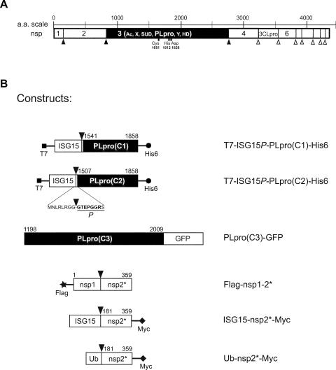
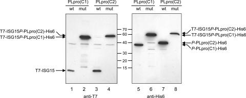
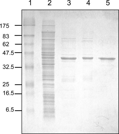
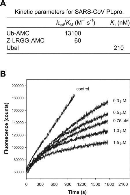
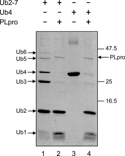
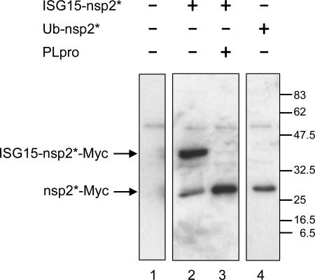
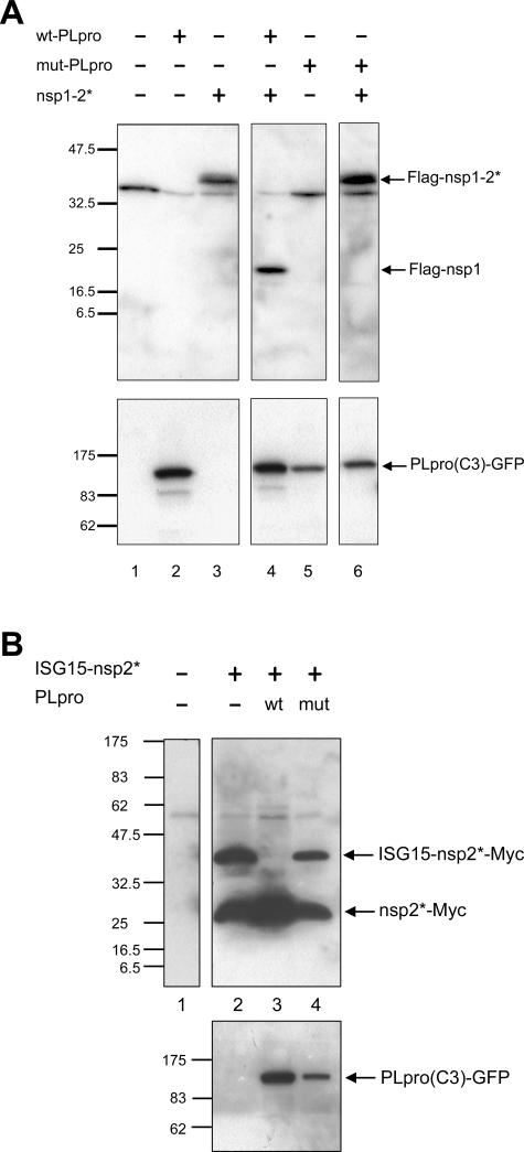
The severe acute respiratory syndrome coronavirus papain-like protease (SARS-CoV PLpro) is involved in the processing of the viral polyprotein and, thereby, contributes to the biogenesis of the virus replication complex. Structural bioinformatics has revealed a relationship for the SARS-CoV PLpro to herpesvirus-associated ubiquitin-specific protease (HAUSP), a ubiquitin-specific protease, indicating potential deubiquitinating activity in addition to its function in polyprotein processing (T. Sulea, H. A. Lindner, E. O. Purisima, and R. Menard, J. Virol. 79:4550-4551, 2005). In order to confirm this prediction, we overexpressed and purified SARS-CoV PLpro (amino acids [aa]1507 to 1858) from Escherichia coli. The purified enzyme hydrolyzed ubiquitin-7-amino-4-methylcoumarin (Ub-AMC), a general deubiquitinating enzyme substrate, with a catalytic efficiency of 13,100 M(-1)s(-1), 220-fold more efficiently than the small synthetic peptide substrate Z-LRGG-AMC, which incorporates the C-terminal four residues of ubiquitin. In addition, SARS-CoV PLpro was inhibited by the specific deubiquitinating enzyme inhibitor ubiquitin aldehyde, with an inhibition constant of 210 nM. The purified SARS-CoV PLpro disassembles branched polyubiquitin chains with lengths of two to seven (Ub2-7) or four (Ub4) units, which involves isopeptide bond cleavage. SARS-CoV PLpro processing activity was also detected against a protein fused to the C terminus of the ubiquitin-like modifier ISG15, both in vitro using the purified enzyme and in HeLa cells by coexpression with SARS-CoV PLpro (aa 1198 to 2009). These results clearly establish that SARS-CoV PLpro is a deubiquitinating enzyme, thereby confirming our earlier prediction. This unexpected activity for a coronavirus papain-like protease suggests a novel viral strategy to modulate the host cell ubiquitination machinery to its advantage.


Figures

FIG. 1.
FIG. 1.
Schematic diagrams of the SARS-CoV replicase polyprotein 1a and fusion protein constructs used in this work. (A) Filled and open arrowheads indicate the positions of the PLpro and 3CLpro processing sites, respectively. nsp3 is shown inverted, and Ac (acidic), X, SUD (SARS unique domain), PLpro, Y, and HD (hydrophobic domain) refer to the putative nsp3 domain organization. The three residues of the putative PLpro catalytic triad are identified. (B) Fusion protein constructs are shown and labeled as described in the text. Filled arrowheads indicate the positions of putative sites for cleavage by SARS-CoV PLpro, one of which was confirmed in this work by N-terminal sequencing and is shown in underlined bold type. Numbers of N- and C-terminal residues of nsp1-, nsp2-, and nsp3-derived sequences are indicated. N- and C-terminal extensions represent accordingly labeled peptide epitopes added for Western blot detection. aa, amino acid.
FIG. 2.
FIG. 2.
Expression of ISG15P-SARS-CoV PLpro fusion proteins in E. coli. The expression of T7-ISG15P-PLpro(C1)-His6 and T7-ISG15P-PLpro(C2)-His6 wild-type (wt) proteins (lanes 1, 3, 5, and 7) and C1651A mutants (mut) (lanes 2, 4, 6, and 8) was analyzed by resolving 5-μl aliquots of cell lysates by 12% SDS-PAGE and Western blot detection with T7- and His6-specific antibodies. Positions of molecular mass standards are indicated between the blots (in kDa).
FIG. 3.
FIG. 3.
SDS-PAGE analysis of SARS-CoV PLpro purified from E. coli. SARS-CoV PLpro (from residue Gly1507 to Thr1858) was expressed as a fusion protein to the precursor of ISG15. SARS-CoV PLpro was purified as a 41-kDa autocleavage product, with the prosequence of ISG15 remaining at the N terminus. Marker proteins (lane 1), 1.5 μl of the soluble fraction from the cell lysate (lane 2), and 1.5 μg (each) of eluate from Ni-NTA (lane 3), PD-10 (lane 4), and MonoQ (lane 5) columns were separated by 12% SDS-PAGE and stained with Coomassie blue. The molecular weights of the marker proteins are indicated on the left of the gel (in kDa).
FIG. 4.
FIG. 4.
Enzymatic characterization of SARS-CoV PLpro purified from E. coli. (A) Kinetic parameters of substrate hydrolysis and of inhibition by Ubal. (B) Fluorescence versus time progress curves for hydrolysis of Ub-AMC (125 nM) in the presence of various amounts of Ubal. Inhibitor concentrations are indicated next to the curves.
FIG. 5.
FIG. 5.
Hydrolysis of branched polyubiquitin chains by SARS-CoV PLpro purified from E. coli. Lys48-linked Ub2-7 or Ub4 chains were incubated with SARS-CoV PLpro (lanes 2 and 4). SARS-CoV PLpro was present at 8 μg/ml, and either substrate was present at 200 μg/ml. Controls were without enzyme (lanes 1 and 3). Proteins were resolved by 10% SDS and revealed by Coomassie blue staining. The molecular weights of the marker proteins are indicated on the right of the gel (in kDa).
FIG. 6.
FIG. 6.
Hydrolysis of an ISG15-nsp2* fusion protein by SARS-CoV PLpro purified from E. coli. ISG15-nsp2*-Myc was expressed in HeLa cells. Aliquots (20 μg) of cell lysate proteins were incubated without enzyme or with 200 ng of purified SARS-CoV PLpro in volumes of 20 μl. Samples were resolved by 10% SDS-PAGE and subjected to anti-Myc Western blotting (lanes 2 and 3). Expression of the corresponding Ub-nsp2* fusion is also shown (lane 4). The molecular weights of the marker proteins are indicated on the right of the gel (in kDa).
FIG. 7.
FIG. 7.
Cell-based trans-cleavage assays of SARS-CoV PLpro activity. (A) Flag-nsp1-2* and either the wild-type (wt) or the C1651A mutant (mut) of PLpro(C3)-GFP were either expressed alone or coexpressed in HeLa cells. Aliquots of 20 μg of cell lysate proteins were resolved by 10% SDS-PAGE and subjected to anti-Flag Western blotting. Expression of the PLpro fusion proteins was confirmed using an anti-GFP antibody. (B) Either PLpro variant was coexpressed with ISG15-nsp2*-Myc, and an anti-Myc antibody was used for Western blot detection. The molecular weights of the marker proteins are indicated on the left of each gel (in kDa).

References

    1. Amerik, A. Y., and M. Hochstrasser. 2004. Mechanism and function of deubiquitinating enzymes. Biochim. Biophys. Acta 1695:189-207. - PubMed
    1. Andres, G., A. Alejo, C. Simon-Mateo, and M. L. Salas. 2001. African swine fever virus protease, a new viral member of the SUMO-1-specific protease family. J. Biol. Chem. 276:780-787. - PubMed
    1. Baker, R. T. 1996. Protein expression using ubiquitin fusion and cleavage. Curr. Opin. Biotechnol. 7:541-546. - PubMed
    1. Balakirev, M. Y., M. Jaquinod, A. L. Haas, and J. Chroboczek. 2002. Deubiquitinating function of adenovirus proteinase. J. Virol. 76:6323-6331. - PMC - PubMed
    1. Bradford, M. M. 1976. A rapid and sensitive method for the quantitation of microgram quantities of protein utilizing the principle of protein-dye binding. Anal. Biochem. 72:248-254. - PubMed

Publication types

MeSH terms