. 2005 Dec;79(24):15302-13.
doi: 10.1128/JVI.79.24.15302-15313.2005.

Respiratory syncytial virus-inducible BCL-3 expression antagonizes the STAT/IRF and NF-kappaB signaling pathways by inducing histone deacetylase 1 recruitment to the interleukin-8 promoter

Affiliations

Respiratory syncytial virus-inducible BCL-3 expression antagonizes the STAT/IRF and NF-kappaB signaling pathways by inducing histone deacetylase 1 recruitment to the interleukin-8 promoter

Mohammad Jamaluddin et al. J Virol. 2005 Dec.

Abstract

Respiratory syncytial virus (RSV) is a paramyxovirus that produces airway inflammation, in part by inducing interleukin-8 (IL-8) expression, a CXC-type chemokine, via the NF-kappaB/RelA and STAT/IRF signaling pathways. In RSV-infected A549 cells, IL-8 transcription attenuates after 24 h in spite of ongoing viral replication and persistence of nuclear RelA, suggesting a mechanism for transcriptional attenuation. RSV infection induces B-cell lymphoma protein -3 (Bcl-3) expression 6 to 12 h after viral infection, at times when IL-8 transcription is inhibited. By contrast, 293 cells, deficient in inducible Bcl-3 expression, show no attenuation of IL-8 transcription. We therefore examined Bcl-3's role in terminating virus-inducible IL-8 transcription. Transient expression of Bcl-3 potently inhibited virus-inducible IL-8 transcription by disrupting both the NF-kappaB and STAT/IRF pathways. Although previously Bcl-3 was thought to capture 50-kDa NF-kappaB1 isoforms in the cytoplasm, immunoprecipitation (IP) and electrophoretic mobility shift assays indicate that nuclear Bcl-3 associates with NF-kappaB1 without affecting DNA binding. Additionally, Bcl-3 potently inhibited the STAT/IRF pathway. Nondenaturing co-IP assays indicate that nuclear Bcl-3 associates with STAT-1 and histone deacetylase 1 (HDAC-1), increasing HDAC-1 recruitment to the IL-8 promoter. Treatment with the HDAC inhibitor trichostatin A blocks attenuation of IL-8 transcription. A nuclear targeting-deficient Bcl-3 is unable to enhance HDAC-1-mediated chemokine repression. Finally, small inhibitory RNA-mediated Bcl-3 "knockdown" resulted in enhanced RSV-induced chemokine expression in A549 cells. These data indicate that Bcl-3 is a virus-inducible inhibitor of chemokine transcription by interfering with the NF-kappaB and STAT/IRF signaling pathways by complexing with them and recruiting HDAC-1 to attenuate target promoter activity.


Figures

FIG. 1.
FIG. 1.
Kinetics of IL-8 promoter activation in A549 cells by RSV is distinct from that of 293 cells. (A) RSV infection induces IL-8 promoter activity in A549 cells in a biphasic manner. A549 cells were transiently transfected with hIL-8 LUC (2 μg) and β-Gal (0.5 μg) plasmids. After 24 h of transfection, cells were infected with RSV (MOI, 1.0) for the indicated times, and reporter activity was measured. Luciferase activity was normalized with β-galactosidase activity and expressed as fold induction relative to zero h. Each bar is the mean ± standard error of the mean (SEM) of luciferase activity measured in triplicate from a representative experiment repeated three times. **, P < 0.001 from 15-h RSV infection, Student's t test. Inset, Western immunoblot for nuclear NF-κB/RelA(p65). (Bottom panel) The blot was probed with β-actin as loading control. (B) RSV infection increases Bcl-3 expression in A549 cells. A 100-μg aliquot of cytoplasmic (Cyto) and nuclear (Nuc) protein from A549 cells infected with RSV for different hours (indicated at top) was fractionated by SDS-PAGE. (Top panel) Bcl-3 was detected by Western immunoblotting. (Bottom panel) The blot was probed with anti-β-actin Ab to show equal protein loading. (C) Bcl-3 expression in RSV-infected 293 cells. Whole-cell extract was prepared from A549 and 293 cells either uninfected or infected with RSV for 15 h. Equal amounts of protein (100 μg) from A549 and 293 cell extracts were fractionated by SDS-PAGE, and Bcl-3 was detected by Western immunoblotting. (Bottom panel) β-Actin stain for loading control. (D) Kinetics of RSV-induced IL-8 promoter activity in 293 cells. 293 cells were transiently transfected with hIL-8/LUC (2 μg) and β-Gal (0.5 μg) plasmids. Cells were infected with RSV (MOI, 1.0) for the indicated times, and luciferase activity was measured 48 h after transfection. Each bar is the mean ± SEM of luciferase activity of triplicate measurements from a representative experiment repeated three times. **, significant difference from 15 h (P < 0.001).
FIG. 2.
FIG. 2.
Bcl-3 expression inhibits IL-8 promoter activity mediated by NF-κB and ISRE. (A) Bcl-3 inhibits RSV-induced IL-8 promoter activity. 293 cells were transfected with hIL-8/LUC reporter plasmid, pcDNA3 empty vector, or pcDNA3-Flag-Bcl-3 expression vectors as indicated. At 24 h later, cells were infected with or without purified RSV (MOI, 1.0) for 24 h, and luciferase reporter activity was measured. Luciferase reporter activity was normalized with an internal control (β-Gal activity) and expressed as the fold induction over the basal activity. Inset, Western immunoblot of cells fractionated into cytoplasmic and nuclear fractions. Molecular mass markers (in kDa) are shown at the left. Top panel, extracts were probed with anti-Flag antibody. Bottom panel, anti-β-actin Ab was used as an internal control. (B) Effect of Bcl-3 on promoter activity driven by NF-κB, ISRE, and AP-1 sites. Luciferase reporters driven by multimeric NF-κB, IRF-1(ISRE), or AP-1 sites were cotransfected with 200 ng empty vector (-Bcl-3) or Bcl-3 expression vector (+ Bcl-3) into 293 cells. Transfected cells were stimulated with TNF-α (20 ng/ml, 5 h), RSV(MOI, 1.0, 24 h), or PMA (0.5 μM, 5 h), and luciferase reporter activity was measured. Luciferase activity was normalized with β-Gal activity. Relative to unstimulated controls, TNF stimulated the NF-κB multimer 200-fold, RSV induced the ISRE multimer 140-fold, and PMA induced the AP-1 multimer by 50-fold. Data are represented as the percentage of maximal reporter activity (set at 100%). Each bar is the mean ± standard error of the mean (SEM) of two experiments done in triplicate. *, P < 0.05; **, P < 0.001; Student's t test. (C) Bcl-3 inhibits ISRE-driven transcriptional activity in HEp-2 cells. HEp-2 cells were transiently cotransfected with 2 μg (ISRE)x3-Luc plasmids and pcDNA3-Flag-Bcl-3 (Bcl-3) or empty vector (Vec) (100 ng). After 24 h, transfected cells were infected with RSV for 24 h and luciferase activity was measured. Results are means ± SEM of two experiments with triplicate deattenuations. Luciferase activity was normalized with β-Gal activity and is expressed as the fold activation over uninfected cells. **, significantly different from RSV-infected cells (P < 0.001).
FIG. 3.
FIG. 3.
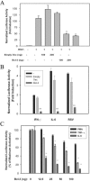
Bcl-3 expression inhibits the STAT/IRF-1 pathway. (A) Bcl-3 inhibits IRF-1 promoter activity. A549 cells were cotransfected with 2 μg hIRF-1/LUC plasmids and either pcDNA3-Flag-Bcl-3 (Bcl-3) or empty vector (Vec). Where indicated, cells were infected with RSV (MOI, 1.0; 24 h). Shown is the normalized luciferase reporter activity. (B) Stimulus type-independent inhibition of STAT-Luc by Bcl-3. 293 cells were transiently transfected with STAT-Luc reporter plasmid with or without Bcl-3 expression vector. Following 24 h of transfection cells were either left untreated (-) or treated with IFN-γ, IL-6, or RSV for 24 h. Each bar is the mean ± standard error of the mean (SEM) of a representative experiment repeated twice. **, significant difference from IFN-γ, IL-6, or RSV alone (P < 0.001). (C) Bcl-3 is a highly potent inhibitor of STAT activity. 293 cells were transfected with STAT-Luc, NF-κB-Luc, or AP-1-Luc and either cotransfected with empty vector or an increasing amount of Bcl-3 expression vector (Bcl-3), and luciferase activity was measured following IL-6 (24 h), TNF (6 h), or PMA (5 h) stimulation. Data are means ± SEM of a representative experiment with triplicate deattenuations. *, P < 0.05 from RSV alone; **, P < 0.001 from RSV alone.
FIG. 4.
FIG. 4.
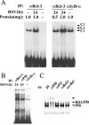
Bcl-3 inhibits NF-κB-dependent IL-8 transcription through NF-κB association. (A) RSV infection increases Bcl-3 association with NF-κB. Immunoprecipitation (IP)-EMSA was done with nuclear extract from uninfected or RSV-infected A549 cells. Nuclear protein (0.5, 1, and 2 mg) was IP with ant-Bcl-3 Ab (αBcl-3) or anti-IκB-α Ab (αIκB-α), dissociated from beads, and used in an EMSA performed with 32P-labeled double-stranded NF-κB binding site from the IL-8 promoter (see Materials and Methods). (B) NF-κB complex associated with IκBα is distinct from that associated with Bcl-3. Nuclear protein (1 mg) from RSV-infected A549 cells was IP with anti-Bcl-3 Ab (αBcl-3) or anti-p50 Ab (αp50). Cytoplasmic protein (1 mg) from uninfected cells was immunoprecipitated with anti-IκBα antibody (αIκBα). The NF-κB site was analyzed by EMSA, as in Fig. 3B. (C) Identification NF-κB subunits by antibody supershift assay. Nuclear proteins obtained by IP using anti-Bcl-3 Ab were incubated with preimmune serum (PI) or antibody to p50, p52, p65, or cRel on ice for 30 min prior to EMSA. The compositions of the DNA binding complex and the supershifted band were indicated by the arrow on the right.
FIG. 5.
FIG. 5.
Bcl-3 interacts with STAT-1 in RSV-infected nuclei. (A) Nondenaturing co-IP of Bcl-3 complexes. Bcl-3-transfected 293 cells were uninfected or infected with RSV for 20 h. A 250-μg aliquot of nuclear extract was IP with anti-Flag Ab. STAT-1 in immune complexes was detected by Western immunoblotting. Load, input sample. (Bottom panel) The membrane was reprobed with anti-Flag Ab to determine Bcl-3 recovery. (B) Nondenaturing co-IP of STAT-1 complexes. Flag-Bcl-3 expression vector (2.5 μg) was transfected into 293 cells. A 250-μg aliquot of nuclear protein was IP using anti-STAT-1 Ab (α-STAT-1) or preimmune rabbit IgG (IgG). Starting material (load) was used as staining control. (Top panel) Flag-Bcl-3 in the immune complex was detected in the Western immunoblot assay using anti-Flag Ab. (Bottom panel) STAT-1 recovery was detected by reprobing the membrane with anti-STAT-1 Ab. (C) Bcl-3 expression does not inhibit RSV-induced STAT-1 and -3 DNA binding, as shown with the EMSA of STAT DNA binding activity. 293 cells were transfected with either 2.5 μg of empty vector (Vec) or 2.5 μg pcDNA3-Flag-Bcl-3 expression vector (Bcl-3) for 24 h. Cells were then RSV infected (MOI, 1) for the indicated times, and nuclear protein was extracted. An EMSA was carried out with 15 μg protein using 32P-labeled SIE probe. STAT, inducible STAT-1 and -3 binding complex; NS, nonspecific binding B. (D) Bcl-3 associates with STAT-3. V5-tagged STAT3 and Flag-Bcl-3 expression vectors were cotransfected. Lysates were immunoprecipitated with anti-Flag Ab or IgG as indicated. STAT-3 in the immune complex was detected with Western blotting using anti-V5 Ab.
FIG. 6.
FIG. 6.
Bcl-3 recruits HDAC-1 to the IL-8 promoter. (A) RSV infection increases Bcl-3 and HDAC-1 association in vivo. Nuclear extracts (1 mg) from control or RSV-infected (15 h) A549 cells were IP with anti-Bcl-3 Ab (α-BCL-3) or IgG. Immune complexes were washed, and HDAC-1 was detected by Western immunoblotting. (Bottom panel) The membrane was reprobed with anti-Bcl-3 Ab. (B) HDAC-1 and Bcl-3 recruitment to the IL-8 promoter in RSV infection, as shown in a ChIP assay with A549 cells infected with RSV for the times indicated. Shown is ethidium bromide-stained agarose gel. (Top panel) IL-8 promoter PCR of chromatin IP with IgG or anti-Bcl-3 Ab (α-Bcl-3). (Middle panel) PCR of chromatin IP with anti-HDAC-1 Ab. Bottom panel, control PCR for equivalent amount of DNA used as input for IP (input). M, DNA size markers; PC, positive control genomic DNA. (C) Bcl-3 expression enhances HDAC-1 recruitment to IL-8 promoter. Empty pcDNA (-) or Flag-Bcl-3 expression vectors (+) were transfected into 293 cells. After 24 h, a ChIP assay was performed using either anti-HDAC-1 Ab (α-HDAC-1; 5 μg; lanes 2 and 3) or IgG (lane 1) as described in Materials and Methods. (Upper panel) IL-8 PCR of HDAC-1 IP chromatin; (lower panel) IL-8 PCR from IP input. (D) An HDAC inhibitor reverses late-phase IL-8 inhibition. A549 cells were transfected with hIL-8/LUC. After 24 h, cells were pretreated with or without TSA (100 nM) for 5 h and infected with RSV for the indicated times. Luciferase activity was normalized with β-galactosidase activity and expressed as the percentage of maximal activation observed at 15 h of RSV infection. *, P < 0.01 at same infection time, +TSA versus -TSA; **, P < 0.001 compared to RSV infection alone. (E) HDAC-1 inhibition of transcription requires nuclear Bcl-3. 293 cells were transfected with 0.5 μg ISRE-Luc or cotransfected with 500 ng of HDAC-1, 200 ng of WT Flag-Bcl-3, or N-terminal deletion mutant (ΔN Flag-Bcl-3) plasmids as indicated. After infection with RSV for 24 h, cells were lysed and luciferase activity was measured. Each bar is the mean ± standard error of the mean of normalized luciferase activity from two experiments done in triplicate. (F) Wild-type but not the N-terminal deletion mutant of Bcl-3 is localized in the nucleus. 293 cells were transiently transfected with 2.5 μg of WT or the N-terminal deletion (ΔN) of Flag-tagged Bcl-3 plasmids. After 24 h, cytoplasmic (Cyt) and nuclear (Nuc) extracts were prepared from the transfected cells as described in Materials and Methods. WT or ΔN Flag Bcl-3 was detected by Western immunoblotting using Flag monoclonal antibody. Numbers on the left indicate molecular weight standards.
FIG. 7.
FIG. 7.
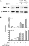
siRNA-mediated down-regulation of endogenous Bcl-3 enhances RSV-induced IL-8 and IRF-1 expression in A549 cells. (A) Down-regulation of Bcl-3 protein expression. (Top panel) Whole-cell lysate (200 μg protein) from A549 cells was transfected either with Bcl-3 siRNA (50 nM) or NS-siRNA (50 nM) and was infected with (+) or without (-) RSV. Bcl-3 was detected by Western immunoblotting. (Bottom panel) The same membrane was reprobed with anti-β-actin Ab to show equal loading. (B) Bcl-3 down-regulation enhances RSV-induced IL-8 and IRF-1 expression. RNA from untransfected NS-siRNA (50 nM) or Bcl-3 siRNA (50 nM) cells with or without RSV infection was subjected to Q-RT-PCR. (Top panel) IL-8 transcript levels; (bottom panel) IRF-1 mRNA. All data presented are normalized to 18S RNA and expressed as the fold induction relative to untransfected and uninfected controls. Results are means ± standard deviations of two experiments run in duplicate. *, P < 0.02; **, P < 0.001.
FIG. 8.
FIG. 8.
Schematic model for Bcl-3-mediated inhibition of chemokine expression. Based on these findings, Bcl-3 inhibits RSV-induced chemokine expression by antagonizing the NF-κB and STAT-1/IRF signaling pathways. Shown is a schematic diagram of activated nuclear factors early (left) in the course of RSV infection (<12 h), and late (right) in the course of RSV infection (>24 h). Early in the course of RSV infection, the IL-8 promoter is occupied by transcription factors corresponding to the IRF-, STAT, and NF-κB families (for simplicity, only IRF-1 and STAT-1 are depicted), and IRF-1 synthesis is STAT-1 dependent. Late in the course of RSV infection, Bcl-3 is synthesized, where it antagonizes the transcriptional activity of NF-κB and STAT-1 by complexing and recruiting HDAC-1 to target genes.

Similar articles

Cited by

References

    1. Aherne, W. T., T. Bird, S. D. B. Court, P. S. Gardner, and J. McQuillin. 1970. Pathological changes in virus infections of the lower respiratory tract in children. J. Clin. Pathol. 23:7-18. - PMC - PubMed
    1. Alexander, W. S., R. Starr, J. E. Fenner, C. L. Scott, E. Handman, N. S. Sprigg, J. E. Corbin, A. L. Cornish, R. Darwiche, C. M. Owczarek, T. W. Kay, N. A. Nicola, P. J. Hertzog, D. Metcalf, and D. J. Hilton. 1999. SOCS1 is a critical inhibitor of interferon gamma signaling and prevents the potentially fatal neonatal actions of this cytokine. Cell 98:597-608. - PubMed
    1. Baggiolini, M., B. Dewald, and B. Moser. 1997. Human chemokines: an update. Annu. Rev. Immunol. 15:675-705. - PubMed
    1. Barnes, P. J., and M. Karin. 1997. Nuclear factor-κB: a pivotal transcription factor in chronic inflammatory diseases. N. Engl. J. Med. 336:1066-1071. - PubMed
    1. Bitko, V., A. Velazquez, L. Yank, Y.-C. Yang, and S. Barik. 1997. Transcriptional induction of multiple cytokines by human respiratory syncytial virus requires activation of NF-κB and is inhibited by sodium salicylate and aspirin. Virology 232:369-378. - PubMed

Publication types

MeSH terms

LinkOut - more resources