. 2005 Dec;79(24):15376-87.
doi: 10.1128/JVI.79.24.15376-15387.2005.

Division of labor within human immunodeficiency virus integrase complexes: determinants of catalysis and target DNA capture

Affiliations

Division of labor within human immunodeficiency virus integrase complexes: determinants of catalysis and target DNA capture

Tracy L Diamond et al. J Virol. 2005 Dec.

Abstract

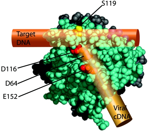
Following the completion of reverse transcription, the human immunodeficiency virus integrase (IN) enzyme covalently links the viral cDNA to a host cell chromosome. An IN multimer carries out this reaction, but the roles of individual monomers within the complex are mostly unknown. Here we analyzed the distribution of functions for target DNA capture and catalysis within the IN multimer. We used forced complementation between pairs of IN deletion derivatives in vitro as a tool for probing cis-trans relationships and analyzed amino acid substitutions affecting either catalysis or target site selection within these complementing complexes. This allowed the demonstration that the IN variant contributing the active catalytic domain was also responsible for recognition of the integration target DNA. We were further able to establish that a single monomer is responsible for both functions by use of assay mixtures containing three different IN genotypes. These data specify the ligands bound at the catalytically relevant IN monomer and allow more-specific modeling of the mechanism of inhibitors that also bind this surface of IN.


Figures

FIG. 1.
FIG. 1.
Integrase and the integration reaction. (A) Diagram of the activities of integrase on oligonucleotide substrates matching the viral DNA end. See text for details. 5′ ends of oligonucleotides are depicted by black dots. (B) Complementation between derivatives of IN (13, 43). Ovals are used to depict the N-terminal (black), catalytic (light gray), and C-terminal (dark gray) domains. The ability to carry out strand transfer, terminal cleavage, and/or disintegration (disint.) is indicated by +. (C) Diagram of the PCR-based in vitro integration assay. Lambda target DNA was mixed with IN and an oligonucleotide matching the precleaved viral DNA end was then added to start the reaction. Integration products were then denatured and amplified using (i) a primer complementary to the donor DNA and (ii) [32P]ATP-labeled primer complementary to the lambda target DNA. Amplified products were analyzed by gel electrophoresis.
FIG. 2.
FIG. 2.
Complementation in vitro among IN mutants analyzed using a PCR-based method. (A) Assays of integration into the top strand of a phage lambda DNA target. (B) Assays of integration into the bottom strand of a phage lambda DNA target. Full-length (1-288), C-terminally truncated (1-212), or N-terminally truncated (50-288) proteins were used in the PCR-based in vitro integration assay either alone or in the mixtures indicated above the autoradiogram. Lane 1 depicts a negative control where all steps were performed but no IN was added. +, wild-type sequence; “N,” D116N catalytic mutant.
FIG. 3.
FIG. 3.
Complementation assays in vitro containing S119A indicate that the 50-288 partner is responsible for both target sequence recognition and catalysis. (A) Assays of integration into the top strand of a phage lambda DNA target. Full-length (1-288), C-terminally truncated (1-212), or N-terminally truncated (50-288) proteins were used in the PCR-based in vitro integration assay either alone or in mixtures. IN derivatives contained the wild-type sequence (+), the D116N catalytic substitution (“N”), the S119A target site specificity substitution (“A”), or both substitutions (“NA”). Lane 1 depicts a negative control in which all steps were performed but no IN was added. Lane 24 depicts a negative control of the PCR only. (B) Quantitation of target site preference in the top strand of a phage lambda DNA target. To determine whether the reactions produced a wild-type or an S119A target site pattern, the ratio between the intensities of bands α and β (marked in panel A) within each lane was determined and graphed on the logarithmic scale underneath the gel lanes. P = 0.0095 for comparison of pooled + to pooled A values; Mann-Whitney test. (C) Assays of integration into the bottom strand of a phage lambda DNA target. Reaction conditions for each lane are marked as described for panel A. Lane 1 depicts a negative control of the PCR only. Lane 2 depicts a negative control in which all reaction steps were performed but no IN was added. (D) Quantitation of target site preference in the bottom strand of a phage lambda DNA target. The ratio between the intensities of bands γ and δ (marked in panel C) was determined and graphed on a logarithmic scale. P = 0.0095 for comparison of pooled + to pooled A values; Mann-Whitney test.
FIG. 3.
FIG. 3.
Complementation assays in vitro containing S119A indicate that the 50-288 partner is responsible for both target sequence recognition and catalysis. (A) Assays of integration into the top strand of a phage lambda DNA target. Full-length (1-288), C-terminally truncated (1-212), or N-terminally truncated (50-288) proteins were used in the PCR-based in vitro integration assay either alone or in mixtures. IN derivatives contained the wild-type sequence (+), the D116N catalytic substitution (“N”), the S119A target site specificity substitution (“A”), or both substitutions (“NA”). Lane 1 depicts a negative control in which all steps were performed but no IN was added. Lane 24 depicts a negative control of the PCR only. (B) Quantitation of target site preference in the top strand of a phage lambda DNA target. To determine whether the reactions produced a wild-type or an S119A target site pattern, the ratio between the intensities of bands α and β (marked in panel A) within each lane was determined and graphed on the logarithmic scale underneath the gel lanes. P = 0.0095 for comparison of pooled + to pooled A values; Mann-Whitney test. (C) Assays of integration into the bottom strand of a phage lambda DNA target. Reaction conditions for each lane are marked as described for panel A. Lane 1 depicts a negative control of the PCR only. Lane 2 depicts a negative control in which all reaction steps were performed but no IN was added. (D) Quantitation of target site preference in the bottom strand of a phage lambda DNA target. The ratio between the intensities of bands γ and δ (marked in panel C) was determined and graphed on a logarithmic scale. P = 0.0095 for comparison of pooled + to pooled A values; Mann-Whitney test.
FIG. 4.
FIG. 4.
Complementation assays in vitro containing S119D indicate that the 50-288 partner is responsible for both target sequence recognition and catalysis. (A) Assays of integration into the top strand of a phage lambda DNA target. The S119D target site specificity substitution (“D”) and both substitutions together (“ND”) are marked; other markings are as described for Fig. 3. Lane 1 depicts a negative control where all reaction steps were performed but no IN was added. Lane 23 depicts a negative control of the PCR only. (B) Quantitation of target site preference in the top strand of a phage lambda DNA target was carried out as described above for the ratio of ɛ to ζ. P = 0.0095 for comparison of pooled + to pooled D values; Mann-Whitney test. (C) Assays of integration into the bottom strand of a phage lambda DNA target. Reaction conditions for each lane are marked as described for panel A. Lanes 1 and 23 depict negative controls as described for panel A. (D) Quantitation of target site preference in the bottom strand of a phage lambda DNA target carried out as described above for the ratio of η to θ. P = 0.0095 for comparison of pooled + to pooled D values; Mann-Whitney test.
FIG. 4.
FIG. 4.
Complementation assays in vitro containing S119D indicate that the 50-288 partner is responsible for both target sequence recognition and catalysis. (A) Assays of integration into the top strand of a phage lambda DNA target. The S119D target site specificity substitution (“D”) and both substitutions together (“ND”) are marked; other markings are as described for Fig. 3. Lane 1 depicts a negative control where all reaction steps were performed but no IN was added. Lane 23 depicts a negative control of the PCR only. (B) Quantitation of target site preference in the top strand of a phage lambda DNA target was carried out as described above for the ratio of ɛ to ζ. P = 0.0095 for comparison of pooled + to pooled D values; Mann-Whitney test. (C) Assays of integration into the bottom strand of a phage lambda DNA target. Reaction conditions for each lane are marked as described for panel A. Lanes 1 and 23 depict negative controls as described for panel A. (D) Quantitation of target site preference in the bottom strand of a phage lambda DNA target carried out as described above for the ratio of η to θ. P = 0.0095 for comparison of pooled + to pooled D values; Mann-Whitney test.
FIG. 5.
FIG. 5.
Complementation assay using oligonucleotide-based substrates in the presence of S119D. Strand transfer activity of full-length (1-288), C-terminally truncated (1-212), or N-terminally truncated (50-288) IN variants containing wild-type sequence (+), D116N (“N”), S119D (“D”), or D116N and S119D (“ND”) was measured with end-labeled substrates. Proteins were analyzed either individually or in double mixtures of 1-212 and 50-288 proteins. The 30-nucleotide substrate DNA and strand transfer products are labeled.
FIG. 6.
FIG. 6.
Model for interaction between the IN catalytic core domain, viral LTR, and target DNA. The structure of a core domain dimer is shown with one core domain monomer (cyan) containing the active catalytic triad (red) and interacting with both the target (copper cylinder) and viral (gold cylinder) DNAs. The S119 residue (yellow) is also marked. Coordinates are from 1BIU.pdb (21). The protein structure in this figure was made using the PyMOL Molecular Graphics System (Delano Scientific LLC).

Similar articles

Cited by

References

    1. Appa, R. S., C. G. Shin, P. Lee, and S. A. Chow. 2001. Role of the nonspecific DNA-binding region and alpha helices within the core domain of retroviral integrases in selecting target DNA sites for integration. J. Biol. Chem. 276:45848-45855. - PubMed
    1. Baker, T. A., M. Mizuuchi, H. Savilahti, and K. Mizuuchi. 1993. Division of labor among monomers within the Mu transposase tetramer. Cell 74:723-733. - PubMed
    1. Bushman, F. D. 2001. Lateral DNA transfer: mechanisms and consequences. Cold Spring Harbor Laboratory Press, Cold Spring Harbor, N.Y.
    1. Bushman, F. D. 1994. Tethering human immunodeficiency virus 1 integrase to a DNA site directs integration to nearby sequences. Proc. Natl. Acad. Sci. USA 91:9233-9237. - PMC - PubMed
    1. Bushman, F. D., A. Engelman, I. Palmer, P. Wingfield, and R. Craigie. 1993. Domains of the integrase protein of human immunodeficiency virus type 1 responsible for polynucleotidyl transfer and zinc binding. Proc. Natl. Acad. Sci. USA 90:3428-3432. - PMC - PubMed

Publication types

LinkOut - more resources