. 2005 Dec;79(24):15467-76.
doi: 10.1128/JVI.79.24.15467-15476.2005.

Recombination in the genesis and evolution of hepatitis B virus genotypes

Affiliations

Recombination in the genesis and evolution of hepatitis B virus genotypes

Peter Simmonds et al. J Virol. 2005 Dec.

Abstract

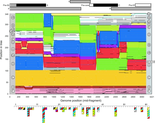
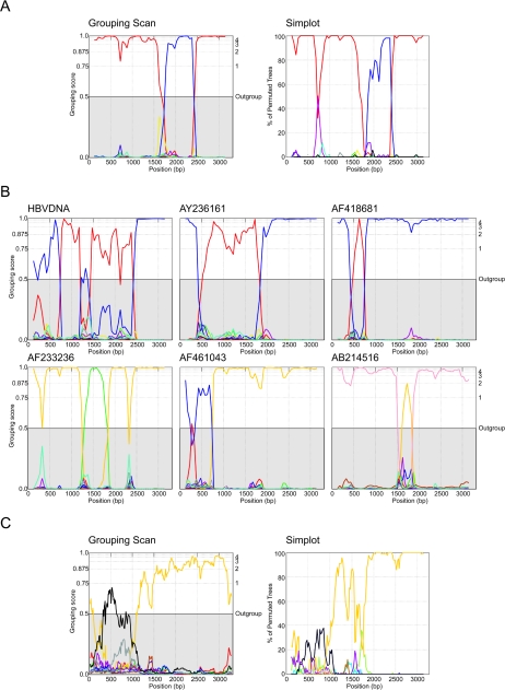
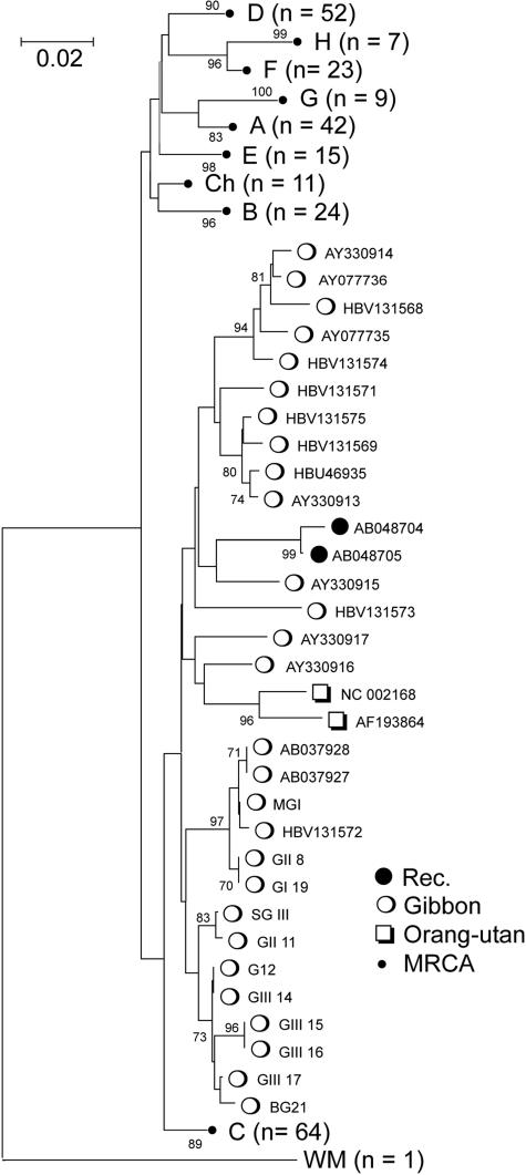
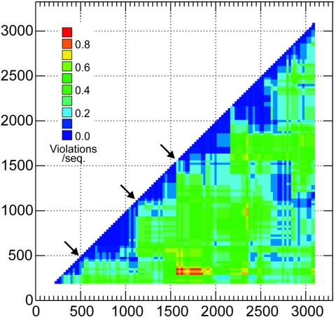
Hepatitis B virus (HBV) infection is widely distributed in both human and ape populations throughout the world and is a major cause of human morbidity and mortality. HBV variants are currently classified into the human genotypes A to H and species-associated chimpanzee and gibbon/orangutan groups. To examine the role of recombination in the evolution of HBV, large-scale data retrieval and automated phylogenetic analysis (TreeOrder scanning) were carried out on all available published complete genome sequences of HBV. We detected a total of 24 phylogenetically independent potential recombinants (different genotype combinations or distinct breakpoints), eight of which were previously undescribed. Instances of intergenotype recombination were observed in all human and ape HBV variants, including evidence for a novel gibbon/genotype C recombinant among HBV variants from Vietnam. By recording sequence positions in trees generated from sequential fragments across the genome, violations of phylogeny between trees also provided evidence for frequent intragenotype recombination between members of genotypes A, D, F/H, and gibbon variants but not in B, C, or the Asian B/C recombinant group. In many cases, favored positions for both inter- and intragenotype recombination matched positions of phylogenetic reorganization between the human and ape genotypes, such as the end of the surface gene and the core gene, where sequence relationships between genotypes changed in the TreeOrder scan. These findings provide evidence for the occurrence of past, extensive recombination events in the evolutionary history of the currently classified genotypes of HBV and potentially in changes in its global epidemiology and associations with human disease.


Figures

FIG. 1.
FIG. 1.
TreeOrder Scan of nonrecombinant HBV and recB sequences, showing positions of individual sequences (y axis) in phylogenetic trees generated from sequential 250-base sequence fragments, incrementing by 25 bases (midpoints indicated in x axis). Changes in sequence orders resulting from changes in phylogeny at the 70% bootstrap level are shown. Sequences are color coded by genotype, as indicated by labels in left and right margins: genotype A, red; B, light green; recB (rB), green; C, yellow; D, blue; E, purple; F, pink; G, pale yellow; H, brown; chimpanzee (Ch), gray; gibbon (Gi), white; orangutan (Or, turquoise; woolly monkey (outgroup on line 1), black. The recombinant sequence, AY161141, is shown as a dotted line. The boundaries of sequence groupings with 70% or greater bootstrap support are indicated with black horizontal lines; for clarity, only clades with six or more members were demarcated. All sequence positions were numbered relative to the HBVADW2 reference sequence (length, 3,221 bp). For comparison, the TreeOrder Scan was aligned with a scale genome diagram of HBV (upper panel) and with positions of breakpoints in recombinant HBV variants (triangles in lower panel). Each independent breakpoint is indicated as a square, localized to one of 61 separate 250-base sequence fragments spanning the genome, with segment 62 representing the junction between the beginning and end of the alignment. Genotype transitions are shown in the upper-left and lower-right quadrants of the square and are color coded as for the TreeOrder Scan; unfilled triangles represent unknown genotypes; gibbon sequences are in black.
FIG. 2.
FIG. 2.
(A) Comparison of Group Scanning and SIMPLOT analysis of the recombinant sequence AY161141. In each case, sequence fragments of 250 bases incrementing by 50 bases, 100 bootstrap replicates, were compared with sequence groups (Group Scan) or 50% consensus sequences of the eight human genotypes and four primate genotypes (color coded as described in the legend of Fig. 1). For the group scanning analysis, only values of >0.5 indicate grouping within a genotype group. (B) Group Scan analysis of examples of newly discovered recombinant HBV variants (listed in Table 1) and of novel recombinant sites in a previously analyzed sequence, HBVDNA. (C) Group Scan and SIMPLOT analysis of the sequence AB048704. Because the recombinant region in the S gene was relatively phylogenetically uninformative, longer sequence fragments of 400 bases incrementing by 15 bases (Group Scan) or 20 bases (SIMPLOT) were analyzed. Sequences are color coded as described in the legend of Fig. 1, except that the gibbon HBV variants are shown in black. The closely related variant, AB048705, produced comparable results (data not shown).
FIG. 3.
FIG. 3.
Phylogenetic analysis of positions 251 to 650 (corresponding to the putative recombinant region in the S gene) (Fig. 2C) of sequences AB048704 and AB048705 (Rec), all available gibbon- and orangutan-derived sequences, and nonrecombinant human- and chimpanzee-derived HBV variants (set A). Since sequences from each genotype were monophyletic, only the most recent common ancestor (MRCA) is shown for non-gibbon genotypes. The tree was constructed by neighbor-joining using Jukes-Cantor corrected distances in the MEGA2 package (19), using 200 bootstrap replicates (values of 70% or greater shown).
FIG. 4.
FIG. 4.
Phylogenetic compatibility matrix of gibbon-derived HBV sequences, showing frequencies of phylogeny violations for each pairwise comparison of sequence fragments. Frequencies are color coded to indicate number of phylogeny violation per sequence (see key). Phylogenetically compatible regions are shown as deep blue. Arrows indicates the main sites where changes in phylogeny of the gibbon sequences lead to regions of incompatibility between fragments. For this analysis, sequence fragments of 400 bases, incrementing by 25 bases across the genome, were compared (117 fragments total), using a bootstrap value of 70% above which phylogeny violations were computed.

Similar articles

Cited by

References

    1. Akuta, N., and H. Kumada. 2005. Influence of hepatitis B virus genotypes on the response to antiviral therapies. J. Antimicrob. Chemother. 55:139-142. - PubMed
    1. Andre, F. 2000. Hepatitis B epidemiology in Asia, the Middle East and Africa. Vaccine 18(Suppl. 1):S20-S22. - PubMed
    1. Bollyky, P. L., and E. C. Holmes. 1999. Reconstructing the complex evolutionary history of hepatitis B virus. J. Mol. Evol. 49:130-141. - PubMed
    1. Bollyky, P. L., A. Rambaut, P. H. Harvey, and E. C. Holmes. 1996. Recombination between sequences of hepatitis B virus from different genotypes. J. Mol. Evol. 42:97-102. - PubMed
    1. Bowyer, S. M., and J. G. Sim. 2000. Relationships within and between genotypes of hepatitis B virus at points across the genome: footprints of recombination in certain isolates. J. Gen. Virol. 81:379-392. - PubMed