. 2005 Dec;79(24):15477-93.
doi: 10.1128/JVI.79.24.15477-15493.2005.

Human cytomegalovirus infection induces specific hyperphosphorylation of the carboxyl-terminal domain of the large subunit of RNA polymerase II that is associated with changes in the abundance, activity, and localization of cdk9 and cdk7

Affiliations

Human cytomegalovirus infection induces specific hyperphosphorylation of the carboxyl-terminal domain of the large subunit of RNA polymerase II that is associated with changes in the abundance, activity, and localization of cdk9 and cdk7

Sama Tamrakar et al. J Virol. 2005 Dec.

Abstract

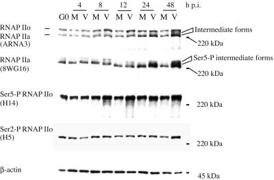
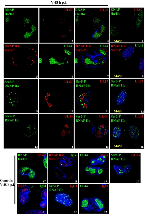
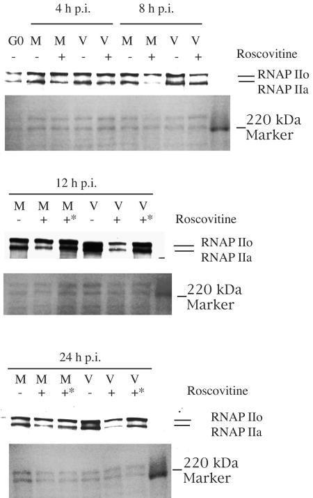
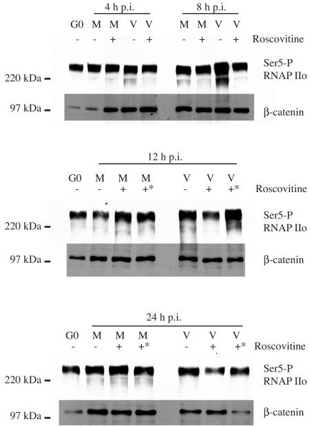
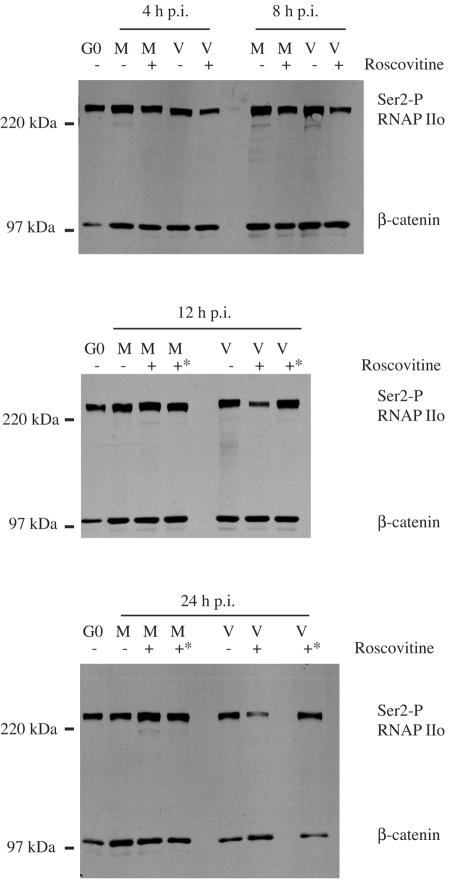
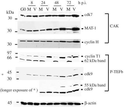
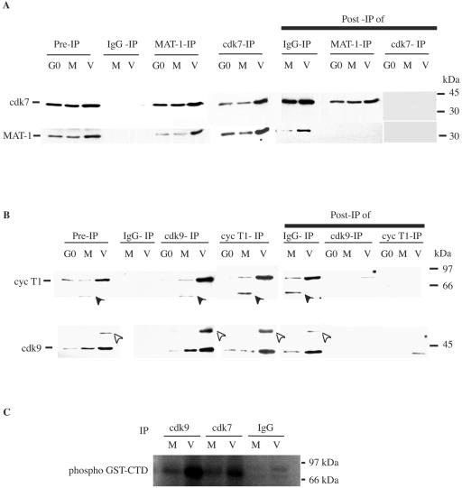
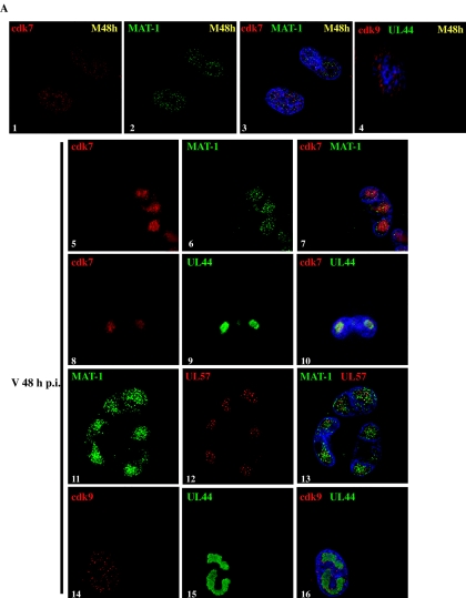
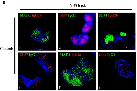
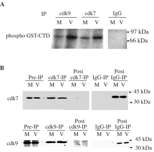
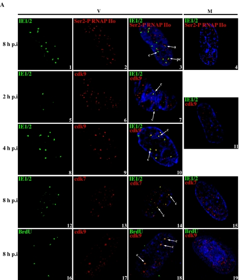
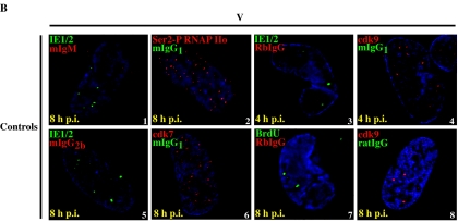
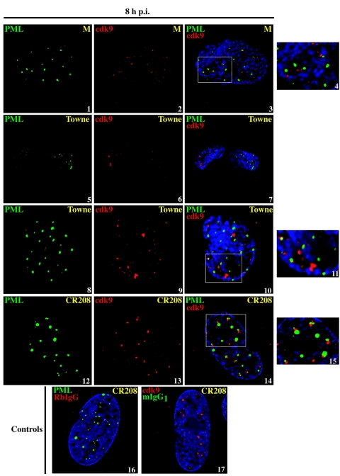
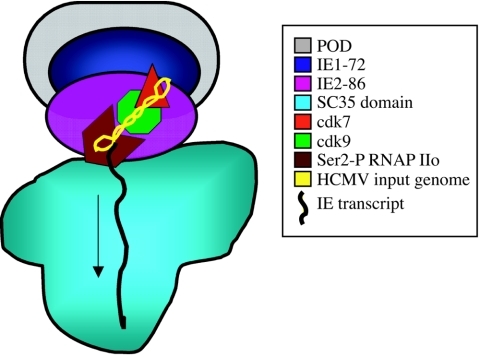
Human cytomegalovirus infection in the presence of the cyclin-dependent kinase (cdk) inhibitor roscovitine leads to changes in differential splicing and the polyadenylation of immediate early IE1/IE2 and UL37 transcripts (V. Sanchez, A. K. McElroy, J. Yen, S. Tamrakar, C. L. Clark, R. A. Schwartz, and D. H. Spector, J. Virol. 78:11219-11232, 2004). To determine if this was associated with specific phosphorylation of the C-terminal domain (CTD) of the RNA polymerase II (RNAP II) large subunit by cdk7/cyclin H and cdk9/cyclin T1, we examined the expression and localization of these kinases and the various phosphorylated forms of RNAP II. Infection resulted in increased RNAP II CTD phosphorylated on serines 2 and 5 and increased levels of activity of cdk7 and cdk9. At early times, cdk9 localizes with input viral DNA, and aggregates of cdk9 and cdk7 and a subset of Ser2-phosphorylated RNAP II colocalize with IE1/IE2 proteins adjacent to promyelocytic leukemia protein oncogenic domains. Later, cdk9 and Ser2-phosphorylated RNAP II form a nuclear punctate pattern; cdk7 resides in replication centers, and Ser5-phosphorylated RNAP II clusters at the peripheries of replication centers. Roscovitine treatment leads to decreased levels of hyperphosphorylated RNAP II (RNAP IIo) in infected cells and of hypophosphorylated RNAP II in mock-infected and infected cells. The RNAP IIo decrease does not occur if roscovitine is added 8 h postinfection, as was previously observed for processing of IE transcripts. These results suggest that accurate IE gene expression requires specific phosphorylation of the RNAP II CTD early in infection.


Figures

FIG. 1.
FIG. 1.
Mobility of RNAP II changes during the infection along with the increases in Ser2 and Ser5 phosphorylation of the CTD. G0-synchronized cells were released into G1, infected with HCMV Towne at an MOI of 5 or mock infected, and harvested at the time intervals indicated. Total cell lysates from an equal number of cells were loaded on 6% polyacrylamide gels and transferred to nitrocellulose membranes in buffer containing 0.1% SDS. Western blotting was carried out using different antibodies against RNAP II. ARNA3 recognizes the region of RNAP II outside of the CTD, 8WG16 is specific for unphosphorylated CTD and the CTD that is phosphorylated only on Ser5, and H5 and H14 are specific for the CTD phosphorylated on Ser2 and Ser5, respectively. β-Actin was used as a loading control.
FIG. 2.
FIG. 2.
Localization of RNAP II in HCMV-infected cells. G0-synchronized cells that were released into G1 and infected with HCMV at an MOI of 5 or mock infected were seeded onto glass coverslips. At 48 h p.i., cells were washed with PBS and fixed in either paraformaldehyde or ice-cold methanol. Paraformaldehyde-fixed cells were permeabilized with 0.1% Triton X-100 or with ice-cold acetone. Cells (acetone-treated) were immunostained with the monoclonal antibody ARNA3 (IgG1) or 8WG16 (IgG2a). Cells permeabilized with Triton X-100 were stained with H5 (IgM) to detect the RNAP IIo with the CTD phosphorylated on Ser2, and the methanol-fixed cells were stained with H14 (IgM) to detect the RNAP IIo with the CTD phosphorylated on Ser5. Cells were costained with a RNAP II antibody and either UL44 (IgG1) or UL57 (IgG2a), both of which localize to the viral replication center. Specific antibodies were detected with FITC- or TRITC-conjugated isotype-specific secondary antibodies. Nuclei were stained with Hoechst dye. For immunostaining controls, one of the specific antibodies in the pair was replaced with a nonspecific immunoglobulin (purified mouse IgM or IgG). The corresponding isotype-specific secondary antibodies matching those in the costaining were then used. Except for those marked M48h, all confocal images are of virus (V) at 48 h p.i. The images are confocal optical sections of 0.2 microns. Magnification, ×600.
FIG. 3.
FIG. 3.
The cdk inhibitor roscovitine affects the virus-induced phosphorylation state of RNAP II. HCMV-infected (lanes V) or mock-infected (lanes M) cells were treated with 20 μM roscovitine at the time of infection or at 8 h p.i. and were harvested at different time points. Total cell lysates from an equal number of cells were run on 6% gels and transferred to nitrocellulose membranes in buffer containing 0.1% SDS. The membranes were subjected to Western blotting using the ARNA3 antibody, which detects all forms of RNAP II. +* indicates that roscovitine was added at 8 h p.i. The membrane stained with amido black is shown below each Western blot to show the protein loading.
FIG. 4.
FIG. 4.
The cdk inhibitor roscovitine affects virus-induced phosphorylation of Ser5 within the RNAP II CTD. Infected (lanes V) and mock-infected (lanes M) cells were treated with roscovitine, and lysates were prepared, subjected to gel electrophoresis, and transferred to nitrocellulose membranes for Western blotting as described in the legend for Fig. 3. The membranes were probed with the antibody H14, which is directed against RNAP IIo with the CTD phosphorylated on Ser5. +* indicates that roscovitine was added at 8 h p.i. The lower portion of the blot was probed for β-catenin as a protein loading control.
FIG. 5.
FIG. 5.
The cdk inhibitor roscovitine affects the virus-induced phosphorylation of Ser2 within the RNAP II CTD. Infected (lanes V) and mock-infected (lanes M) cells were treated with roscovitine, and lysates were prepared, subjected to gel electrophoresis and transferred to nitrocellulose membranes for Western blotting as described in the legend for Fig. 3. The membranes were probed with the antibody H5, which is directed against RNAP IIo with the CTD phosphorylated on Ser2. +* indicates that roscovitine was added at 8 h p.i. The lower portion of the blot was probed for β-catenin as a protein loading control.
FIG. 6.
FIG. 6.
Levels of cdk-activating kinase (CAK) components and P-TEFb increase during HCMV infection. Upon release from G0, synchronized HFFs were infected with HCMV Towne (lanes V) at an MOI of 5 or mock infected (lanes M). Cells were harvested at the indicated time points. Lysates from an equal number of cells were loaded onto gels, electrophoresed, and subjected to Western blotting with antibodies against cdk7, MAT-1, cyclin H, cyclin T1, and cdk9. β-Actin was used as a protein loading control.
FIG. 7.
FIG. 7.
The levels of cdk7/MAT-1 and cdk9/cyclin T1 complexes and the kinase activities of cdk7 and cdk9 increase during the HCMV infection. G0-synchronized HFFs were mock infected (lanes M) or HCMV infected (lanes V) and harvested at 48 h p.i. (A) Lysates from an equal number of cells were immunodepleted of cdk7 or MAT-1 using rabbit polyclonal antibodies. The immunoprecipitates and immunodepleted lysates were subjected to gel electrophoresis and Western blotting with cdk7- or MAT-1-specific monoclonal antibodies. A control immunoprecipitation with normal rabbit IgG was also included. (B) Equal numbers of mock-infected or HCMV-infected cells were lysed and immunodepleted of cdk9 or cyclin T1 using rabbit polyclonal antibodies. Western blotting of the immunoprecipitates and the depleted lysates was carried out using monoclonal antibodies against cdk9 and cyclin T1. A control immunoprecipitation with normal rabbit IgG was also included. The filled arrowhead indicates the 62-kDa band recognized by the cyclin T1 monoclonal antibody, and the open arrowhead indicates the 55-kDa band recognized by the cdk9 antibody. (C) Equal numbers of mock-infected or HCMV-infected cells were lysed and immunoprecipitated with cdk7 or cdk9 polyclonal antibodies or normal rabbit IgG coupled to the agarose beads. Purified GST-CTD (0.1 μg) was used as the substrate in each reaction as described in Materials and Methods. A mixture of 2 μCi of [γ-32P]ATP, kinase assay buffer, and substrate was added to the immunocomplex, followed by incubation at 37°C. The protein was separated by electrophoresis and exposed to X-ray film. The faint band that appears in the IP of the viral lysate at 48 h p.i. with the normal rabbit IgG is nonspecific and does not run at the same position as the phospho GST-CTD.
FIG.8.
FIG.8.
Localization of cdk7, MAT-1 and cdk9 in HCMV-infected cells. (A) G0-synchronized cells were released in G1, infected with HCMV at an MOI of 5 or mock infected, and seeded onto the glass coverslips. At 48 h p.i., cells were washed with PBS, fixed in paraformaldehyde, permeabilized, and immunostained with monoclonal antibodies against cdk7 (IgG2b), MAT-1 (IgG1), or cdk9 (IgG2b) and a viral replication center protein UL44 (IgG1) or UL57 (IgG2a) as described in Materials and Methods. Specific antibody staining was detected with FITC- or TRITC-conjugated isotype-specific secondary antibodies. (B) For controls, one of the specific antibodies of the pair was replaced with purified normal mouse IgG, and the corresponding pair of isotype-specific secondary antibodies was used. Nuclei were stained with Hoechst dye. Except for those marked M48h, all of the images are of infected cells at 48 h p.i. All of the images except image 4 in panel A are confocal optical sections of 0.2 μm. Magnification, ×600.
FIG.8.
FIG.8.
Localization of cdk7, MAT-1 and cdk9 in HCMV-infected cells. (A) G0-synchronized cells were released in G1, infected with HCMV at an MOI of 5 or mock infected, and seeded onto the glass coverslips. At 48 h p.i., cells were washed with PBS, fixed in paraformaldehyde, permeabilized, and immunostained with monoclonal antibodies against cdk7 (IgG2b), MAT-1 (IgG1), or cdk9 (IgG2b) and a viral replication center protein UL44 (IgG1) or UL57 (IgG2a) as described in Materials and Methods. Specific antibody staining was detected with FITC- or TRITC-conjugated isotype-specific secondary antibodies. (B) For controls, one of the specific antibodies of the pair was replaced with purified normal mouse IgG, and the corresponding pair of isotype-specific secondary antibodies was used. Nuclei were stained with Hoechst dye. Except for those marked M48h, all of the images are of infected cells at 48 h p.i. All of the images except image 4 in panel A are confocal optical sections of 0.2 μm. Magnification, ×600.
FIG. 9.
FIG. 9.
cdk7- and cdk9-associated kinase activities towards the RNAP II CTD are increased in infected cells at early times. Cells infected with HCMV (lanes V) or mock infected (lanes M) were harvested at 8 h p.i. (A) Equivalent numbers of cells were lysed and immunoprecipitated with cdk7 or cdk9 polyclonal antibodies, or normal rabbit IgG coupled to the agarose beads. Purified GST-CTD (0.1 μg) was used as the substrate in each reaction as described in Materials and Methods. A mixture of 2 μCi of [γ-32P]ATP, kinase assay buffer, and substrate was added to the immunocomplex, followed by incubation at 37°C. The electrophoresed protein was exposed to an X-ray film. (B) Western blots of the corresponding immunoprecipitates probed for cdk7 or cdk9 with monoclonal antibodies.
FIG. 10.
FIG. 10.
Ser2-phosphorylated RNAP IIo, cdk7, and cdk9 localize with IE protein domains. (A) G0-synchronized cells were released into G1 and infected at an MOI of 3 or 5 or mock infected and seeded onto coverslips. The cells were washed with PBS and fixed with either 2% paraformaldehyde or ice-cold methanol between 2 to 8 h p.i. Cells were coimmunostained with the appropriate combinations of CH16.0 MAb (IgG1) to detect IE1/IE2, H5 MAb (IgM) to detect Ser2-phosphorylated RNAP IIo, and cdk9 rabbit polyclonal antibody, cdk7 MAb (IgG2b), and BrdU rat polyclonal antibody. Specific antibodies were detected with FITC- or TRITC-conjugated isotype-specific anti-mouse Ab, goat anti-rat FITC-IgG, or goat anti-rabbit Cy3-IgG secondary antibodies. Nuclei were stained with Hoechst dye. (B) For immunostaining controls, one of the specific antibodies in the pair was replaced with a nonspecific immunoglobulin (purified mouse IgM, isotype-specific mouse IgG, rabbit IgG, or rat IgG). The corresponding isotype-specific secondary antibodies matching those in the costaining were then used. Arrows denote examples exhibiting some degree of colocalization: a, adjacent; pc, partial colocalization; c, completely colocalized. All the images are 0.2-μm sections that were obtained by confocal microscopy. Magnification is ×1,000 under oil immersion.
FIG. 10.
FIG. 10.
Ser2-phosphorylated RNAP IIo, cdk7, and cdk9 localize with IE protein domains. (A) G0-synchronized cells were released into G1 and infected at an MOI of 3 or 5 or mock infected and seeded onto coverslips. The cells were washed with PBS and fixed with either 2% paraformaldehyde or ice-cold methanol between 2 to 8 h p.i. Cells were coimmunostained with the appropriate combinations of CH16.0 MAb (IgG1) to detect IE1/IE2, H5 MAb (IgM) to detect Ser2-phosphorylated RNAP IIo, and cdk9 rabbit polyclonal antibody, cdk7 MAb (IgG2b), and BrdU rat polyclonal antibody. Specific antibodies were detected with FITC- or TRITC-conjugated isotype-specific anti-mouse Ab, goat anti-rat FITC-IgG, or goat anti-rabbit Cy3-IgG secondary antibodies. Nuclei were stained with Hoechst dye. (B) For immunostaining controls, one of the specific antibodies in the pair was replaced with a nonspecific immunoglobulin (purified mouse IgM, isotype-specific mouse IgG, rabbit IgG, or rat IgG). The corresponding isotype-specific secondary antibodies matching those in the costaining were then used. Arrows denote examples exhibiting some degree of colocalization: a, adjacent; pc, partial colocalization; c, completely colocalized. All the images are 0.2-μm sections that were obtained by confocal microscopy. Magnification is ×1,000 under oil immersion.
FIG. 11.
FIG. 11.
Cdk9 localizes adjacent to sites that correspond to the PODS in infected cells prior to their dispersal. G0-synchronized cells were released into G1 and infected with HCMV Towne or HCMV CR208 virus at an MOI of 5 or mock infected and seeded onto coverslips. The cells were washed with PBS and fixed with 2% paraformaldehyde at 8 h p.i. Cells were coimmunostained with the appropriate combinations of PML MAb (IgG1) and cdk9 rabbit polyclonal antibody. Specific antibodies were detected with goat anti-mouse isotype-specific FITC-conjugated or goat anti-rabbit Cy3 secondary antibodies. Nuclei were stained with Hoechst dye. For immunostaining controls, one of the specific antibodies in the pair was replaced with a normal isotype-specific mouse IgG1 or rabbit IgG. The corresponding isotype-specific secondary antibodies matching those in the costaining were then used. Panels 4, 11, and 15 are insets depicting an enlarged view of the region within the white box from the previous panel. All of the images are 0.2-μm sections that were obtained by confocal microscopy. Panels 5 to 7 and 8 to 11 are different cells from the same experiment. Magnification is ×600 under oil immersion for panels 5 to 7 and ×1,000 under oil immersion for all other panels. Inset panels 4, 11, and 15 were magnified 200% using Adobe Photoshop v. 7.0.
FIG. 12.
FIG. 12.
Model for an IE transcription site in HCMV-infected cells. The input genome that is deposited at the POD structure functions as a template for IE RNA synthesis. Cellular hypophosphorylated RNAP IIa is recruited to the site and is phosphorylated by cdk7 and cdk9 to the hyperphosphorylated and transcriptionally active RNAP IIo. The IE transcripts move towards the SC35 domains, probably for further processing. The newly synthesized major IE proteins also localize to this region. IE1-72 eventually causes POD dispersal, but the IE2-86 protein remains at the established transcription site. The color key to structures is shown at the right.

Similar articles

Cited by

References

    1. Adair, R., G. W. Liebisch, and A. M. Colberg-Poley. 2003. Complex alternative processing of human cytomegalovirus UL37 pre-mRNA. J. Gen. Virol. 84:3353-3358. - PubMed
    1. Adair, R., G. W. Liebisch, Y. Su, and A. M. Colberg-Poley. 2004. Alteration of cellular RNA splicing and polyadenylation machineries during productive human cytomegalovirus infection. J. Gen. Virol. 85:3542-3553. - PubMed
    1. Ahn, J.-H., W.-J. Jang, and G. S. Hayward. 1999. The human cytomegalovirus IE2 and UL112-113 proteins accumulate in viral DNA replication compartments that initiate from the periphery of promyelocytic leukemia protein-associated nuclear bodies (PODs or ND10). J. Virol. 73:10458-10471. - PMC - PubMed
    1. Ahn, J. H., E. R. Brignole, and G. S. Hayward. 1998. Disruption of PML subnuclear domains by the acidic IE1 protein of human cytomegalovirus is mediated through interaction with PML and may modulate a RING finger-dependent cryptic transactivator function of PML. Mol. Cell. Biol. 18:4899-4913. - PMC - PubMed
    1. Ahn, J. H., and G. S. Hayward. 1997. The major immediate-early proteins IE1 and IE2 of human cytomegalovirus colocalize with and disrupt PML-associated nuclear bodies at very early times in infected permissive cells. J. Virol. 71:4599-4613. - PMC - PubMed

Publication types

MeSH terms

Substances