. 2005 Dec;79(24):15573-7.
doi: 10.1128/JVI.79.24.15573-15577.2005.

Identification of a functional envelope protein from the HERV-K family of human endogenous retroviruses

Affiliations

Identification of a functional envelope protein from the HERV-K family of human endogenous retroviruses

Marie Dewannieux et al. J Virol. 2005 Dec.

Abstract

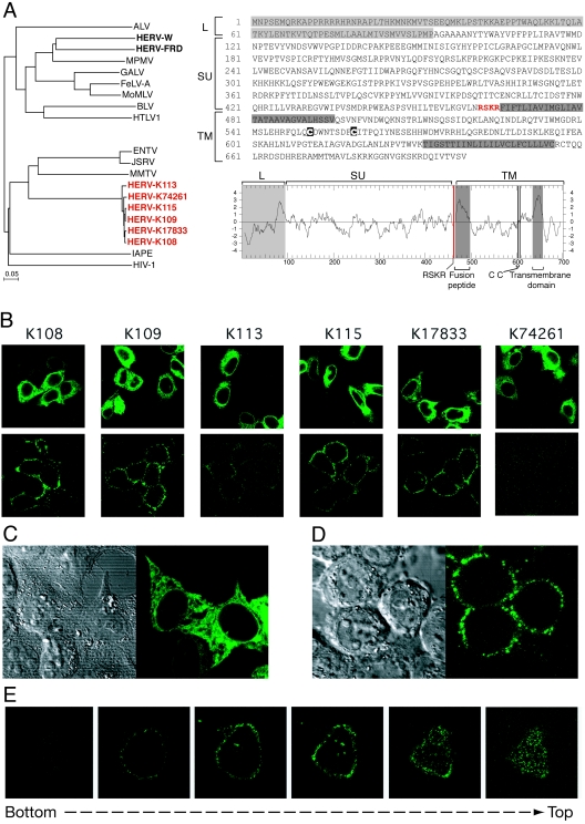
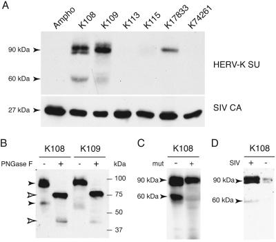
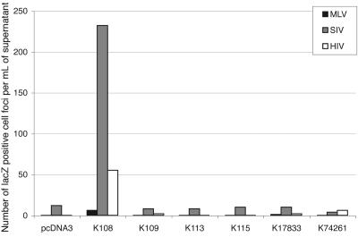
Genome-wide screening of sequence databases for human endogenous retroviruses (HERVs) has led to the identification of 18 coding env genes, among which two-the syncytin genes-encode fusogenic ENV proteins possibly involved in placenta physiology. Here we show that a third ENV, originating from the most "recent" HERV-K(HML2) family, is functional. Immunofluorescence analysis of env-transduced cells demonstrates expression of the protein at the cell surface, and we show that the protein confers infectivity to simian immunodeficiency virus pseudotypes. Western blot analysis of the pseudotyped virions further discloses the expected specific cleavage of the ENV precursor protein. This functional ENV could play a role in the amplification--via infection of the germ line--of the HERV-K genomic copies, all the more as coding HERV-K gag and pol genes can similarly be found in the human genome, which could therefore generate infectious virions of a fully endogenous origin.


Figures

FIG. 1.
FIG. 1.
Structure and expression of the HERV-K ENV proteins. (A, left) Phylogenetic tree based on the ENV TM domain showing the clustering of the six HERV-K copies with the betaretrovirus group. Protein sequences were aligned using ClustalW, and the resulting alignment was manually refined before the tree was calculated using the neighbor-joining method (PHYLIP software). Nucleotide sequences of the six env genes are as follows (accession number, position in the sequence entry, orientation): for K108, AC072054, 30,365 to 32,464 (minus); for K109, AF164615, 6,412 to 8,508 (plus); for K113, AY037928, 6,451 to 8,550 (plus); for K115, AY037929, 6,442 to 8,541 (plus); for K17833, Y17833, 5,581 to 7,680 (plus); and for K74261, AC074261, 93,508 to 95,604 (plus). (Right) Sequence and hydrophobicity profile of the HERV-K108 ENV highlighting the canonical ENV features (see the text). ALV, avian leukemia virus; MPMV, Mason-Pfizer monkey virus; GALV, gibbon ape leukemia virus; FeLV-A, feline leukemia virus A; MoMLV, Moloney MLV; BLV, bovine leukemia virus; HTLV1, human T-cell leukemia virus type 1; ENTV, enzootic nasal tumor virus; JSRV, Jaagsiekte sheep retrovirus; MMTV, mouse mammary tumor virus; IAPE, intracisternal A-particle-related envelope-encoding element. (B-E) Immunofluorescence analysis of HeLa cells transiently transfected with expression vectors of each of the six fully coding HERV-K env genes. (B) In the upper panels, cells were fixed, permeabilized, and stained for HERV-K env expression (whole-cell staining); in the lower panels, living cells were observed directly after staining without prior fixation or permeabilization (cell surface staining). (C, D) Higher magnification of representative images of fixed and permeabilized cells (C) or of living cells (D) transfected previously with the HERV-K108 env gene. (Left) Image of the cells under phase-contrast microscopy; (right) image of the same cells stained for the HERV-K ENV by immunofluorescence. (E) Successive confocal images of a living cell stained for the HERV-K108 ENV, demonstrating cell surface localization. In all experiments, HERV-K ENV detection was performed on HeLa cells grown on glass coverslips approximately 24 h posttransfection. HERV-K ENVs were detected using a mouse monoclonal antibody (raised by us against a recombinant His-tagged protein corresponding to amino acids 223 to 437 of K109 ENV, using the pET-28 vector from Novagen), and an Alexa Fluor 488-conjugated anti-mouse secondary antibody (Molecular Probes). No signal was detected with cells transfected with an irrelevant expression vector (not shown). Observations were made under a Zeiss LSM 510 laser scanning confocal microscope.
FIG. 2.
FIG. 2.
Western blot analysis of the incorporation of HERV-K ENVs into SIV pseudotypes. (A) Virions contained in the supernatant of human 293T cells cotransfected with an expression vector for the SIV core proteins, a corresponding lacZ gene-marked defective retroviral vector, and an expression vector for the env gene to be tested (or a vector for the amphotropic MLV envelope as a control [Ampho]) were assayed for the presence of the HERV-K ENV protein. Forty-eight hours posttransfection, supernatants of the 293T cells were harvested, pelleted by ultracentrifugation onto a 20% sucrose cushion, and recovered for SDS-PAGE analysis. (B) SIV particles pseudotyped with K108 and K109 ENVs were purified as described for panel A and treated or not treated with peptide-N-glycosidase F (PNGase F; NEB Biolabs) as recommended by the manufacturer before SDS-PAGE. (C) SIV particles were pseudotyped with the K108 ENV protein or a K108 ENV protein mutated at the furin cleavage site (RSKR to ASAR) (mut) and analyzed as described for panel A. As the K108 mutant protein is less expressed than its wild-type counterpart, twice the volume of cell medium was used for the purification step. (D) The supernatants of 293T cells transfected with the K108 ENV expression vector and either a control plasmid (CMV β, without SIV) or the SIV plasmids (see panel A; with SIV) were concentrated and analyzed by SDS-PAGE as described for panel A. All blots were first used for the detection of HERV-K ENV proteins, using the same monoclonal antibody as in Fig. 1. The membrane in panel A was thereafter tested for SIV p27 capsid using a mouse monoclonal antibody (2F12; National Institutes of Health AIDS Research and Reference Reagent Program), to check for equal loadings of the lanes (lower panel). The membrane in panel D was also incubated with an antibody against actin (Santa Cruz) to exclude the possibility of contamination by cell proteins in the course of virion purification (not shown).
FIG. 3.
FIG. 3.
Assay for infectivity of the viral particles pseudotyped with the proteins encoded by the six fully coding HERV-K env genes. Human 293T cells were cotransfected with an expression vector for the retroviral proteins—except ENV—from a gammaretrovirus (MLV) or lentiviruses (HIV-1 and SIV), a corresponding lacZ gene-marked defective retroviral vector, and an expression vector for the env gene to be tested (or an empty vector [pcDNA3] as a negative control and a vector for the amphotropic MLV ENV [not shown] as a positive control). Viral supernatants were collected 36 h after transfection of 293T cells, filtered through 0.45-μm-pore-size membranes, and added to the target feline G355.5 cells in 24-well plates that were then subjected to spinoculation (12). After an additional 60 h of incubation, the lacZ-positive cell colonies were counted following in situ histochemical staining for β-galactosidase activity. A positive control performed with the amphotropic MLV ENV gave titers of 8 × 10⁵, 4 × 10⁵, and 6 × 10⁴ infectious particles per ml for the MLV, SIV, and HIV cores, respectively. This graph gives the results obtained in an experiment representative of two to five independent assays performed under the same conditions (the mean viral titer observed for K108 ENV with the SIV core is 150, with variations ranging from 60 to 400 depending on the overall efficiency of each infection assay).

References

    1. An, D. S., Y. Xie, and I. S. Chen. 2001. Envelope gene of the human endogenous retrovirus HERV-W encodes a functional retrovirus envelope. J. Virol. 75:3488-3489. - PMC - PubMed
    1. Bannert, N., and R. Kurth. 2004. Retroelements and the human genome: new perspectives on an old relation. Proc. Natl. Acad. Sci. USA 13:14572-14579. - PMC - PubMed
    1. Barbulescu, M., G. Turner, M. I. Seaman, A. S. Deinard, K. K. Kidd, and J. Lenz. 1999. Many human endogenous retrovirus K (HERV-K) proviruses are unique to humans. Curr. Biol. 9:861-868. - PubMed
    1. Belshaw, R., V. Pereira, A. Katzourakis, G. Talbot, J. Paces, A. Burt, and M. Tristem. 2004. Long-term reinfection of the human genome by endogenous retroviruses. Proc. Natl. Acad. Sci. USA 25:25. - PMC - PubMed
    1. Benit, L., P. Dessen, and T. Heidmann. 2001. Identification, phylogeny, and evolution of retroviral elements based on their envelope genes. J. Virol. 75:11709-11719. - PMC - PubMed

Publication types

Substances

LinkOut - more resources