. 2005 Nov 24;438(7067):454-9.
doi: 10.1038/nature04150.

Isolation and characterization of a protochordate histocompatibility locus

Affiliations

Isolation and characterization of a protochordate histocompatibility locus

Anthony W De Tomaso et al. Nature. .

Abstract

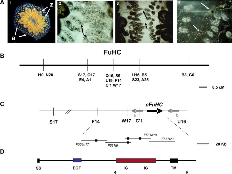
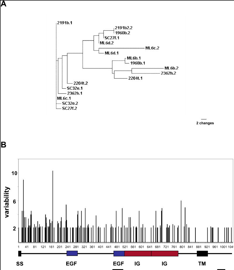
Histocompatibility--the ability of an organism to distinguish its own cells and tissue from those of another--is a universal phenomenon in the Metazoa. In vertebrates, histocompatibility is a function of the immune system controlled by a highly polymorphic major histocompatibility complex (MHC), which encodes proteins that target foreign molecules for immune cell recognition. The association of the MHC and immune function suggests an evolutionary relationship between metazoan histocompatibility and the origins of vertebrate immunity. However, the MHC of vertebrates is the only functionally characterized histocompatibility system; the mechanisms underlying this process in non-vertebrates are unknown. A primitive chordate, the ascidian Botryllus schlosseri, also undergoes a histocompatibility reaction controlled by a highly polymorphic locus. Here we describe the isolation of a candidate gene encoding an immunoglobulin superfamily member that, by itself, predicts the outcome of histocompatibility reactions. This is the first non-vertebrate histocompatibility gene described, and may provide insights into the evolution of vertebrate adaptive immunity.


Figures

Figure 1
Figure 1
Histocompatibility and positional cloning of the FuHC locus in Botryllus schlosseri. A. An adult B. schlosseri colony (panel 1) consists of asexually-derived individuals called zooids (z) united by a common blood supply (colors are caused by pigment cells in the blood). The vasculature stops at the periphery of the colony in small protrusions called ampullae (a), where the histocompatibility reaction takes place. When two colonies grow close together the ampullae reach out and interact (panel 2), and this will result in one of two outcomes. Either the two ampullae will fuse (panel 3), allowing the circulation of the two colonies to interconnect, or they will reject each other (panel 4). Rejection is a localized inflammatory reaction where blood cells leak from the ampullae and begin killing each other, resulting in dark spots called points of rejection (bottom panel, arrows) where some melanization occurs during the reaction. Histocompatibility takes ca. 24 hrs to occur and is controlled by a single, highly polymorphic locus called the FuHC (for fusion/hisftocompatibility). Colonies will fuse if they share one or both alleles, and will reject if no alleles are shared. B. Genetic map of the FuHC region from one backcross population. Genetic markers (AFLPs) are represented by a letter and number. In one F2 cross (Table 1, F2 cross AY x AY), C’1, was found to physically lie within a small 18Kb double crossover in one individual, this crossover did not include the cFuHC C. Physical map surrounding the candidate FuHC locus. Fosmid clones from the minimal tiling path of this region are shown on the bottom; small black boxes represent the physical location of AFLP markers, and the thick black arrow represents the location of the cFuHC locus and direction of transcription. Gray arrows show the flanking genes, a. Phospholipase A2-like, b. retinoblastoma binding protein-like D. Predicted structure of the FuHC protein, ss-signal sequence, EGF-epidermal growth factor repeat, Ig-immunoglobulin domain, TM-transmembrane domain. Black arrows indicate the location of two alternative splices, one which creates a secreted form of the protein, and another which shortens the cytoplasmic tail (see text).
Figure 2
Figure 2
Graphic representation of the overall polymorphism of the cFuHC A A phylogram of 18 cFuHC alleles isolated from wild-type animals collected in Monterey (Numbers), Santa Cruz (SC) and Moss Landing (ML) marinas (see methods) B. the domain structure of the protein (below) is overlayed with a graphic representation of amino acid variability along the protein. The same 18 wild type alleles used in A were aligned, and the variability is defined as the ratio of the number of different amino acids to the frequency of the most common amino acid at each position. The secreted exons and new COOH tail exons are also shown (underlined). There are no hypervariable regions, and the majority of the variability (i.e., the shortest vertical line) consists of single amino acid changes spread throughout the ectodomain.
Figure 3
Figure 3
Expression analysis of the cFuHC. A. RT-PCR of cDNA isolated from ampullae (a), blood (b), tadpoles (t), eggs (e) and sperm (s), Top gel, 600 bp fragment including the transmembrane domain (tm); bottom gel, 350 bp fragment which includes the secreted 3′ UTR (sec). Both primer sets straddle a large intron, preventing amplification from genomic DNA B-I. Localization of cFuHC expression via in situ hybridization in eggs, tadpoles and oozooids (described in Methods). Panels B, D and G are sense controls. Eggs (Panel C) were isolated immediately prior to fertilization but show no cFuHC expression when compared to controls. In the tadpole, expression is restricted to the anterior of the larvae, and outlines the nascent ampullae (Panel E, arrows). Panel F shows a larvae in the middle of metamorphosis, with strong expression seen at the tips of the extending ampullae (arrow). Expression in the adult body is seen in the epithelia of the ampullae (Panel H). A close-up of an ampullae is shown in Panel I, with expression seen in both a subset of blood cells and the epithelium of the ampullae and blood vessel. Light blue staining is vector blue, dark purple is NBT/BCIP. The punctate, light blue staining seen in B-E is a result of vector blue precipitation that often occurs during color development.

Comment in

References

    1. Flajnik, M.F., Miller, K. & Dupasquier, L. Evolution of the immune system in: W.E. Paul, Editors, Fundamental Immunology (5th ed), Lippincott, Williams & Wilkins, Philadelphia pp. 519–570. (2003),
    1. Dupasquier L, Zucchetti I, De Santis R. Immunoglobulin superfamily receptors in protochordates : before RAG time. Immunol Rev. 2004;198:233–248. - PubMed
    1. Oka H, Watanabe H. Problems of colony specificity in compound ascidians. Bull Biol Stn Asamushi. 1960;10:153–155.
    1. Sabbadin A. Le basi genetiche dell capacita di fusione fra colonie in Botryllus schlosseri (Ascidiacea) Atti Accad Na Lincei Rend. 1962;32:1031–1035.
    1. Scofield VL, Schlumpberger JM, West LA, Weissman IL. Protochordate allorecognition is controlled by an MHC-like gene system. Nature. 1982;295:499–502. - PubMed

Publication types

Associated data