. 2005 Nov 23;20(4):513-24.
doi: 10.1016/j.molcel.2005.09.028.

Structural basis of interdomain communication in the Hsc70 chaperone

Affiliations

Structural basis of interdomain communication in the Hsc70 chaperone

Jianwen Jiang et al. Mol Cell. .

Abstract

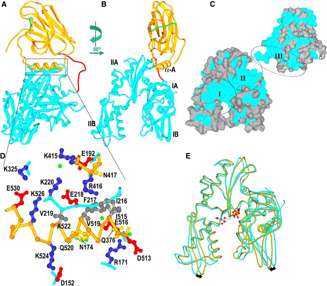
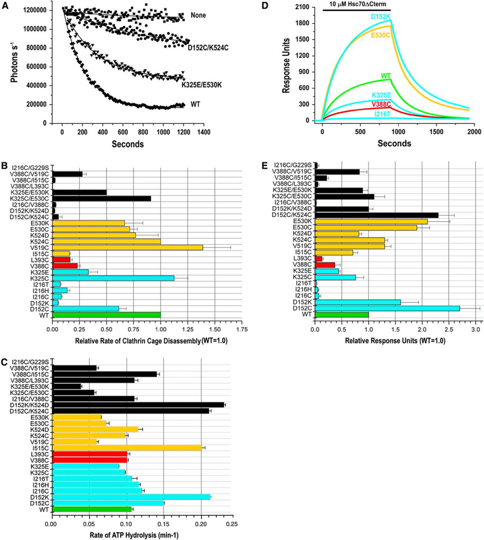
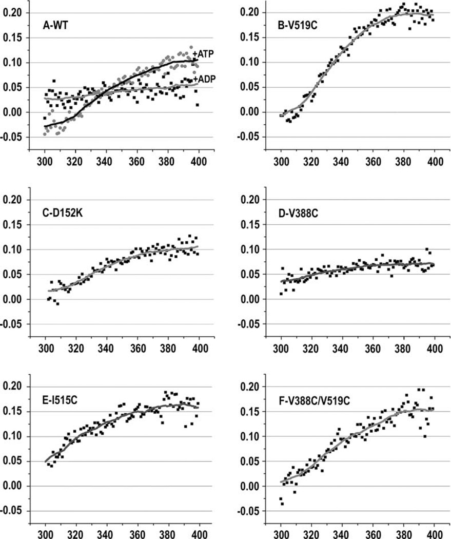
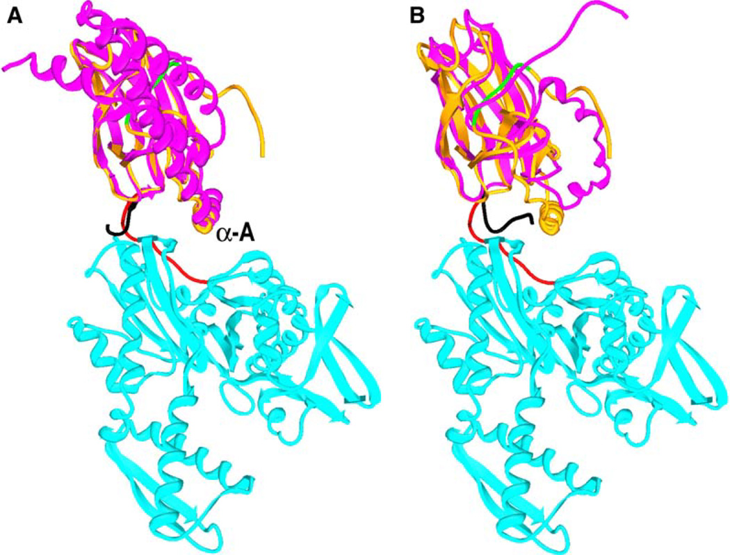
Hsp70 family proteins are highly conserved chaperones involved in protein folding, degradation, targeting and translocation, and protein complex remodeling. They are comprised of an N-terminal nucleotide binding domain (NBD) and a C-terminal protein substrate binding domain (SBD). ATP binding to the NBD alters SBD conformation and substrate binding kinetics, but an understanding of the mechanism of interdomain communication has been hampered by the lack of a crystal structure of an intact chaperone. We report here the 2.6 angstroms structure of a functionally intact bovine Hsc70 (bHsc70) and a mutational analysis of the observed interdomain interface and the immediately adjacent interdomain linker. This analysis identifies interdomain interactions critical for chaperone function and supports an allosteric mechanism in which the interdomain linker invades and disrupts the interdomain interface when ATP binds.


Figures

Figure 1
Figure 1. bHsc70ΔCterm Structure
(A) Ribbons representation of bHsc70 with the NBD (aa 1–383) in cyan, the SBD (aa 395–554) in orange, the linker (aa 384–394) in red, and aa 539– 544, which occupy the substrate binding site, in green. (B) As in (A), but with the molecule rotated by 90° as indicated. (C) Solvent-accessible surface of bHsc70 with residues that are identical in bHsc70 and the DnaK NBD and SBD colored blue and other residues in gray. The SBD has been broken away from the NBD and rotated to expose its interface with the NBD. The two largest regions of conserved surface in the NBD are around the ATP binding site (“I”) and the interface with the SBD (“II”). The interdomain linker and the interface with the NBD on the SBD (“III”) are also well conserved. (D) Expanded view of interactions between the SBD (C-α trace in orange) and NBD (C-α trace in cyan; side chains in blue, red, yellow, or dark gray for +, −, polar, or nonpolar, respectively; waters in green). Interdomain interactions include the following: salt bridges between K325 and E530 and between K524 and D152; longer-range ionic interactions between E218 and both R426 and K526 as well as between R171 and D513; a cluster of six hydrophobes (I216, F217, V219, I515, V519, A522); one direct (N417:E192) and five water-mediated H bonds (between Q520 and N174, K220 and K415, E516 and Q376, and R376 and the F217 amide). (E) Superposition of the NBD from the bHsc70 structure (cyan), with structure of an isolated bHsc70 NBD (1kax) crystallized with bound ATP (olive). ATP binding may induce the closing suggested by the arrows.
Figure 2
Figure 2. Effects of Interdomain Interface and Linker Mutations on Clathrin Cage Disassembly, ATPase Rates, and Auxilin Binding
(A) Clathrin cage disassembly measured by number of photons scattered per second versus time for the wt and two mutant bHsc70Δcterm enzymes. “None” is a reaction with no chaperone. Data are fit to single exponential decays; error bars show ranges from two experiments. (B) Relative rates of clathrin cage disassembly (wt = 1.0) for all enzymes. Data bars in all panels are colored green for wt, cyan for single mutants in the NBD, red for single mutants in the linker, orange for single mutants in the SBD, and black for all double mutants. Error bars are ±SE for at least two experiments. (C) Single-turnover ATPase rates for all enzymes; error bars show ranges from two experiments. (D) Representative SPR experiment showing wt and mutant enzyme binding to immobilized auxilin with 1 mM ATP and an ATP-regenerating system (no binding is observed in the absence of ATP or coupled auxilin). (E) Summary of binding for all enzymes. Binding is relative to wt controls, which were run in parallel for every experiment. Error bars are ±SE for two to four determinations.
Figure 3
Figure 3. Effects of Interdomain Interface and Linker Mutations on ATP-Induced Fluorescence Changes
Fractional differences in W fluorescence spectra with and without ATP ([fluorescence - ATP] - [fluorescence + ATP])/(fluorescence -ATP]) are plotted. In (A), spectral differences due to ADP are also plotted for comparison.
Figure 4
Figure 4. Alternate Conformations of the Interdomain Linker
(A) bHsc70ΔCterm structure colored as in Figure 1A with DnaK SBD (pdb 1dkx in magenta with the interdomain linker in black) superimposed. (B) bHsc70ΔCterm with the rat Hsc70 SBD (pdb 1bpr in magenta with the interdomain linker in black) superimposed.
Figure 5
Figure 5. Effects of Interdomain Interface and Linker Mutations on ATP-Induced Protection of the Linker from Proteolysis
(A–E) Proteolysis of indicated bHsc70ΔCterm enzymes in the absence (−) or presence (+) of ATP. Cleavage in the linker generates the 44 kDa NBD fragment. Densitometric traces are shown below their respective lanes. (F) Degree of protease resistance conferred by ATP for all mutants is presented as the ratio of the fraction of 44 kDa fragment in the absence of ATP to the fraction in the presence of ATP ([%44 kDa(−ATP)]/[%44 kDa(+ATP)]). The horizontal line corresponds to a 1.5-fold decrease in linker protease sensitivity.
Figure 6
Figure 6. Accessibility and Side Chain Proximity in Singly and Doubly Cys-Substituted Enzymes
(A) (Top two panels) UV illumination of SDS-PAGE of singly Cys-substituted enzymes reacted with monochlorobimane in either the absence (“−ATP”) or presence (“+ATP”) of ATP. Lower two panels show Coomassie blue staining of the same gels. (B) Nonreducing Coomassie-stained SDS-PAGE of enzymes treated with 3 mM oxidized glutathione in either the absence (“−”) or presence (“+”) of ATP.
Figure 7
Figure 7
Effects of Mutations of Different Amino Acids in the Interdomain Interface or Linker on Cage Disassembly, ATP Quenching of W Fluorescence, ATP Protection of the Linker from Proteolysis, ATPase Rates, and Auxilin Binding

Comment in

References

    1. Barouch W, Prasad K, Greene LE, Eisenberg E. ATPase activity associated with the uncoating of clathrin baskets by Hsp70. J. Biol. Chem. 1994;269:28563–28568. - PubMed
    1. Brünger AT, Adams PD, Clore GM, DeLano WL, Gros P, Grosse-Kunstleve RW, Jiang JS, Kuszewski J, Nilges M, Pannu NS, et al. Crystallography & NMR system: a new software suite for macromolecular structure determination. Acta Crystallogr. D Biol. Crystallogr. 1998;54:905–921. - PubMed
    1. Buchberger A, Theyssen H, Schroder H, McCarty JS, Virgallita G, Milkereit P, Reinstein J, Bukau B. Nucleotide-induced conformational changes in the ATPase and substrate binding domains of the DnaK chaperone provide evidence for interdo-main communication. J. Biol. Chem. 1995;270:16903–16910. - PubMed
    1. Bukau B, Horwich AL. The Hsp70 and Hsp60 chaper-one machines. Cell. 1998;92:351–359. - PubMed
    1. Cyr D, Langer T, Douglas M. DnaJ-like proteins: molecular chaperones and specific regulators of Hsp70. Trends Biochem. Sci. 1994;19:176–181. - PubMed

Publication types

Associated data