. 2006 Feb 17;281(7):4364-70.
doi: 10.1074/jbc.M504363200. Epub 2005 Nov 23.

Molecular structure of a 9-MDa icosahedral pyruvate dehydrogenase subcomplex containing the E2 and E3 enzymes using cryoelectron microscopy

Affiliations

Molecular structure of a 9-MDa icosahedral pyruvate dehydrogenase subcomplex containing the E2 and E3 enzymes using cryoelectron microscopy

Jacqueline L S Milne et al. J Biol Chem. .

Abstract

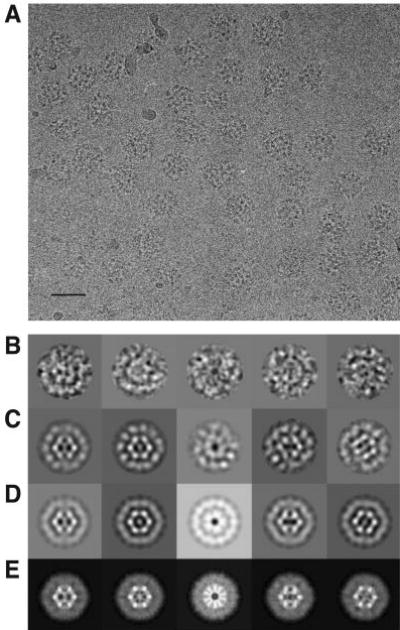
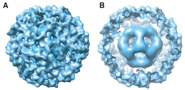
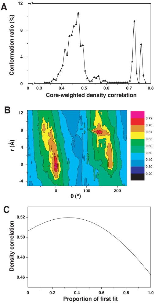
The pyruvate dehydrogenase multienzyme complexes are among the largest multifunctional catalytic machines in cells, catalyzing the production of acetyl CoA from pyruvate. We have previously reported the molecular architecture of an 11-MDa subcomplex comprising the 60-mer icosahedral dihydrolipoyl acetyltransferase (E2) decorated with 60 copies of the heterotetrameric (alpha(2)beta(2)) 153-kDa pyruvate decarboxylase (E1) from Bacillus stearothermophilus (Milne, J. L. S., Shi, D., Rosenthal, P. B., Sunshine, J. S., Domingo, G. J., Wu, X., Brooks, B. R., Perham, R. N., Henderson, R., and Subramaniam, S. (2002) EMBO J. 21, 5587-5598). An annular gap of approximately 90 A separates the acetyltransferase catalytic domains of the E2 from an outer shell formed of E1 tetramers. Using cryoelectron microscopy, we present here a three-dimensional reconstruction of the E2 core decorated with 60 copies of the homodimeric 100-kDa dihydrolipoyl dehydrogenase (E3). The E2E3 complex has a similar annular gap of approximately 75 A between the inner icosahedral assembly of acetyltransferase domains and the outer shell of E3 homodimers. Automated fitting of the E3 coordinates into the map suggests excellent correspondence between the density of the outer shell map and the positions of the two best fitting orientations of E3. As in the case of E1 in the E1E2 complex, the central 2-fold axis of the E3 homodimer is roughly oriented along the periphery of the shell, making the active sites of the enzyme accessible from the annular gap between the E2 core and the outer shell. The similarities in architecture of the E1E2 and E2E3 complexes indicate fundamental similarities in the mechanism of active site coupling involved in the two key stages requiring motion of the swinging lipoyl domain across the annular gap, namely the synthesis of acetyl CoA and regeneration of the dithiolane ring of the lipoyl domain.


Figures

FIGURE 1
FIGURE 1
A, a representative micrograph recorded at 300 kV from a frozen hydrated specimen of the fully occupied E2E3 complex. The scale bar equals 500 Å. B, gallery of molecular images of the E2E3 complex after filtering to suppress noise and inverting the density as an initial correction for the effects of the microscope contrast transfer function. Class-averaged views (C) of the set of images, two-dimensional projections (D) of the initial model, and two-dimensional projections (E) of the refined three-dimensional model, each shown in about the same orientation as seen in the molecular images in A.
FIGURE 2
FIGURE 2
A, surface representation of the refined three-dimensional model of E2E3 viewed along a 3-fold axis of symmetry. B, the same representation with a portion of the outer protein shell removed to aid visualization of the inner E2 core. Both panels were generated using the program Chimera (43).
FIGURE 3
FIGURE 3
A, angular distribution of the 6054 molecular images represented within the icosahedral asymmetric triangle. Each point represents the angular parameters derived by the refinement procedure of the projection calculated from the reference density map that has the highest correspondence to the experimentally observed projection image. The 2-fold apex (oval), the 3-fold apex (triangle), and the 5-fold apices (pentagons) correspond to angular values for φ and θ of 0° and 90°, 0° and 69.1°, 31.8° and 90°, and −31.8° and 90°, respectively. B, plot of the Fourier shell correlation (triangles) and the corresponding Fourier shell phase residual (circles) at different resolutions. The values in the plot reflect the resolution-dependence of the agreement between two halves of the set of 6054 molecular images used to construct the refined model. The resolution (~27 Å) at which the Fourier shell correlation drops to 0.5, and the Fourier shell phase residual increases to 45° is taken to represent the resolution limit of the reconstruction.
FIGURE 4
FIGURE 4
A, core-weighted density distribution of the 729 local maximums obtained from fitting the homodimeric E3·PSBD complex to the density of the E2E3 map using an automated Monte Carlo search performed as described under “Experimental Procedures.” B, contour plot of the core-weighted correlation function describing the locations of the two best fits of the homodimeric E3·PSBD complex, which are represented by the peaks with correlation values above 0.7. The spatial translation in x, y, z (r) and angular rotation (φ) from the best fit (global minimum) to the second best fit is indicated. C, plot describing the variation in correlation coefficients with change in relative proportion of the occupancies of the two best fit orientations. The fractional occupancy of the best fit was varied from 0 to 1 in increments of 0.01.
FIGURE 5
FIGURE 5. Superposition of the atomic coordinates of the homodimeric E3·PSBD complex into the E2E3 density map generated by electron microscopy (blue mesh)
Visualization of the map from the 3-fold axis sectioned to include density contributed by only one layer of the outer E3 protein shell (A and C) or the central section of density from the E2 core and the E3 protein shell (B and D). The complementary locations of the E3·PSBD complex present in the best fitting (A and C) and second-best fitting (B and D) orientations identified by the core-weighted Monte Carlo search are illustrated. The E3 dimers and PSBD are represented as red and black ribbons, respectively. For visual clarity, a thicker ribbon is used to represent the PSBD.
FIGURE 6
FIGURE 6
A, atomic representation of a hypothetical native PDH complex consisting of 50 E1 heterotetramers and 10 E3 dimers randomly distributed around the central E2 core. The Protein Data Bank coordinates for E1 (1QS0), E3 complexed to the PSBD (1EBD), and E2 catalytic domain (1B5S) were used. Positions of the E1 heterotetramer (purple) are based on the best fit orientation as described in Ref. and also represent the fit that has a higher occupancy evaluated using the criterion (X. Wu, J. L. S. Milne, and S. Subramaniam, unpublished observations) shown in Fig. 4C. Positions of the E3 homodimer complexed with the E2 PSBDs (yellow) are based on the fit shown in Fig. 5B, which represents the fit with the higher E3 occupancy. Positions of the E2 catalytic domain (gray) were generated by application of icosahedral symmetry to the coordinates of the monomer derived by x-ray crystallography (17).
FIGURE 7
FIGURE 7. Model for active-site coupling in a hypothetical E1E2E3 complex
Three E1 tetramers (purple) and three E3 dimers (yellow) are shown located in the outer protein shell above the inner icosahedron (gray) formed by 60 E2 catalytic domains. Six full-length E2 molecules are highlighted in red, green, or yellow, to illustrate in each the catalytic domain, PSBD, and lipoyl domain that are separated by interdomain linkers. The E1 and E3 densities are shown as separate trimers to facilitate comparison; under physiological conditions, the E1 and E3 molecules are expected to be randomly distributed around the core. The inner linkers are shown to originate from the region of the density map that is near residue 204, as deduced from the map derived using cryoelectron microscopy by Borgnia et al. (44). The lipoyl domains are shown in a range of possible positions inside the annular gap. Selected active sites of E1, E2, and E3 are depicted in white. The lipoyl domain of each E2 molecule is expected to shuttle between the active sites of E3 and E1 molecules located in the outer shell and those of E2 located in the inner core and are long enough to reach across one trimer in the outer shell to another, as indicated schematically.

References

    1. Patel MS, Harris RA. FASEB J. 1995;9:1164–1172. - PubMed
    1. Yeaman SJ, Kirby JA, Jones DEJ. Immunol Rev. 2000;174:238–249. - PubMed
    1. Reed LJ, Hackert ML. J Biol Chem. 1990;265:8971–8974. - PubMed
    1. Perham RN. Biochemistry. 1991;30:8501–8512. - PubMed
    1. Perham RN. Annu Rev Biochem. 2000;69:961–1004. - PubMed

Publication types

MeSH terms

Substances