. 2006 Jan 15;570(Pt 2):309-23.
doi: 10.1113/jphysiol.2005.100800. Epub 2005 Nov 24.

A role for 5,6-epoxyeicosatrienoic acid in calcium entry by de novo conformational coupling in human platelets

Affiliations

A role for 5,6-epoxyeicosatrienoic acid in calcium entry by de novo conformational coupling in human platelets

Nidhal Ben-Amor et al. J Physiol. .

Abstract

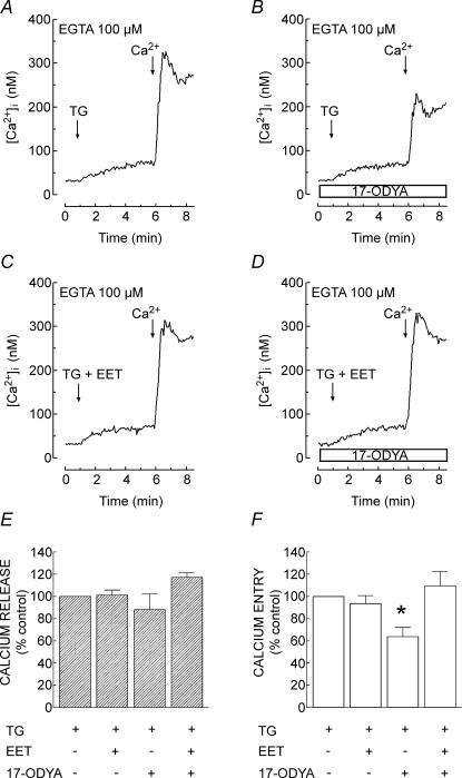
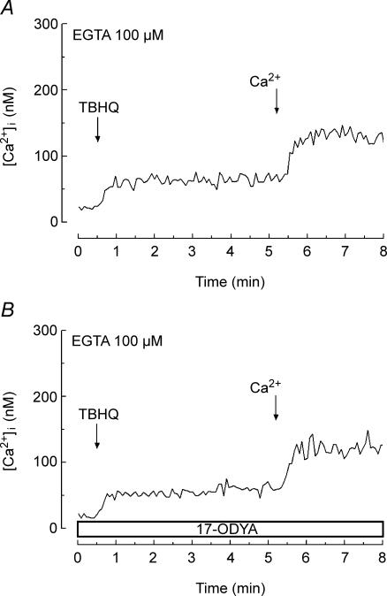
A major pathway for Ca(2+) entry in non-excitable cells is activated following depletion of intracellular Ca(2+) stores. A de novo conformational coupling between elements in the plasma membrane (PM) and Ca(2+) stores has been proposed as the most likely mechanism to activate this capacitative Ca(2+) entry (CCE) in several cell types, including platelets. Here we report that a cytochrome P450 metabolite, 5,6-EET, might be a component of the de novo conformational coupling in human platelets. In these cells, 5,6-EET induces divalent cation entry without having any detectable effect on Ca(2+) store depletion. 5,6-EET-induced Ca(2+) entry was sensitive to the CCE blockers 2-APB, lanthanum, SKF-96365 and nickel and impaired by incubation with anti-hTRPC1 antibody. Ca(2+) entry stimulated by low concentrations of thapsigargin, which selectively depletes the dense tubular system and induces EET production, was impaired by the cytochrome P450 inhibitor 17-ODYA, which has no effect on CCE mediated by depletion of the acidic stores using 2,5-di-(tert-butyl)-1,4-hydroquinone. We have found that 5,6-EET-induced Ca(2+) entry requires basal levels of H(2)O(2), which might maintain a redox state favourable for this event. Finally, our results indicate that 5,6-EET induces the activation of tyrosine kinase proteins and the reorganization of the actin cytoskeleton, which might provide a support for the transport of portions of the Ca(2+) store towards the PM to facilitate de novo coupling between IP(3)R type II and hTRPC1 detected by coimmunoprecipitation. We propose that the involvement of 5,6-EET in TG-induced coupling between IP(3)R type II and hTRPC1 and subsequently CCE is compatible with the de novo conformational coupling in human platelets.


Figures

Figure 1
Figure 1. Effect of 5,6-EET on Ca2+ release and entry in human platelets
A, fura-2-loaded human platelets were treated in a Ca2+-free medium (100 μm EGTA was added) with various concentrations of 5,6-EET (0.01–3 μm) or left untreated (dotted trace) followed by addition of CaCl2 (final concentration 300 μm) to initiate Ca2+ entry. Modifications in [Ca2+]i were monitored using the 340/380 nm ratio and traces were calibrated in terms of [Ca2+]i. B, bars indicate the amount of Ca2+ entry and release in the presence of different concentrations of 5,6-EET. Ca2+ entry and release were estimated using the integral of the rise in [Ca2+]i for 3 min after addition of CaCl2 or 5,6-EET, respectively, and expressed as nanomolar seconds (nm s) as described in Methods. Values are expressed as the mean ± s.e.m. from six independent experiments. *P < 0.05 compared to the resting level.
Figure 2
Figure 2. Effect of 5,6-EET on extracellular Mn2+ entry
Human platelets were loaded with fura-2 and resuspended in a Ca2+-free medium as described in Methods. Fura-2-fluorescence was measured with an excitation wavelength of 360 nm, the isoemissive wavelength. Platelets were stimulated with different concentrations of 5,6-EET (0.01–3 μm, traces a–d) 5 min before addition of MnCl2 (final concentration 300 μm). Mn2+ was added to untreated control cells (trace e) or cells treated with 5,6-EET. Traces are representative of seven separate experiments.
Figure 3
Figure 3. 5,6-EET-induced Ca2+ entry is inhibited by 2-APB, SKF 96365, LaCl3, NiCl2 and the anti-hTRPC1 antibody
Fura-2-loaded human platelets were preincubated at 37°C with 100 μm 2-APB (A) or 10 μm SKF 96365 for 10 min (B), or with 10 μm NiCl2 (C) or 100 μm LaCl3 for 1 min (D), or with 15 μg ml−1 anti-hTRPC1 antibody (αTRPC1 ab) for 10 min (E) or the vehicles as Control (bold traces) and then treated in a Ca2+-free medium (100 μm EGTA was added) for 5 min with 1 μm 5,6-EET followed by addition of CaCl2 (300 μm) to initiate Ca2+ entry. F, bars indicate the percentage of Ca2+ entry under the different experimental conditions relative to their respective control. Ca2+ entry was determined as described in Methods and values are expressed as the mean ± s.e.m. from five to nine independent experiments. ***P < 0.001 compared to control.
Figure 4
Figure 4. Role of cytochrome P450 enzymes on CCE induced by low concentrations of TG
Fura-2-loaded human platelets were preincubated for 10 min at 37°C with 10 μm 17-ODYA (B and D) or the vehicle as Control (A and C) and then treated in a Ca2+-free medium (100 μm EGTA was added) for 5 min with TG (10 nm) in the absence or presence of 1 μm 5,6-EET, as indicated, followed by addition of CaCl2 (300 μm) to initiate Ca2+ entry. E and F, bars indicate the percentage of Ca2+ release (E) and entry (F) under the different experimental conditions relative to their control (vehicle was added). Ca2+ release and entry were determined as described in Methods and values are expressed as the mean ± s.e.m. from nine independent experiments. *P < 0.05 compared to control.
Figure 5
Figure 5. Role of cytochrome P450 enzymes on CCE induced by treatment with TBHQ
Fura-2-loaded human platelets were preincubated for 10 min at 37°C with 10 μm 17-ODYA (B) or the vehicle as Control (A) and then treated in a Ca2+-free medium (100 μm EGTA was added) for 5 min with TBHQ (20 μm) followed by addition of CaCl2 (300 μm) to initiate Ca2+ entry. Changes in [Ca2+]i were monitored using the 340/380 nm ratio and traces were calibrated in terms of [Ca2+]i. Traces are representative of 11 separate experiments.
Figure 6
Figure 6. Effect of Cytochalasin D on 5,6-EET-induced Ca2+ entry
Fura-2-loaded human platelets were preincubated for 40 min at 37°C with 10 μm Cyt D or the vehicle as Control and then treated in a Ca2+-free medium (100 μm EGTA was added) for 5 min with 1 μm 5,6-EET (A) or 3 μm BN (B) followed by addition of CaCl2 (300 μm) to initiate Ca2+ entry. Changes in [Ca2+]i were monitored using the 340/380 nm ratio and traces were calibrated in terms of [Ca2+]i. Traces are representative of six separate experiments.
Figure 7
Figure 7. Effect of 5,6-EET on actin polymerization in platelets
Human platelets were treated with 1 μm 5,6-EET in a Ca2+-free medium (100 μm EGTA was added). Samples were removed 5 s before and 5, 10, 30 and 60 s after the addition of 5,6-EET and the actin filament content was determined as described in Methods. Values given are 5,6-EET-evoked actin filament formation as a percentage of control and results are expressed as mean ± s.e.m. of nine separate determinations. *P < 0.05, **P < 0.01 compared to control (resting cells).
Figure 8
Figure 8. Effect of catalase on 5,6-EET-induced Ca2+ entry
A, human platelets loaded with CM-H2DCFDA were stimulated with 1 μm 5,6-EET in a Ca2+-free medium (100 μm EGTA was added). Traces are representative of eight independent experiments. B–D, fura-2-loaded human platelets were preincubated for 10 min at 37°C with 300 U ml−1 catalase or the vehicle as Control and then treated in a Ca2+-free medium (100 μm EGTA was added) for 5 min with 10 μm H2O2 (B), 1 μm 5,6-EET (C) or 1 μm BN (D) followed by addition of CaCl2 (300 μm) to initiate Ca2+ entry. Changes in [Ca2+]i were monitored using the 340/380 nm ratio and traces were calibrated in terms of [Ca2+]i. Traces are representative of 10 separate experiments.
Figure 9
Figure 9. Role for protein tyrosine phosphorylation on 5,6-EET-induced Ca2+ entry in platelets
A, human platelets were treated with different concentrations of 5,6-EET (0.01–3 μm) in a Ca2+-free medium (100 μm EGTA was added). Samples were taken from the platelet suspension 5 s before and 5 min after the addition of different concentrations of 5,6-EET. Platelet proteins were analysed by 10% SDS-PAGE and subsequent Western blotting with a specific anti-phosphotyrosine antibody, and the presence of phosphotyrosine residues quantified by densitometry in Western blots as described. Molecular masses (M) indicated on the right were determined using molecular-mass markers run in the same gel. B, bars represent the integrated optical density for entire lanes under each condition. Results are expressed as fold-increases (mean ± s.e.m. of four separate experiments) over the integrated optical density of resting platelet. *P < 0.05, **P < 0.01 compared to control (resting cells). C, fura-2-loaded human platelets were incubated at 37°C with 1 μg ml−1 M-2,5-DHC or the vehicle for 30 min. At the time of the experiment 100 μm EGTA was added. Cells were then stimulated with 5,6-EET (1 μm) and, 5 min later, CaCl2 (final concentration 300 μm) was added to the medium to initiate Ca2+ entry. Elevations in [Ca2+]i were monitored using the 340/380 nm ratio as described in Methods. Traces shown are representative of four others.
Figure 10
Figure 10. Role of 5,6-EET on TG-induced coupling between IP3R II and hTRPC1 in human platelets
Platelets were preincubated for 10 min at 37°C in the presence (lane 4) or absence (lanes 1–3) of 10 μm 17-ODYA. Cells were then stimulated with either 5,6-EET (1 μm) or TG (10 nm). Samples were taken 5 s before and 3 min after the addition of 5,6-EET or TG and lysed. Whole cell lysates were immunoprecipitated with anti-hTRPC1 antibody. Immunoprecipitates were analysed by Western blotting using either anti-IP3R type II polyclonal antibody (upper panel) or anti-hTRPC1 antibody (lower panel) as described in Methods. These results are representative of four independent experiments.

Similar articles

Cited by

References

    1. Alonso-Torre SR, Alvarez J, Montero M, Sanchez A, Garcia-Sancho J. Control of Ca2+ entry into HL60 and U937 human leukaemia cells by the filling state of the intracellular Ca2+ stores. Biochem J. 1993;289:761–766. - PMC - PubMed
    1. Alvarez DF, Gjerde EA, Townsley MI. Role of EETs in regulation of endothelial permeability in rat lung. Am J Physiol Lung Cell Mol Physiol. 2004;286:445–451. - PubMed
    1. Beech DJ, Muraki K, Flemming R. Non-selective cationic channels of smooth muscle and the mammalian homologues of Drosophila TRP. J Physiol. 2004;559:685–706. - PMC - PubMed
    1. Bergdhal A, Gómez MF, Dreja K, Xu SZ, Adner M, Beech DJ, Broman J, Hellstrand P, Sward K. Cholesterol depletion impairs vascular reactivity to endothelin-1 by reducing store-operated Ca2+ entry dependent on TRPC1. Circ Res. 2003;93:839–847. - PubMed
    1. Bird GS, Putney JW. Inhibition of thapsigargin-induced calcium entry by microinjected guanine nucleotide analogues. Evidence for the involvement of a small G-protein in capacitative calcium entry. J Biol Chem. 1993;268:21486–21488. - PubMed

Publication types

MeSH terms

Substances

LinkOut - more resources