. 2005 Dec 21;24(24):4415-24.
doi: 10.1038/sj.emboj.7600885. Epub 2005 Nov 24.

Phosphorylation by casein kinase 2 induces PACS-1 binding of nephrocystin and targeting to cilia

Affiliations

Phosphorylation by casein kinase 2 induces PACS-1 binding of nephrocystin and targeting to cilia

Bernhard Schermer et al. EMBO J. .

Abstract

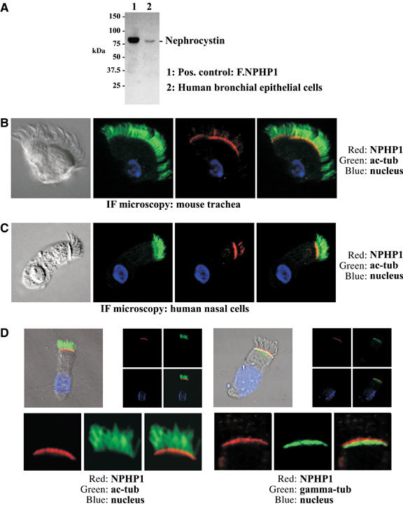
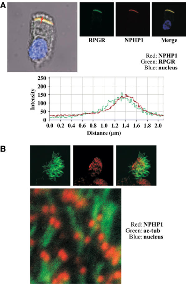
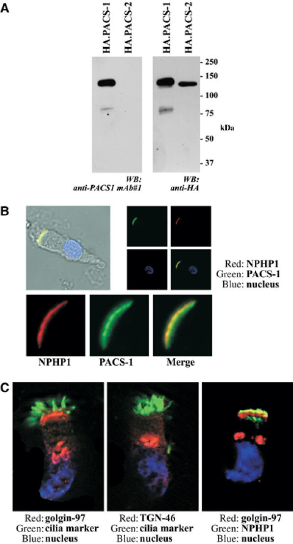
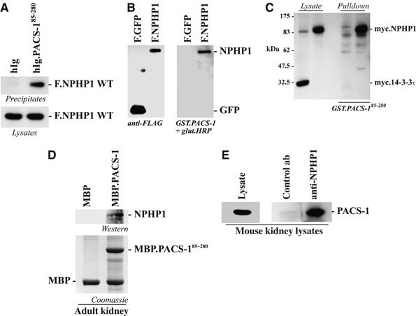
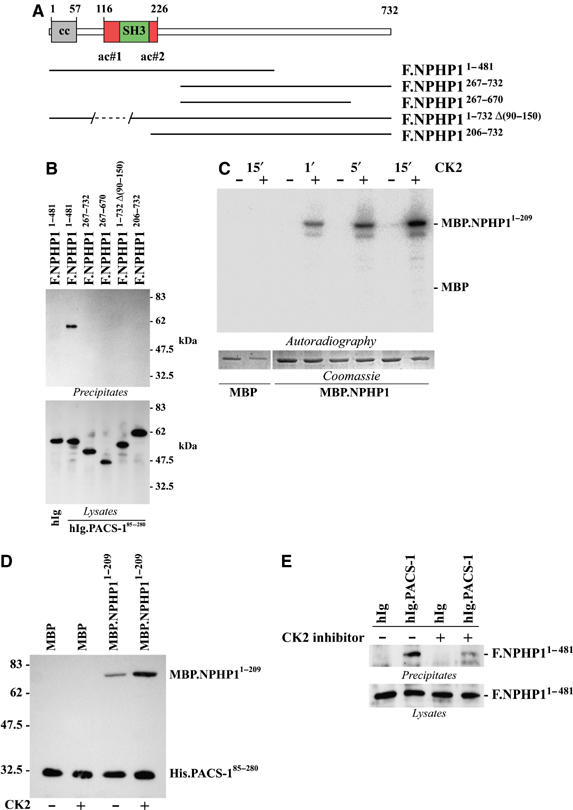
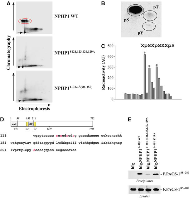
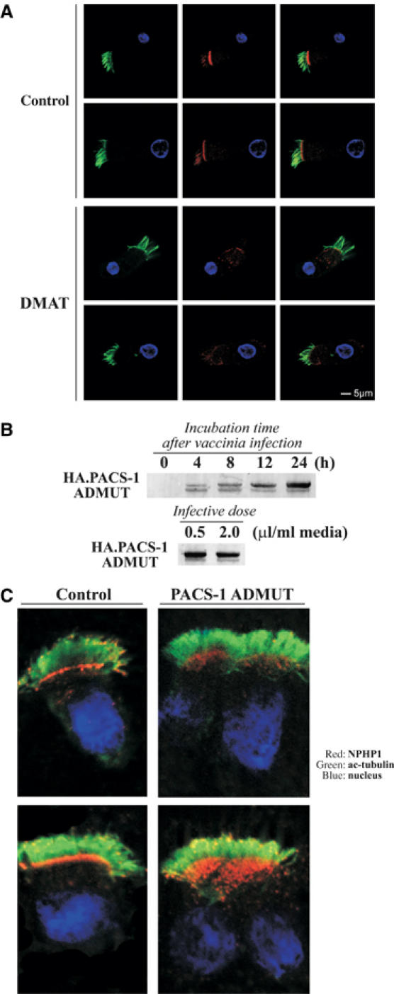
Mutations in proteins localized to cilia and basal bodies have been implicated in a growing number of human diseases. Access of these proteins to the ciliary compartment requires targeting to the base of the cilia. However, the mechanisms involved in transport of cilia proteins to this transitional zone are elusive. Here we show that nephrocystin, a ciliary protein mutated in the most prevalent form of cystic kidney disease in childhood, is expressed in respiratory epithelial cells and accumulates at the base of cilia, overlapping with markers of the basal body area and the transition zone. Nephrocystin interacts with the phosphofurin acidic cluster sorting protein (PACS)-1. Casein kinase 2 (CK2)-mediated phosphorylation of three critical serine residues within a cluster of acidic amino acids in nephrocystin mediates PACS-1 binding, and is essential for colocalization of nephrocystin with PACS-1 at the base of cilia. Inhibition of CK2 activity abrogates this interaction and results in the loss of correct nephrocystin targeting. These data suggest that CK2-dependent transport processes represent a novel pathway of targeting proteins to the cilia.


Figures

Figure 1
Figure 1
Nephrocystin is expressed in respiratory epithelial cells and accumulates at the base of cilia. (A) Lysates of HEK 293T cells transfected with FLAG-tagged NPHP1 (left lane) and human tracheal epithelial cells (right lane) were immunoblotted with an affinity-purified specific anti-nephrocystin antibody (Benzing et al, 2001) demonstrating nephrocystin expression in respiratory epithelial cells. (B, C) Conventional immunofluorescence microscopy of mouse tracheal cells (B) and human nasal epithelial cells (C) stained with a specific anti-nephrocystin antibody (Benzing et al, 2001) demonstrates distinct and punctate nephrocystin staining in the ciliary axoneme and a strong signal at the base of motile cilia of the respiratory tract (red). Cilia were visualized with antiacetylated tubulin antibody (green); blue, nuclei. (D) Confocal microscopy confirms intense staining for endogenous nephrocystin (red) at the base of cilia in human nasal epithelial cells. Staining for acetylated tubulin (green) is used to highlight the cilium. Nuclei are stained in blue (left panel). Double labelling for gamma-tubulin (green), a marker of the basal bodies, and nephrocystin (red) confirms accumulation of nephrocystin protein at the transition zone/basal body area (right panel).
Figure 2
Figure 2
Colocalization of nephrocystin with RPGR at the transition zone/basal body area of cilia in human respiratory epithelial cells. (A) Staining of endogenous nephrocystin (red) and RGPR (green) in respiratory epithelial cells reveals colocalization of both proteins at the transition zone/basal body area of motile cilia. Nuclei (blue). Fluorescence signal intensities of nephrocystin (red) and RPGR (green), generated from a scanned horizontal line shown as red arrow in the merged image, are shown in the bottom panel. (B) Nephrocystin staining is confined to cilia and does not derive from other parts of the apical membrane. Cilia were disintegrated mechanically to show localization of nephrocystin at the base of each disintegrated cilium. Nephrocystin (red), acetylated tubulin (green), and nuclei (blue).
Figure 3
Figure 3
Nephrocystin colocalizes with the transport protein PACS-1 at the base of cilia of respiratory epithelial cells. (A) Monoclonal anti-PACS-1 antibody mAb#1 specifically recognizes HA-tagged PACS-1, but does not show crossreactivity with the related protein HA.PACS-2. HEK 293T cells were transfected with the plasmids as indicated. Lysates were prepared and immunoblotted with anti-PACS-1 antibody mAb#1 (left panel) as well as anti-HA antibody (right panel). (B) Colocalization of PACS-1 (green) with nephrocystin (red) at the base of the cilia. Nuclei (blue). Human nasal epithelial cells were isolated by brush biopsy and stained with anti-nephrocystin and anti-PACS-1 antibodies. (C) Native nasal epithelial cells were stained with anti-golgin-97 (Molecular Probes; left panel) and anti-TGN-46 antibody (Serotec; middle panel). Costaining with anti-NPHP1 antibody demonstrates that the base of the cilia is partially overlapping with a golgin-97-positive distinct domain of the secretory pathway (right panel).
Figure 4
Figure 4
Nephrocystin interacts with acidic cluster sorting protein PACS-1. (A) Nephrocystin co-precipitates with PACS-1. Ig-tagged PACS-1 (amino acids 85–280) or a control protein and Flag-tagged nephrocystin (F.NPHP1) were expressed in HEK 293T cells and precipitated with protein G. Western blot analysis was performed with a FLAG-specific antibody (top). Expression levels of F.NPHP1 in the lysates are shown (bottom). (B) Lysates of cells expressing FLAG-tagged green fluorescent protein (F.GFP) or nephrocystin (F.NPHP1) were analyzed on Western blots with anti-FLAG antibody (left) or by overlay assays using a GST fusion protein of PACS-1 coupled to horseradish peroxidase as detection reagent (right panel). (C) Pulldown assays using a recombinant GST fusion protein of PACS-1 confirm specific interaction with nephrocystin. (D) Pulldown of endogenous nephrocystin from mouse kidney lysates (top). Protein lysates from mouse kidneys were incubated with recombinant MBP fusion proteins immobilized on beads. After incubation at 4°C, the beads were washed extensively and bound proteins were analyzed by immunoblotting. Co-precipitating endogenous nephrocystin protein is shown in the upper panel, expression of recombinant MBP fusion proteins is shown in the lower panel. (E) Endogenous nephrocystin interacts with PACS-1 in the kidney, suggesting that the interaction occurs in vivo. Protein lysates from mouse kidneys were subjected to immunoprecipitation with a control antibody (second lane) or specific anti-nephrocystin antibody (third lane), washed extensively and immunoblotted with specific anti-PACS-1 antibody. The first lane depicts PACS-1 expression in the kidney lysate.
Figure 5
Figure 5
The interaction between nephrocystin and PACS-1 is phosphorylation-dependent and maps to a cluster of acidic residues in nephrocystin. (A) Several Flag-tagged nephrocystin truncations were generated to analyze the interaction with PACS-1. The SH3 domain in nephrocystin (green) is flanked by two clusters of acidic residues (red). (B) PACS-1 interaction requires a region of nephrocystin that contains the first acidic cluster (top). HEK 293T cells were cotransfected with Ig-tagged PACS-1 (amino acids 85–280) and FLAG-tagged nephrocystin constructs as indicated. Ig-tagged PACS-1 was precipitated with protein G and the precipitates were analyzed for co-precipitating nephrocystin truncations (upper panel). Expression levels of FLAG-tagged nephrocystin constructs are shown (bottom). Expression level of hIg.PACS-1 was identical in all cell lysates (not shown). (C) A bacterially expressed recombinant fusion protein of nephrocystin (MBP.NPHP1) containing the first acidic cluster (amino acids 1–209) is time-dependently phosphorylated by CK2. Autoradiograph after in vitro phosphorylation of MBP or MBP.NPHP1 with CK2 (top). Expression of recombinant fusion proteins is shown (bottom). (D) In vitro interaction of His.PACS-185−285 with MBP (before and after treatment with CK2, lanes 1 and 2) and MBP.NPHP11−209 (before and after treatment with CK2, lanes 3 and 4) shows dependence of the interaction on CK2 activity. His-tagged PACS-1 (amino acids 85–280) was bound to Ni+ beads and incubated with equal amounts of MBP (first two lanes) or MBP.NPHP1 (amino acids 1–209) preincubated or not preincubated with CK2. Ni+ beads were washed extensively and analyzed for co-precipitating MBP fusion proteins with anti-MBP antibody. His-tagged PACS-1 was visualized by reprobing the blot with anti-His antibody. (E) Treatment of cells with the CK2 inhibitor TBB (20 μM, 4 h) prior to cell lysis inhibits the interaction of PACS-1 with nephrocystin (upper panel). Expression levels are shown (lower panel). Equal expression of Ig-tagged proteins was confirmed by reprobing with anti-human-IgG antibody (not shown).
Figure 6
Figure 6
Requisite phosphorylation of serines 121, 123, 126, but not serine 129, of nephrocystin mediates interaction with PACS-1. (A) Radioactive labelling, followed by precipitation of nephrocystin, completes tryptic digest of the protein, and two-dimensional separation of the peptide fragments reveals a major phosphopeptide in wild-type nephrocystin that is absent in the serine-to-alanine mutants lacking serine 121, 123, 126, and 129, as well as in the deletion mutant of the first acidic cluster of nephrocystin. (B) The indicated peptide was eluted and a fraction was hydrolyzed and subjected to phosphoamino-acid analysis (locations of standard phosphoamino acids are indicated by black circles, pS-phospho-serine, pT-phospho-threonine, pY-phospho-tyrosine). (C) The remaining portion of the eluted phosphopeptide was subjected to 20 cycles of Edman degradation and cleaved amino acids were collected and analyzed using a PhosphorImager to locate the position of the phosphorylation site(s). The content of 32P radioactivity of each sequencing cycle is expressed in arbitrary units (AU). (D) The SH3 domain of nephrocystin (highlighted in gray) is flanked by two acidic clusters (yellow) containing putative CK2 phosphorylation sites (red). (E) Mutation of the CK2 phosphorylation sites in nephrocystin to alanines prevents binding of PACS-1. HEK 293T cells were transfected with the plasmids as indicated and subjected to precipitation with protein G, followed by immunoblotting with anti-FLAG antibody. The lower panel shows expression in the lysates.
Figure 7
Figure 7
Targeting of nephrocystin to the transition zone/basal body area requires CK2 activity and is dependent on PACS-1. (A) Native human respiratory epithelial cells, freshly isolated by brush biopsy, were incubated with solvent (upper panels) or the highly selective CK2 inhibitor DMAT (1.5 μM, 2 h at 37°C) (lower panels), fixated in paraformaldehyde (4%) and stained for nephrocystin (red) and acetylated tubulin (green). Nuclei (blue). Instead of localizing to the transition zone/basal body area as shown in solvent-treated cells (upper panel), nephrocystin assumes a vesicle-like subcellular distribution (lower panel). (B) Vaccinia virus-mediated expression of a dominant-negative mutant of PACS-1 (PACS-1 ADMUT) in mouse trachea. Time- and dose-dependent expression of PACS-1 ADMUT was analyzed on immunoblots of mouse tracheal cells with anti-HA antibody. (C) Expression of PACS-1 ADMUT results in loss of nephrocystin from the ciliary base. Respiratory epithelial cells were harvested 16 h after the infection with either control vaccinia virus (Control) or PACS-1 ADMUT vaccinia virus (PACS-1 ADMUT) and stained with antiacetylated tubulin and anti-nephrocystin antibody.

References

    1. Ansley SJ, Badano JL, Blacque OE, Hill J, Hoskins BE, Leitch CC, Kim JC, Ross AJ, Eichers ER, Teslovich TM, Mah AK, Johnsen RC, Cavender JC, Lewis RA, Leroux MR, Beales PL, Katsanis N (2003) Basal body dysfunction is a likely cause of pleiotropic Bardet–Biedl syndrome. Nature 425: 628–633 - PubMed
    1. Benzing T, Brandes R, Sellin L, Schermer B, Lecker S, Walz G, Kim E (1999) Upregulation of RGS7 may contribute to tumor necrosis factor-induced changes in central nervous function. Nat Med 5: 913–918 - PubMed
    1. Benzing T, Gerke P, Hopker K, Hildebrandt F, Kim E, Walz G (2001) Nephrocystin interacts with Pyk2, p130(Cas), and tensin and triggers phosphorylation of Pyk2. Proc Natl Acad Sci USA 98: 9784–9789 - PMC - PubMed
    1. Benzing T, Yaffe MB, Arnould T, Sellin L, Schermer B, Schilling B, Schreiber R, Kunzelmann K, Leparc GG, Kim E, Walz G (2000) 14-3-3 interacts with regulator of G protein signaling proteins and modulates their activity. J Biol Chem 275: 28167–28172 - PubMed
    1. Blagoveshchenskaya AD, Thomas L, Feliciangeli SF, Hung CH, Thomas G (2002) HIV-1 Nef downregulates MHC-I by a PACS-1- and PI3K-regulated ARF6 endocytic pathway. Cell 111: 853–866 - PubMed

Publication types

MeSH terms