. 2005 Dec 7;24(23):4198-208.
doi: 10.1038/sj.emboj.7600880. Epub 2005 Nov 24.

A view of consecutive binding events from structures of tetrameric endonuclease SfiI bound to DNA

Affiliations

A view of consecutive binding events from structures of tetrameric endonuclease SfiI bound to DNA

Eva Scheuring Vanamee et al. EMBO J. .

Abstract

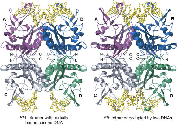
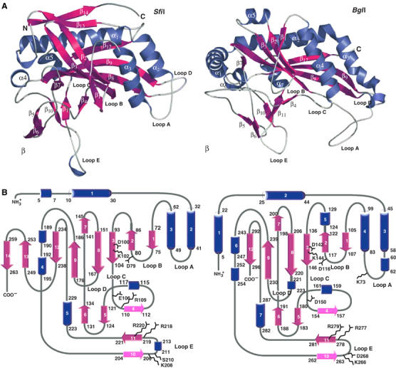
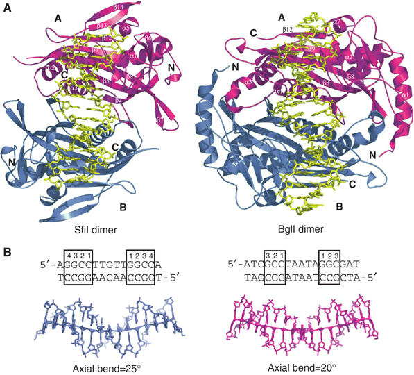
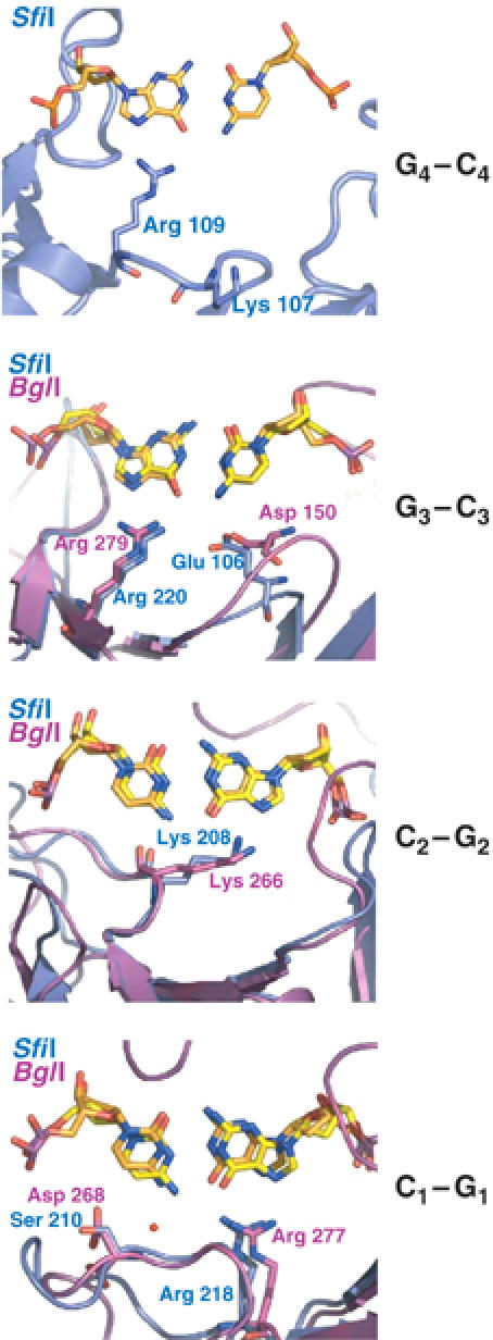
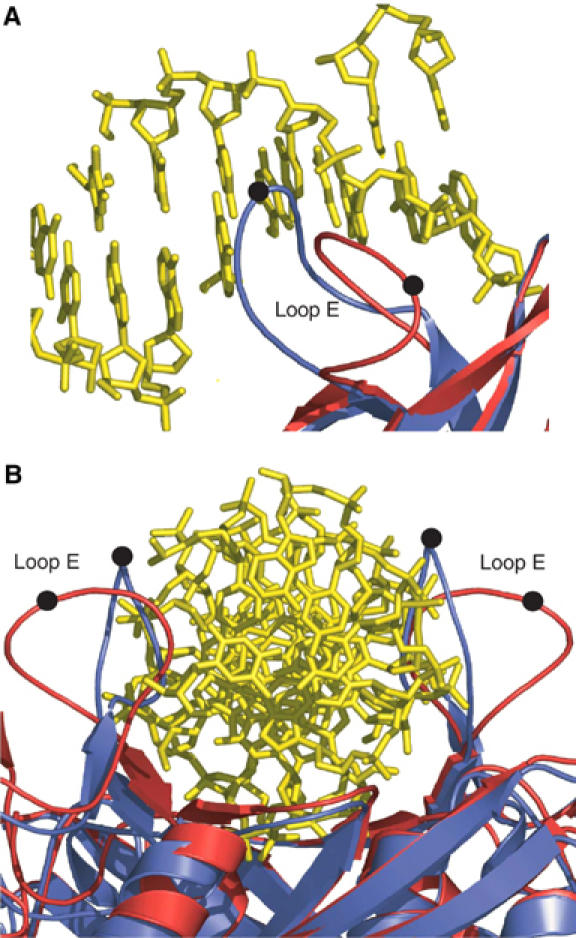
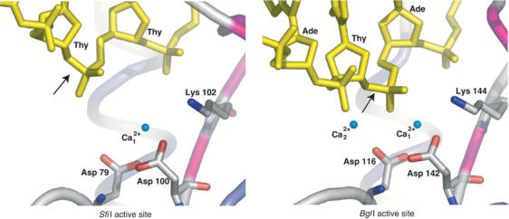
Many reactions in cells proceed via the sequestration of two DNA molecules in a synaptic complex. SfiI is a member of a growing family of restriction enzymes that can bind and cleave two DNA sites simultaneously. We present here the structures of tetrameric SfiI in complex with cognate DNA. The structures reveal two different binding states of SfiI: one with both DNA-binding sites fully occupied and the other with fully and partially occupied sites. These two states provide details on how SfiI recognizes and cleaves its target DNA sites, and gives insight into sequential binding events. The SfiI recognition sequence (GGCCNNNN[downward arrow]NGGCC) is a subset of the recognition sequence of BglI (GCCNNNN[downward arrow]NGGC), and both enzymes cleave their target DNAs to leave 3-base 3' overhangs. We show that even though SfiI is a tetramer and BglI is a dimer, and there is little sequence similarity between the two enzymes, their modes of DNA recognition are unusually similar.


Figures

Figure 1
Figure 1
Schematic representation of SfiI–DNA complexes. On the right is shown the native structure with SfiI tetramer bound to two DNA molecules. Each subunit is color coded as follows: (A) magenta, (B) blue, (C) light gray, (D) (green); the DNA is shown in yellow. On the left is shown the Se–Met structure, wherein the A/B dimer is bound to fully occupied DNA and the C/D dimer is bound to partially occupied DNA. The nonspecific middle bases in the partially occupied DNA could not be built due to the lack of electron density. Note also that the loop contacting the partially occupied DNA in the C/D dimer adopts a different conformation.
Figure 2
Figure 2
Comparisons between SfiI and BglI subunits. (A) Structure of SfiI (left) and BglI (right) monomers. The secondary structural elements are labeled and colored as follows: α helices are blue, β strands are magenta, and loops are white. The 3/10 helices are unlabeled. (B) Topology diagram of SfiI (left) and BglI (right) subunit. The color scheme is the same as in (A).
Figure 3
Figure 3
(A) Comparison between SfiI and BglI dimers. SfiI (left) and BglI (right) dimers bound to DNA. The monomers are colored magenta (A) and blue (B), and the DNA is shown in yellow. Both enzymes bind DNA in a similar fashion, with each monomer contacting one half site. (B) The structure of the DNA in SfiI (left) and BglI (right). The DNA molecules are represented in ‘stick' (SfiI in blue and BglI in magenta). The DNA sequences are listed on top, with the recognition sequences highlighted with a box around the base pairs. The cognate base-pairs are numbered from inside out. The SfiI and BglI DNAs are bent 25° and 20°, respectively, as calculated by CURVES (Lavery and Sklenar, 1988), and highlighted by the lines through the DNAs. In SfiI, electron density is seen for only 15 out of the 21 nucleotide residues.
Figure 4
Figure 4
Comparison of base-specific DNA interactions in SfiI and BglI. The proteins are shown in ribbon representation (SfiI in blue and BglI in magenta). The nucleotides (SfiI DNA in orange and BglI DNA in yellow) and the base-specific residues are shown in ‘stick' representation. In each picture, only the base pairs in question are shown; other nucleotides are omitted for clarity.
Figure 5
Figure 5
Change in loop E conformation. (A) Loop E enters the DNA major groove in the native complex (blue), but in the C/D dimer of the Se–Met complex (red) it packs away from the DNA. Black spheres mark the position of Arg 213. (B) View of C/D dimer looking down the DNA axis. In the native complex (blue), loop E's bracket the DNA and help to hold it in place, while in the Se–Met complex (red) the loops are positioned away from the DNA.
Figure 6
Figure 6
The active sites of SfiI (left) and BglI (right). The DNA and the active site residues of SfiI (Asp 79, Asp100, and Lys102) and BglI (Asp116, Asp142, and Lys144) are shown in ‘stick' representation. The scissile phosphodiester is indicated by arrows. The Ca2+ ions are shown as nonbonded spheres and colored aqua, water molecules are not shown. In SfiI, the second Ca2+ is missing and the scissile phosphodiester is ∼3 Å further away than in BglI, indicating that the enzyme is in an inactive state.

Similar articles

Cited by

References

    1. Aggarwal AK (1995) Structure and function of restriction endonucleases. Curr Opin Struct Biol 5: 11–19 - PubMed
    1. Aggarwal AK, Rodgers DW, Drottar M, Ptashne M, Harrison SC (1988) Recognition of a DNA operator by the repressor of phage 434: a view at high resolution. Science 242: 899–907 - PubMed
    1. Alves J, Ruter T, Geiger R, Fliess A, Maass G, Pingoud A (1989) Changing the hydrogen-bonding potential in the DNA binding site of EcoRI by site-directed mutagenesis drastically reduces the enzymatic activity, not, however, the preference of this restriction endonuclease for cleavage within the site-GAATTC. Biochemistry 28: 2678–2684 - PubMed
    1. Alves J, Vennekohl P (2004) Protein engineering of restriction enzymes. In Restriction Endonucleases, Pingoud A (ed), Vol 14, pp 393–411. Berlin, Heidelberg: Springer-Verlag
    1. Bath AJ, Milsom SE, Gormley NA, Halford SE (2002) Many type IIs restriction endonucleases interact with two recognition sites before cleaving DNA. J Biol Chem 277: 4024–4033 - PubMed

Publication types

MeSH terms

Substances