. 2005 Dec 7;24(23):4166-75.
doi: 10.1038/sj.emboj.7600877. Epub 2005 Nov 24.

3-D structural and functional characterization of the purified KATP channel complex Kir6.2-SUR1

Affiliations

3-D structural and functional characterization of the purified KATP channel complex Kir6.2-SUR1

Michael V Mikhailov et al. EMBO J. .

Abstract

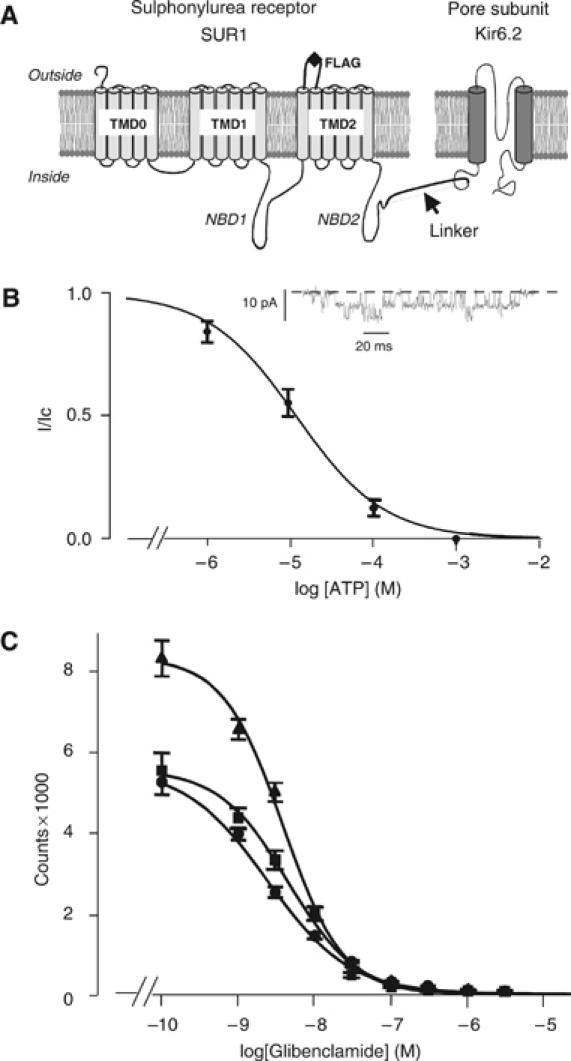
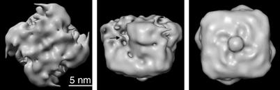
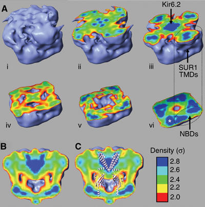
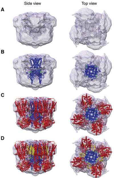
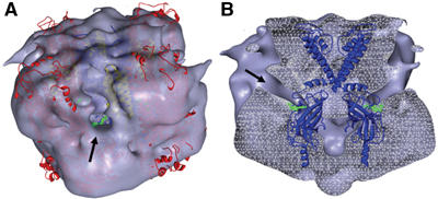
ATP-sensitive potassium (K(ATP)) channels conduct potassium ions across cell membranes and thereby couple cellular energy metabolism to membrane electrical activity. Here, we report the heterologous expression and purification of a functionally active K(ATP) channel complex composed of pore-forming Kir6.2 and regulatory SUR1 subunits, and determination of its structure at 18 A resolution by single-particle electron microscopy. The purified channel shows ATP-ase activity similar to that of ATP-binding cassette proteins related to SUR1, and supports Rb(+) fluxes when reconstituted into liposomes. It has a compact structure, with four SUR1 subunits embracing a central Kir6.2 tetramer in both transmembrane and cytosolic domains. A cleft between adjacent SUR1s provides a route by which ATP may access its binding site on Kir6.2. The nucleotide-binding domains of adjacent SUR1 appear to interact, and form a large docking platform for cytosolic proteins. The structure, in combination with molecular modelling, suggests how SUR1 interacts with Kir6.2.


Figures

Figure 1
Figure 1
Construction and analysis of an SUR1F–Kir6.2 fusion protein. (A) Schematic of the membrane topology of SUR1 and Kir6.2 showing the location of the FLAG tag used for purification and the linker between SUR1 and Kir6.2. (B) Relationship between [ATP] and KATP current (I), expressed relative to the current in the absence of nucleotide (Ic) for SUR1F–Kir6.2 channels (n=8). The curve is the best fit of the Hill equation to the data with an IC50 11 μM and h=0.8. Inset: Single KATP channel currents at −60 mV in the absence of nucleotide. (C) Displacement of binding of [3H]glibenclamide (5 nM) to Sf9 cells expressing SUR1 (▴, n=3), SUR1–Kir6.2 (i.e. without a FLAG tag; ▪, n=3), or SUR1F–Kir6.2 (•, n=3) by unlabelled glibenclamide. The curves are fit to equation (2) and Km values are given in the text. Data points are shown as mean±s.e.m.
Figure 2
Figure 2
Protein purification and functional analysis. (A) Coomassie-stained SDS–PAGE (8%) gels of molecular weight markers (lane 1), solubilized Sf9 cells expressing SUR1F–Kir6.2 (lane 2) and purified SUR1F–Kir6.2 (lane 3). The purity of the protein was ∼80% as assessed using Gel Pro Analyser following digital scanning of the gel. The molecular masses calculated from the migration of molecular standards are indicated. (B) Native PFO-PAGE (5%) gel of molecular mass markers (left lane) and purified SUR1F–Kir6.2 (right lane). The high molecular mass complex (KATP, arrowed) has a molecular mass of ∼900 kDa when estimated by extrapolation from a linear plot (not shown) of log molecular mass versus migration. (C) ATPase activity of SUR1F–Kir6.2 (n=3). The line is the best fit of the Michaelis–Menten equation to the mean data and has a Vmax of 110 nmol Pi/min/mg protein and a Ki of 0.4 mM. Data points are shown as mean±s.e.m of three separate protein preparations. (D) Time course of 86Rb+ uptake into liposomes reconstituted with 2–3 μg SUR1F–Kir6.2/mg lipid (formula image
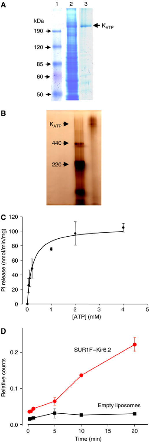
) or into control liposomes containing no protein (▪). Data shown are the mean±s.e.m. of three separate experiments (one protein preparation; similar results were obtained with a second protein preparation).
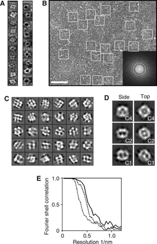
Figure 3
Figure 3
Electron microscopy of the KATP channel. (A) Single SUR1F–Kir6.2 particles unlabelled (left) or labelled with 5-nm-diameter immunogold (right). Particles were negatively stained with uranyl acetate and examined at room temperature. (B) Area of an electron micrograph recorded under cryo conditions, showing trehalose-molybdate-embedded SUR1F–Kir6.2 particles (enclosed by squares). Scale bar (white), 50 nm. Inset: Fourier transform of the micrograph, with the distinctive rings representing crossover points in the contrast transfer function. The edge of the transform corresponds to 1/6.7 Å. (C) Typical projection classes with square-shaped classes displaying roughly four-fold symmetry (‘top/bottom views', upper row), hexagonal-shaped classes (‘partial side views', centre rows), and rectangular-shaped classes with some bilateral symmetry (‘side views', lowest rows). Each box is 270 Å across. (D) Preliminary 3D models calculated from the classes in panel C. Side views (left column) and top views (right column) for C4 symmetry (top row), C2 symmetry (middle row), or no symmetry (C1, bottom row) are shown. Each box is 270 Å across. (E) Fourier shell correlation versus resolution for the final 3D models generated with C1 symmetry (diamond symbols and solid line), C2 symmetry (dashed line), and C4 symmetry (solid line). The line for C4 symmetry passes below a correlation of 0.5 at a resolution of ∼18 Å.
Figure 4
Figure 4
Surface representation of the KATP channel. Top (left), side (middle), and bottom (right) views of the KATP channel complex after refinement of the structure with C4 symmetry, rendered at 2σ. Arrow indicates crevice in the upper part of the structure. The identity of the ‘button' feature in the right panel is not known.
Figure 5
Figure 5
Slices through the 3D structure. The outer surface is rendered at 2σ (light blue). Colours in the interior indicate increasing density: σ=2 (red), 2.2 (yellow), 2.4 (green), 2.6 (cyan), and 2.8 (dark blue). (A) Sequential slices (i–vi) from the presumed extracellular surface. The white asterisk (vi) indicates the bilobed densities presumed to be the NBDs. The position of TMDs 1 and 2 assumed to correspond to the NBDs is indicated. (B, C) Slice perpendicular to the presumed plane of the membrane without (B) and with (C) a molecular model of Kir6.2 positioned in the density. For clarity, only the TMDs of two Kir6.2 subunits, and the cytosolic domains of the two other subunits, are shown.
Figure 6
Figure 6
Location of molecular models within the 3D structure. (A–D) Top and side views, respectively, of the EM density with models of Kir6.2 (blue), SUR1 minus TMD0 (red), and TMD0 (yellow) inserted sequentially from (B) to (D). ATP molecules are shown in green.
Figure 7
Figure 7
Location of molecular models within the 3D structure. (A, B) Surface and transverse section views, respectively, of the EM density with models of Kir6.2 (blue), TMD0 (yellow), and SUR1 minus TMD0 (red) inserted, showing clefts (arrows) through which ATP (green) could access its binding site on Kir6.2. The Kir6.2 model only is shown in (B).

Similar articles

Cited by

References

    1. Antcliff JF, Haider S, Proks P, Sansom MS, Ashcroft FM (2005) Functional analysis of a structural model of the ATP-binding site of the KATP channel Kir6.2 subunit. EMBO J 24: 229–239 - PMC - PubMed
    1. Ashcroft FM (2005) ATP-sensitive potassium channelopathies: focus on insulin secretion. J Clin Invest 115: 2047–2058 - PMC - PubMed
    1. Ashcroft FM, Gribble FM (2000) New windows on the mechanism of action of KATP channel openers. Trends Pharmacol Sci 21: 439–445 - PubMed
    1. Babenko AP, Bryan J (2002) SUR-dependent modulation of KATP channels by an N-terminal KIR6.2 peptide. Defining intersubunit gating interactions. J Biol Chem 277: 43997–44004 - PubMed
    1. Beis K, Collins RF, Ford RC, Kamis AB, Whitfield C, Naismith JH (2004) Three-dimensional structure of Wza, the protein required for translocation of group 1 capsular polysaccharide across the outer membrane of Escherichia coli. J Biol Chem 279: 28227–28232 - PubMed

Publication types

MeSH terms