. 2005 Dec 21;24(24):4260-70.
doi: 10.1038/sj.emboj.7600874. Epub 2005 Nov 24.

A promiscuous liaison between IL-15 receptor and Axl receptor tyrosine kinase in cell death control

Affiliations

A promiscuous liaison between IL-15 receptor and Axl receptor tyrosine kinase in cell death control

Vadim Budagian et al. EMBO J. .

Retraction in

Abstract

Discrimination between cytokine receptor and receptor tyrosine kinase (RTK) signaling pathways is a central paradigm in signal transduction research. Here, we report a 'promiscuous liaison' between both receptors that enables interleukin (IL)-15 to transactivate the signaling pathway of a tyrosine kinase. IL-15 protects murine L929 fibroblasts from tumor necrosis factor alpha (TNFalpha)-induced cell death, but fails to rescue them upon targeted depletion of the RTK, Axl; however, Axl-overexpressing fibroblasts are TNFalpha-resistant. IL-15Ralpha and Axl colocalize on the cell membrane and co-immunoprecipitate even in the absence of IL-15, whereby the extracellular part of Axl proved to be essential for Axl/IL-15Ralpha interaction. Most strikingly, IL-15 treatment mimics stimulation by the Axl ligand, Gas6, resulting in a rapid tyrosine phosphorylation of both Axl and IL-15Ralpha, and activation of the phosphatidylinositol 3-kinase/Akt pathway. This is also seen in mouse embryonic fibroblasts from wild-type but not Axl-/- or IL-15Ralpha-/- mice. Thus, IL-15-induced protection from TNFalpha-mediated cell death involves a hitherto unknown IL-15 receptor complex, consisting of IL-15Ralpha and Axl RTK, and requires their reciprocal activation initiated by ligand-induced IL-15Ralpha.


Figures

Figure 1
Figure 1
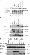
Axl is required for resistance to TNFα cytotoxicity. L929 and L929R cells were transfected with Axl siRNA. (A) Axl mRNA expression was analyzed by RT–PCR (upper panel). β-Actin is used as an internal control (lower panel). (B) WB analysis of Axl protein expression (upper panel). IL-15Rα expression served as a control (lower panel). (C) Cells were treated for 18 h with TNFα (5 ng/ml), IL-15 (100 ng/ml), or both, and cell viability was assessed by PI and annexin V staining and FACS analysis. (D) L929, L929R, and Axl-overexpressing L929 cells were treated or not with TNFα (100 ng/ml) alone or together with 20 mM zVAD-fmk and ceramide generation was determined. All ceramide determinations were performed in duplicate.
Figure 2
Figure 2
IL-15 induces phosphorylation of IL-15Rα and Axl, which constitutively associate. (A) L929 cells were stimulated with IL-15 (100 ng/ml). Cell lysates were precipitated with specific Abs against Axl, IL-15Rα, or IL-2Rγ and analyzed by WB. (B) Axl/IL-15Rα coassociation was detected in a two-site ELISA. Plates were coated with anti-Axl Abs. Axl (white bars) or Axl/IL-15Rα complexes (gray bars) were detected in cell lysates using biotinylated anti-Axl or anti-IL-15Rα Abs, respectively. COS-7 cells served as a negative control. (C) L929 cells were treated for 15 min with IL-15, TNFα, or both. Axl was precipitated from lysates and its phosphorylation was assessed with anti-pTyr Abs (upper panel). Axl detection served as a loading control (lower panel). (D) Axl and IL-15Rα colocalization analysis by confocal microscopy. Cells were stained with anti-IL-15Rα Abs (red), anti-Axl Abs (green), and TOTO-3 for nuclei (blue). Yellow color indicates IL-15Rα and Axl colocalization (right panels).
Figure 3
Figure 3
Mapping analysis of IL-15Rα/Axl interaction. COS-7 cells were transfected with IL-15Rα, Axl, Axl-Bek, and Axl-DN expression constructs in different combinations. IL-15Rα (A) or Axl (B) was precipitated from the cell lysates using specific Abs and analyzed by WB using anti-IL-15Rα or Axl Abs (upper two panels). Transfection efficiency was proved by protein expression analysis in lysates (lower two panels). (C) Cells were stimulated with IL-15 or Gas6 for 15 min. IL-15Rα or Axl was precipitated from the cell lysates and its phosphorylation was analyzed by WB. IL-15Rα or Axl was detected as a control for transfection and loading.
Figure 4
Figure 4
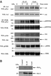
IL-15 mimics Gas6-mediated signaling. (A) L929 cells were activated with IL-15 or Gas6. Axl (first panel) and PI3K (third panel) were precipitated from the cell lysates and analyzed by WB using anti-pTyr Abs. Phosphorylation of Akt (fifth panel) and ERK (seventh panel) was analyzed using phospho-specific Abs. For loading control, Axl, PI3K, Akt, or ERK was detected after stripping (even panels). (B) Bcl-xL and Bcl-2 were detected in cells stimulated for 24 h.
Figure 5
Figure 5
Axl-mediated signaling is impaired in L929 cells upon Axl depletion. L929 cells were transfected with Axl siRNA. After 48 h, cells were harvested, serum-starved for 4 h, and activated with IL-15 or Gas6. Axl (first panel) and PI3K (third panel) were precipitated from the cell lysates and analyzed by WB using anti-pTyr Abs. Phosphorylation of Akt (fifth panel) and ERK (seventh panel) was analyzed by phospho-specific Abs. For loading control, Axl, PI3K, Akt, or ERK was detected on the same blots after stripping (even panels).
Figure 6
Figure 6
Gas6 does not affect IL-15-mediated Axl transactivation. (A) Cells were incubated for 10 min with 100 ng/ml Axl-Fc or control IL-3R-Fc proteins and then IL-15 or Gas6 (100 ng/ml) was added for another 5 or 15 min. Axl phosphorylation was detected by WB. (B) L929 cells were cultured in the presence of warfarin (100 μg/ml) for 24 h and stimulated with IL-15 or Gas6. Axl phosphorylation was detected by WB. (C) Recombinant Gas6 or IL-15 (10 nM) was incubated with 10 nM of Axl-Fc , IL-15Rα-Fc, or control IL-3R-Fc proteins. Complexes were captured on protein A-agarose and analyzed by WB using anti-Gas6 (upper panel) or anti-IL-15 (lower panel) Abs.
Figure 7
Figure 7
IL-15 and Gas6 signal in WT but not in Axl−/−, whereas Gas6 but not IL-15 induces signaling in IL-15Rα−/− MEFs. (A) WB analysis of Axl, IL-15Rα, and IL-2Rγ protein expression in WT, Axl−/−, and IL-15Rα−/− MEFs. (B) IL-15Rα expression was analyzed by FACS on cells labeled with IL-15Rα-IgG2b. (C) MEFs were stimulated with IL-15 or Gas6. Axl was precipitated from the lysates and phosphorylation was analyzed using anti-pTyr Abs (first panel). Same blots were analyzed for the association of Axl with IL-15Rα (third panel). Equal loading was confirmed by re-probing of blots with anti-Axl Abs (second panel). PI3K (fourth and fifth panels) and IL-15Rα (last two panels) were precipitated and their phosphorylation was detected using anti-pTyr Abs. Phosphorylation of Akt and ERK was detected using phospho-specific Abs. Blots were re-probed with PI3K, IL-15Rα, Akt, or ERK Abs for loading control. (D) Bcl-xL and Bcl-2 were detected in MEFs stimulated with IL-15 or Gas6 for 24 h. (E) Influence of IL-15 or Gas6 on serum deprivation-induced apoptosis in WT, Axl−/−, and IL-15Rα−/− MEFs. Apoptosis was induced by 48 h incubation in serum-free medium. IL-15 or Gas6 was added at a concentration of 100 ng/ml. Cells cultured in serum-free or serum-supplemented media were used as controls. Percentage of apoptotic cells was determined by PI and annexin V staining and FACS analysis. Data represent the mean±s.d. from duplicate samples.
Figure 8
Figure 8
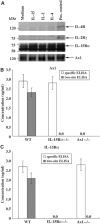
Axl and IL-15Rα coassociate in mouse DCs. (A) DCs were stimulated with cytokines for 15 min. Axl was precipitated from the cell lysates and precipitates were analyzed by WB using anti-IL-4Rα (upper panel), anti-IL-2Rγ (second panel), and anti-IL-15Rα (third panel) Abs. IP using specific Abs served as a positive control. For loading control, blots were developed with anti-Axl Abs (lower panel). (B) Plates were coated with anti-Axl Abs, and biotinylated anti-Axl (specific) or anti-IL-15Rα (two-site) Abs were used as detection Abs. (C) Plates were coated with anti-IL-15Rα Abs, and biotinylated anti-IL-15Rα (specific) or anti-Axl (two-site) Abs served as detection Abs.
Figure 9
Figure 9
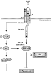
Hypothetical model of cross-talk between IL-15 and Axl signaling pathways suggested by the current study.

Similar articles

Cited by

References

    1. Allen MP, Zeng C, Schneider K, Xiong X, Meintzer MK, Bellosta P, Basilico C, Varnum B, Heidenreich KA, Wiermann ME (1999) Growth arrest-specific gene 6 (Gas6)/adhesion related kinase (Ark) signaling promotes gonadotropin-releasing hormone neuronal survival via extracellular signal-regulated kinase (ERK) and Akt. Mol Endocrinol 13: 191–201 - PubMed
    1. Bellosta P, Costa M, Lin DA, Basilico C (1995) The receptor tyrosine kinase ARK mediates cell aggregation by homophilic binding. Mol Cell Biol 15: 614–625 - PMC - PubMed
    1. Bellosta P, Zhang Q, Goff SP, Basilico C (1997) Signaling through the Ark tyrosine kinase receptor protects from apoptosis in the absence of growth stimulation. Oncogene 15: 2387–2397 - PubMed
    1. Bennasroune A, Gardin A, Aunis D, Cremel G, Hubert P (2004) Tyrosine kinase receptors as attractive targets of cancer therapy. Crit Rev Oncol Hematol 50: 23–38 - PubMed
    1. Brandt K, Bulfone-Paus S, Foster DC, Rückert R (2003) Interleukin-21 inhibits dendritic cell activation and maturation. Blood 102: 4090–4098 - PubMed

Publication types

MeSH terms