. 2005 Dec;115(12):3602-12.
doi: 10.1172/JCI25078. Epub 2005 Nov 23.

Cross-reactive influenza virus-specific CD8+ T cells contribute to lymphoproliferation in Epstein-Barr virus-associated infectious mononucleosis

Affiliations

Cross-reactive influenza virus-specific CD8+ T cells contribute to lymphoproliferation in Epstein-Barr virus-associated infectious mononucleosis

Shalyn C Clute et al. J Clin Invest. 2005 Dec.

Abstract

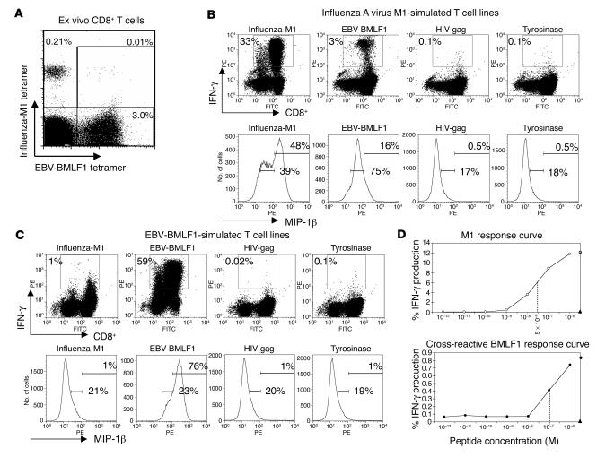
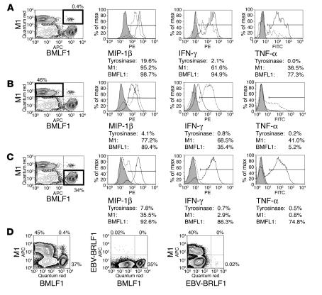
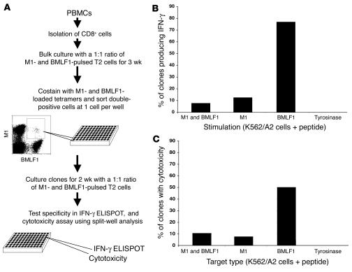
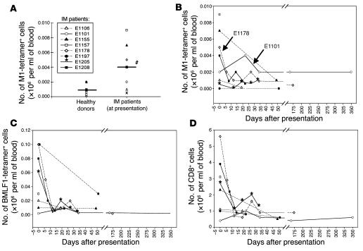
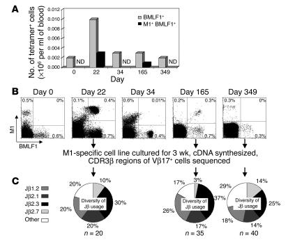
The marked proliferation of activated CD8+ T cells is pathognomonic of EBV-associated infectious mononucleosis (IM), common in young adults. Since the diversity and size of the memory CD8+ T cell population increase with age, we questioned whether IM was mediated by the reactivation of memory CD8+ T cells specific to previously encountered pathogens but cross-reactive with EBV. Of 8 HLA-A2+ IM patients, 5 had activated T cells specific to another common virus, as evidenced by a significantly higher number of peripheral blood influenza A virus M1(58-66)-specific T cells compared with healthy immune donors. Two patients with an augmented M1 response had tetramer-defined cross-reactive cells recognizing influenza M1 and EBV-BMLF1(280-288), which accounted for up to one-third of their BMLF1-specific population and likely contributed to a skewed M1-specific T cell receptor repertoire. These epitopes, with only 33% sequence similarity, mediated differential effects on the function of the cross-reactive T cells, which may contribute to alterations in disease outcome. EBV could potentially encode an extensive pool of T cell epitopes that activate other cross-reactive memory T cells. Our results support the concept that cross-reactive memory CD8+ T cells activated by EBV contribute to the characteristic lymphoproliferation of IM.


Figures

Figure 1
Figure 1
T cell lines grown in the presence of 1 peptide can respond to stimulation with a second unrelated peptide. (A) CD8+ T cells were isolated ex vivo from healthy donor D-002 and costained with M1- and BMLF1-loaded tetramers; 106 events were collected. (B and C) Fresh CD8+ T cell lines derived from donor D-002 were grown for 3–4 weeks in the presence of (B) M1 peptide–pulsed or (C) BMLF1 peptide–pulsed T2 cells and then stained intracellularly for the production of IFN-γ or MIP-1β following 5 hours of stimulation with various HLA-A2–restricted peptides at a 5 μM final concentration. Percentages of CD8+ T cells producing each cytokine are shown. (D) Titration of peptide concentrations in an intracellular IFN-γ assay using an M1-specific T cell line derived from donor D-002 demonstrated a slight difference in avidity for M1 versus BMLF1. Filled triangles, tyrosinase; open circles, M1; and filled circles, BMLF1 stimulation.
Figure 2
Figure 2
Culturing with M1 and BMLF1 peptides simultaneously promotes the growth of cross-reactive cells. A CD8+ T cell line derived from healthy donor D-002 was grown for 4 weeks in the presence of both M1 and BMLF1 peptide–pulsed T2 cells. Cells were stimulated for 5 hours with various peptides and then stained extracellularly with tetramers and intracellularly for the production of MIP-1β, IFN-γ, and TNF-α. We gated (indicated by a bold box) on (A) the percentage of CD8+ T cells that costained with both M1- and BMLF1-loaded tetramers, (B) the percentage of CD8+ T cells that stained with only M1-loaded tetramer, and (C) the percentage of CD8+ T cells that stained with only BMLF1-loaded tetramer. We then assessed the cytokine production of those cells in response to the following peptide stimulations: tyrosinase (gray profiles), M1 (dotted lines), and BMLF1 (solid lines). The percentage of CD8+ T cells producing each cytokine within the positive gate (horizontal lines) drawn is shown below each of the corresponding histograms. (D) This T cell line was stained extracellularly with EBV-BRLF1–loaded tetramer as a control.
Figure 3
Figure 3
Tetramer-defined subsets of cross-reactive T cells differ in their avidity for the 2 epitopes. A similar intracellular IFN-γ assay was performed on the same T cell line described in Figure 2, which had been grown in the presence of M1 and BMLF1 peptides for 4 weeks, using a titration of peptide concentrations. Filled triangles, tyrosinase; open circles, M1; and filled circles, BMLF1 stimulation. We assessed the IFN-γ production of gated, tetramer-defined subsets of the T cell line: (A) M1+ BMLF1+ and (B) M1 BMLF1+.
Figure 4
Figure 4
Cross-reactive clones are heterogeneous in their response to M1 versus BMLF1. (A) An outline of the experimental design used to clone cross-reactive T cells from healthy donor D-002 is shown. (B) CD8+ T cells incubated for 20 hours with either M1-, BMLF1-, or tyrosinase-pulsed K562/HLA-A2 cells. The percentages shown represent the number of wells harboring cells that produced IFN-γ following stimulation with both M1 and BMLF1 or following stimulation with only 1 of the peptides. (C) CD8+ T cells incubated for 8 hours with either M1-, BMLF1-, or tyrosinase-pulsed K562/A2 cells. The percentages shown represent the number of wells harboring cells that killed both M1- and BMLF1-pulsed target cells or that killed only 1 target cell type in a 51chromium release assay. Data presented here accurately represent the trends observed in 3 separate experiments.
Figure 5
Figure 5
IM patients have an augmented number of M1-specific cells in their bloodstream. PBMCs were isolated from 8 healthy donors or from 8 patients experiencing IM. Blood from IM patients was collected at various points after presentation with symptoms of IM. Please note that the number of data points for each patient is variable. (AC) CD8+ cells were first isolated from PBMCs and then immediately stained with tetramer. The percentages of tetramer-positive cells were used to calculate the total number of either M1- or BMLF1-specific cells per ml of blood. (A) The difference between the means of the 2 subject groups was determined to be statistically significant using an unpaired, 2-tailed Student’s t test. #P = 0.02. Patient E1197 had an M1-specific memory population that grew out in culture but was undetectable ex vivo; therefore, this patient was excluded from calculation of the mean. (D) PBMCs were used to costain with CD3 and CD8+ antibodies and calculate the total number of CD8+ T cells per ml of blood.
Figure 6
Figure 6
Acute EBV infection augments the number of cross-reactive cells that recognize M1 and BMLF1. CD8+ T cells were isolated ex vivo from patient E1101 at various time points after presentation with symptoms of IM. (A) The total number of antigen-specific T cells per ml of blood was calculated using the frequencies of tetramer-positive cells. BMLF1+, tetramer positive; M1+ BMLF1+, double-tetramer positive; ND, not determined because the frequency was below the limit of detection using this technique. (B) The percentages of CD8+ T cells staining positive when costained with M1- and BMLF1-loaded tetramers are shown. The number of events shown is variable because the maximum number possible was collected for each sample. (C) CD8+ T cells isolated at days 22, 165, and 349, were cultured for 3 weeks in the presence of M1 peptide–pulsed T2 cells. Following the RNA isolation and cDNA synthesis of those T cell lines, the CDR3β region of Vβ17+ subclones was sequenced. The pie charts illustrate the percentages of unique Vβ17+ subclones using each Jβ family, where n = the total number of unique subclones. The complete CDR3 sequences of all the subclones analyzed are displayed in Supplemental Table 1, structured according to Chothia et al. (57).

Similar articles

Cited by

References

    1. Hoagland RJ. Infectious mononucleosis. Prim. Care. 1975;2:295–307. - PubMed
    1. Young LS, Rickinson AB. Epstein-Barr virus: 40 years on. Nat. Rev. Cancer. 2004;4:757–768. - PubMed
    1. Rea TD, Russo JE, Katon W, Ashley RL, Buchwald DS. Prospective study of the natural history of infectious mononucleosis caused by Epstein-Barr virus. J. Am. Board Fam. Pract. 2001;14:234–242. - PubMed
    1. Auwaerter PG. Infectious mononucleosis in middle age. JAMA. 1999;281:454–459. - PubMed
    1. Axelrod P, Finestone AJ. Infectious mononucleosis in older adults. Am. Fam. Physician. 1990;42:1599–1606. - PubMed

Publication types

MeSH terms