

Review
doi: 10.1002/ar.b.20084.
Eye on regeneration
Affiliations
- PMID: 16308862
- PMCID: PMC2556859
- DOI: 10.1002/ar.b.20084
Item in Clipboard
Review
Eye on regeneration
Anat Rec B New Anat.
2005 Nov.
Abstract
Lens regeneration in newts is a remarkable process, whereby a lost tissue is replaced by transdifferentiation of adult tissues that only a few organisms possess. In this review, we will touch on the approaches being used to study this phenomenon, recent advances in the field of lens regeneration, similarities and differences between development and regeneration, as well as the potential role stem cells may play in understanding this process.
Figures
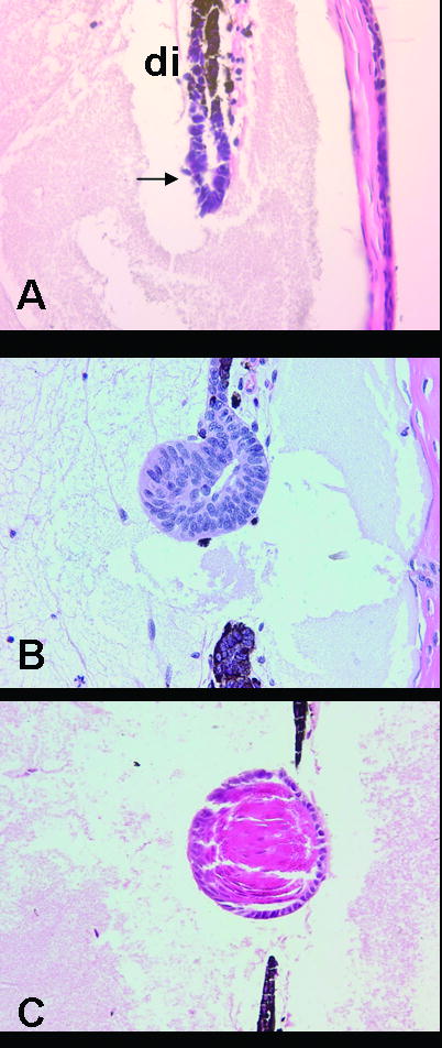
Lens regeneration in the newt stemming from the PECs of the dorsal iris (di). A. 10 days post-lentectomy. Note the formation of a lens vesicle (arrow). B. 15 days post-lentectomy. Cells are elongating into lens fibers. C. 20 days post-lentectomy. The lens is well differentiated with lens fibers.
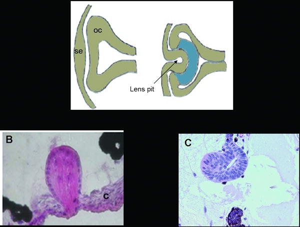
Comparison of lens development with two methods of lens regeneration. A. Schematic showing lens development, which involves a series of inductive interactions between the surface ectoderm and the optic cup. The lens pit eventually gives rise to the lens. se: surface ectoderm, oc: optic cup. B. Lens regeneration in Xenopus laevis. The regenerated lens comes from transdifferentiation of cells in the outer cornea (c). C. Lens regeneration in the newt. The regenerated lens comes from transdifferentiation of cells in the dorsal iris.
References
-
- Amano U, Sato J. Uber die xenoplastische implantation der larvalen des Triturus pyrrhogaster in das entlinste Auge der Larven des Hynobius nebulosus. Jpn J Med Sci I Anat. 1940;8:75–81.
-
- Call MK, Grogg MW, Del Rio-Tsonis K, Tsonis PA. Lens regeneration in mice: implications in cataracts. Exp Eye Res. 2004;78:297–299. - PubMed
-
- Chen S, Zhang Q, Wu X, Schultz PG, Ding S. Dedifferentiation of lineage-committed cells by a small molecule. J Am Chem Soc. 2004;126:410–411. - PubMed
-
- Colucci VL. Sulla rigenereazione parziale dell'occhio nei Tritoni-Istogenesi e sviluppo: Studio sperimentale. Mem R Acad Sci Ist Bologna. 1891;Ser 51:593–629.
Publication types
MeSH terms
Grants and funding
LinkOut - more resources
Full Text Sources
