

Surface ultrastructure of SARS coronavirus revealed by atomic force microscopy
- PMID: 16309462
- PMCID: PMC7162285
- DOI: 10.1111/j.1462-5822.2005.00593.x
Surface ultrastructure of SARS coronavirus revealed by atomic force microscopy
Abstract
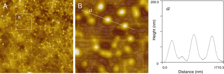
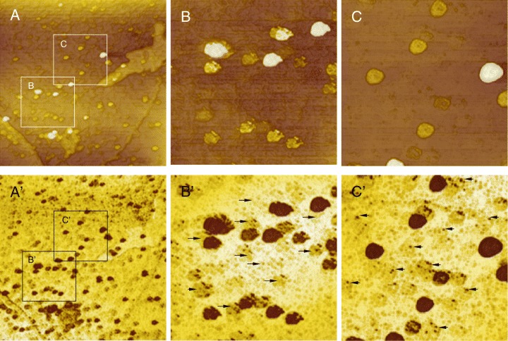
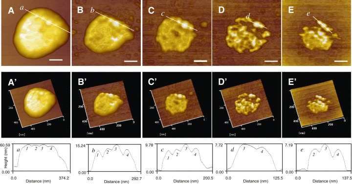
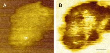
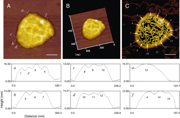
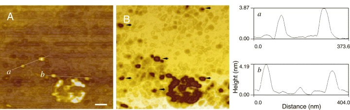
Atomic force microscopy has been used to probe the surface nanostructures of severe acute respiratory syndrome coronavirus (SARS-CoV). Single crown-like virion was directly visualized and quantitative measurements of the dimensions for the structural proteins were provided. A corona of large, distinctive spikes in the envelope was measured after treatment with hydroxyoctanoic acid. High-resolution images revealed that the surface of each single SARS-CoV was surrounded with at least 15 spherical spikes having a diameter of 7.29 +/- 0.73 nm, which is in close agreement with that of S glycoproteins earlier predicted through the genomes of SARS-CoV. This study represents the first direct characterization of the surface ultrastructures of SARS-CoV particles at the nanometre scale and offers new prospects for mapping viral surface properties.
Figures
Similar articles
-
Assembly of human severe acute respiratory syndrome coronavirus-like particles.Biochem Biophys Res Commun. 2004 Jun 11;318(4):833-8. doi: 10.1016/j.bbrc.2004.04.111. Biochem Biophys Res Commun. 2004. PMID: 15147946 Free PMC article.
-
Probing the structure of the SARS coronavirus using scanning electron microscopy.Antivir Ther. 2004 Apr;9(2):287-9. Antivir Ther. 2004. PMID: 15134191
-
Architecture of the SARS coronavirus prefusion spike.Nat Struct Mol Biol. 2006 Aug;13(8):751-2. doi: 10.1038/nsmb1123. Epub 2006 Jul 16. Nat Struct Mol Biol. 2006. PMID: 16845391 Free PMC article.
-
Coronavirus Spike Protein and Tropism Changes.Adv Virus Res. 2016;96:29-57. doi: 10.1016/bs.aivir.2016.08.004. Epub 2016 Sep 13. Adv Virus Res. 2016. PMID: 27712627 Free PMC article. Review.
-
Properties of Coronavirus and SARS-CoV-2.Malays J Pathol. 2020 Apr;42(1):3-11. Malays J Pathol. 2020. PMID: 32342926 Review.
Cited by
-
The emergence of novel coronavirus disease (COVID-19) in Bangladesh: Present status, challenges, and future management.J Adv Vet Anim Res. 2020 Mar 22;7(2):198-208. doi: 10.5455/javar.2020.g410. eCollection 2020 Jun. J Adv Vet Anim Res. 2020. PMID: 32607350 Free PMC article. Review.
-
Interpretation of SARS-CoV-2 behaviour on different substrates and denaturation of virions using ethanol: an atomic force microscopy study.RSC Adv. 2020 Dec 14;10(72):44079-44086. doi: 10.1039/d0ra09083b. eCollection 2020 Dec 9. RSC Adv. 2020. PMID: 35517177 Free PMC article.
-
Single-particle virology.Biophys Rev. 2020 Oct;12(5):1141-1154. doi: 10.1007/s12551-020-00747-9. Epub 2020 Sep 3. Biophys Rev. 2020. PMID: 32880826 Free PMC article. Review.
-
Severe Acute Respiratory Syndrome Coronavirus (SARS-CoV).Perspect Med Virol. 2006;16:43-95. doi: 10.1016/S0168-7069(06)16004-8. Epub 2006 Nov 28. Perspect Med Virol. 2006. PMID: 32287586 Free PMC article. Review.
-
COVID-19-A Theory of Autoimmunity Against ACE-2 Explained.Front Immunol. 2021 Mar 23;12:582166. doi: 10.3389/fimmu.2021.582166. eCollection 2021. Front Immunol. 2021. PMID: 33833750 Free PMC article. Review.
References
-
- Binning, G. , Quate, C.F. (1986) Atomic force microscope. Phys Rev Lett 56: 930–933. - PubMed
-
- Binning, G. , Rohrer, G.H. , Gerber, C.H. , Weibel, E. (1982) Surface studies by scanning tunneling microscopy. Phys Rev Lett 49: 57–61.
-
- Butt, H.J. , Seifert, K. , Bamberg, E. (1993) Imaging molecular defects in alkanethiol monolayers with an atomic force microscope. J Phys Chem 97: 7316–7320.
-
- Drosten, C. , Gunther, S. , Preiser, W. , Werf, S. , Brodt, H.R. , Becker, S. , et al. (2003) Identification of a novel coronavirus in patients with severe acute respiratory syndrome. N Engl J Med 348: 1967–1976. - PubMed
-
- Holmes, K.V. , Enjuanes, L. (2003) The SARS coronavirus: a postgenomic era. Science 300: 1377–1378. - PubMed
Publication types
MeSH terms
Substances
LinkOut - more resources
Full Text Sources
Miscellaneous
