

The haemotoxicity of mitomycin in a repeat dose study in the female CD-1 mouse
- PMID: 16309546
- PMCID: PMC2517448
- DOI: 10.1111/j.0959-9673.2005.00452.x
The haemotoxicity of mitomycin in a repeat dose study in the female CD-1 mouse
Abstract
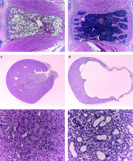
Mitomycin (MMC), like many antineoplastic drugs, induces a predictable, dose-related, bone marrow depression in man and laboratory animals; this change is generally reversible. However, there is evidence that MMC may also cause a late-stage or residual bone marrow injury. The present study in female CD-1 mice investigated the haematological and bone marrow changes induced by MMC in a repeat dose study lasting 50 days. Control and MMC-treated mice were dosed intraperitoneally on eight occasions over 18 days with vehicle, or MMC at 2.5 mg/kg, autopsied (n = 6-12) at 1, 7, 14, 28, 42 and 50 days after the final dose and haematological changes investigated. Femoral nucleated bone marrow cell counts and levels of apoptosis were also evaluated and clonogenic assays carried out; serum levels of FLT3 ligand (FL) were assessed. At day 1 post-dosing, MMC induced significant reductions in RBC, Hb and haematocrit (HCT) values, and there were decreases in reticulocyte, platelet, and femoral nucleated cell counts (FNCC); neutrophil, lymphocyte and monocyte values were also significantly reduced. On days 7 and 14 post-dosing, all haematological parameters showed evidence of a return towards normal values, but at these times, and at day 28, values for RBC and FNCC remained significantly reduced in comparison with controls. At days 42 and 50 post-dosing, many haematological parameters in MMC-treated mice had returned to control levels; however, there remained evidence of late-stage effects on RBC, Hb and HCT values, and FNCC also continued to be significantly decreased. Results for granulocyte-macrophage colony-forming units and erythroid colonies showed a profound decrease immediately post-dosing, but a return to normal values was evident at day 50. Serum FL concentrations demonstrated very significant increases in the immediate post-dosing period, but a return to normal was seen at day 50 post-dosing; a relatively similar pattern was seen in the number of apoptotic femoral marrow nucleated cells. The histopathological examination of kidney tissues from MMC animals at day 42 and 50 post-dosing showed evidence of hydronephrosis with cortical glomerular/tubular atrophy and degeneration. It is therefore concluded that MMC administered on eight occasions over 18 days to female CD-1 mice at 2.5 mg/kg induced profound changes in haematological and bone marrow parameters in the immediate post-dosing period with a return to normal levels at day 50 post-dosing; however, there was evidence of mild but significant late-stage/residual effects on RBC and FNCC, and on cells of the erythroid lineage in the bone marrow.
Figures
References
-
- Alter BP, Potter NU, Li FP. Classification and aetiology of the aplastic anaemias. ClinHaematol. 1978;7:431–465. - PubMed
-
- Appelbaum FR, Fefer A. The pathogenesis of aplastic anaemia. SeminHematol. 1981;18:241–257. - PubMed
-
- Benested HB. Drug mechanisms in marrow aplasia. In: Geary CG, editor. Aplastic Anaemia. London: Ballière-Tindall; 1979. pp. 26–42.
-
- Benning VM, Maratrat MB, Fournier EC, Melcion CP, Cordier AC. Flow cytometric detection of erythropoietic cytotoxicity in mouse bone marrow. JHistochem Cytochem. 1991;39:15–21. - PubMed
-
- Bertho JM, Demarquay C, Frick J, et al. Level of Flt3-ligand in plasma: a possible new bio-indicator for radiation-induced aplasia. Int. J. RadiatBiol. 2001;77:702–712. - PubMed
Publication types
MeSH terms
Substances
LinkOut - more resources
Full Text Sources
Medical
Miscellaneous
