. 2005 Dec;116(4):429-42.
doi: 10.1111/j.1365-2567.2005.02242.x.

CD27+ B cells in human lymphatic organs: re-evaluating the splenic marginal zone

Affiliations

CD27+ B cells in human lymphatic organs: re-evaluating the splenic marginal zone

Birte Steiniger et al. Immunology. 2005 Dec.

Abstract

The marginal zone of human spleens is regarded as an organ-specific region harbouring sessile memory B cells. This opinion has arisen by extrapolating from results obtained in mice and rats. Detection of CD27(+) B cells in situ now revealed similarities among the most superficial region of B-cell follicles in human spleens, reactive lymph nodes, inflamed appendices, tonsils and terminal ilea. The follicular surface in these organs consists of small naïve immunoglobulin D (IgD)(+) CD27(-) B cells predominating in an inner area and larger IgD(+/-) CD27(+) B cells prevailing in a more superficial position. CD27(+) B cells may, however, also occupy the entire follicular periphery around the germinal centre. Together with additional peculiarities this distribution indicates a fundamental microanatomical difference among the human and rodent splenic white pulp. We hypothesize that the follicular periphery represents a recirculation compartment both for naïve and memory/natural reactive B cells in all human secondary lymphatic organs. This assumption implies a difference in recirculation behaviour among human and rodent B memory cells.


Figures
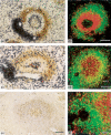
Figure 1
Figure 1
CD27+ B cells in small secondary follicles of human spleens. (a) Subtractive double-staining for CD3 (blue-black) and CD27 (brown). CD27+ B cells are present at the follicular surface, but do not occur at the periphery of the PALS. Seventeen-year-old patient with cranio-cerebral trauma. Bar = 150 µm. (b) Co-expression of CD27 (green) and IgD (red). Co-expression is only minimal. CD27 IgD+ B cells occur in the inner part of the mantle zone and as scattered cells in a superficial follicular position. Seventy-four-year-old patient with traumatic splenic rupture during operation of malignant suprarenal tumour. Bar = 100 µm. (c) Subtractive double-staining for CD3 (blue-black) and CD27 (brown). A section with reduced staining intensity for CD27 was chosen to show CD27++ plasmablasts/plasma cells in the GC. Twenty-one-year-old patient with polytrauma. Bar = 100 µm. (d) Co-expression of CD27 (green) and IgM (red). The level of the section does not hit the GC. CD27+ IgM+ B cells occupy the inner part of the CD27+ area, while CD27+ IgM switched memory B cells are located more superficially. Single CD27 IgM+ B cells are seen in the outermost periphery of the follicle. Same patient as in (c). Bar = 100 µm. (e) Single staining for surface IgA by ABC-tyramide method. Cells of the superficial follicular periphery show increased positivity for sIgA. IgA immune-complex-positive follicular dendritic cells are located in the GC. Single intracellular IgA+ plasma cells occur in a perifollicular position. Same patient as in (c). Haemalum counterstain. Bar = 150 µm. (f) Co-expression of CD27 (green) and IgM (red) in the PALS. CD27+ IgM+ B cells are scattered in the T-cell zone, part of a follicle is located at the right margin. Same patient as (b). Bar = 40 µm.
Figure 2
Figure 2
CD27+ B cells in large secondary follicles and in apparent primary follicles of human spleens. (a) Subtractive double-staining for CD3 (blue-black) and CD27 (brown). The entire follicular periphery outside the GC contains CD27+ B cells. Seventy-nine-year-old patient with gastric carcinoma. Bar = 150 µm. (b) Same section and staining method as (a). The density of CD27+ B cells varies among different follicles. In this follicle CD27+ B cells are less numerous in the inner part of the mantle zone. Bar = 200 µm. (c) Single staining for IgD. IgD+ B cells occur in the mantle zone and overlap into the superficial follicular periphery. IgD+ B cells are also more densely arranged in the most superficial part of the follicle and along the PALS. Same patient as (a). Haemalum counterstain. Bar = 150 µm. (d) Subtractive double-staining for CD3 (blue-black) and CD27 (brown). A follicle with fully developed GC appears to be embedded in an extension of the PALS: The light zone of the GC is indicated by an accumulation of blue CD3+ GC T cells. An area with few CD27+ B cells follows outside the GC. The outermost follicular surface is occupied by a dense layer of CD27+ B cells containing a crescent-shaped row of CD3+ T cells. These T cells are far away from the GC, but in apparent continuity with the PALS located at the lower pole of the follicle. Six-year-old patient with traumatic splenic rupture. Bar = 150 µm. (e) Single-staining for IgD. The follicular mantle zone is occupied by IgD+ B cells, while the more superficial part of the follicle harbours IgD+/– B cells. IgD+ B cells also occur at the outermost follicular periphery and at the surface of the PALS. Same patient as (d). Haemalum counterstain. Bar = 300 µm. (f) Co-expression of CD27 (green) and IgD (red). CD27+ IgD+ (or IgD+/–) B cells occur in the superficial part of the follicle. CD27 IgD+ B cells are located even more superficially. Same patient as (d). Bar = 100 µm. (g) Subtractive double-staining for CD3 (blue-black) and CD27 (brown). This apparent primary follicle shows many CD27+ B cells. The density of these cells decreases towards the follicle interior. Seventy-three-year-old patient with cerebral haemorrhage. Bar = 150 µm. (h) Subtractive double-staining for CD3 (blue-black) and CD27 (brown). The apparent primary follicle contains a highly degenerated GC with accumulation of T cells. The number of CD27+ B cells is relatively high in the mantle zone and increases towards the follicular periphery. Less intensely CD27+ B cells occur in the most superficial position. Twenty-two-year-old suicide patient. Bar = 100 µm.
Figure 3
Figure 3
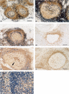
CD27+ B cells in reactive lymph nodes, appendices, follicles of terminal ileum and tonsils. (a–c, e): Subtractive double-staining for CD3 (blue-black) and CD27 (brown). (a) CD27+ B cells in the mantle zones of cortical follicles outside the GCs. GCs do not contain dark and light zones and do not stain for CD27. Tumour-free lymph node from bile duct of a 73-year-old patient with bile duct carcinoma. (b) CD27+ B cells in the mantle zone of a follicle of the appendix. Centroblasts are CD27+, while centrocytes exhibit reduced CD27 expression. CD27++ plasmablasts/plasma cells occur between dark and light zone of the GC. Forty-six-year-old patient with ovarial carcinoma and peritoneal metastases. (c) CD27+ B cells in the mantle zone of a follicle in the lamina propria of the terminal ileum. The GC is also slightly CD27+. Non-involved part of ileum in a 48-year-old patient with resection of ileum and caecum because of perforation of appendix, periappendicitis and purulent peritonitis. (d) Same follicle as (b) at another level. Single staining for IgD shows extension of the mantle zone. Haemalum counterstain. (e) CD27+ B cells in tonsillar follicle with reduced staining intensity chosen to reveal CD27++ plasmablasts/plasma cells. CD27+ B cells are found in the mantle zone. GC B cells are faintly CD27+. CD27++ B cells are located between dark and light zone of the GC and at the basal circumference of the dark zone (arrow). Thirty-nine-year-old patient with chronic-hyperplastic tonsillitis. (f) Single staining for IgD in a tonsillar follicle. IgD+ B cells occur in the mantle zone. In addition, IgD+ B cells of different staining intensity also lodge in the GC. Eleven-year-old patient with chronic hyperplastic tonsillitis. Haemalum counterstain. (g) Subtractive double-staining for CD3 plus Ki67 (first step, blue-black) and CD27 (second step, brown) in a tonsillar follicle. A major number of the CD27++ B cells in the centre of the GC is Ki67. White arrow indicates surface of the GC light zone, asterisk is placed into mantle zone. Interfollicular T-cell zone is located at the left margin. Same patient as (d). (a–f) Bars = 150 µm; (g) bar = 50 µm.

References

    1. MacLennan ICM, Liu Y-J. Marginal zone B cells respond both to polysaccharide antigens and protein antigens. Res Immunol. 1991;142:346–51. - PubMed
    1. Martin F, Kearney JF. Marginal zone B cells. Nature Rev Immunol. 2002;2:323–35. - PubMed
    1. Kanayama N, Cascalho M, Ohmori H. Analysis of marginal zone B cell development in the mouse with limited B cell diversity. role of the antigen receptor signals in the recruitment of B cells to the marginal zone. J Immunol. 2005;174:1438–45. - PubMed
    1. Kumararatne DS, Bazin H, MacLennan ICM. Marginal zones: the major B cell compartment of rat spleens. Eur J Immunol. 1981;11:858–64. - PubMed
    1. Kraal G. Cells in the marginal zone of the spleen. Int Rev Cytol. 1992;132:31–74. - PubMed

Publication types

Substances