Review
. 2005 Nov;207(5):575-601.
doi: 10.1111/j.1469-7580.2005.00473.x.

Relations and interactions between cranial mesoderm and neural crest populations

Affiliations
Review

Relations and interactions between cranial mesoderm and neural crest populations

Drew M Noden et al. J Anat. 2005 Nov.

Abstract

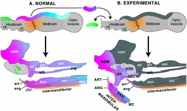
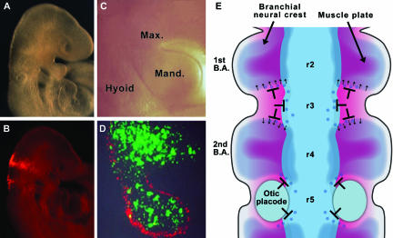
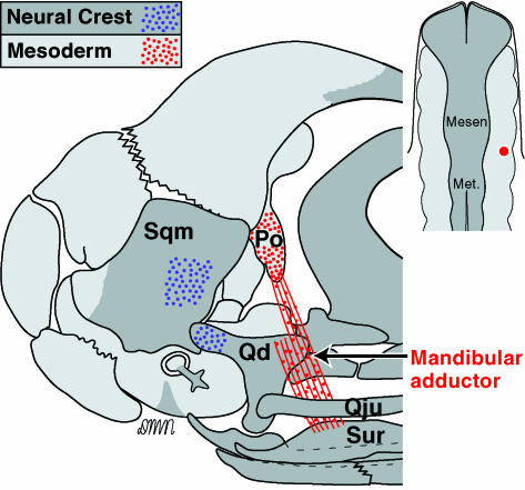
The embryonic head is populated by two robust mesenchymal populations, paraxial mesoderm and neural crest cells. Although the developmental histories of each are distinct and separate, they quickly establish intimate relations that are variably important for the histogenesis and morphogenesis of musculoskeletal components of the calvaria, midface and branchial regions. This review will focus first on the genesis and organization within nascent mesodermal and crest populations, emphasizing interactions that probably initiate or augment the establishment of lineages within each. The principal goal is an analysis of the interactions between crest and mesoderm populations, from their first contacts through their concerted movements into peripheral domains, particularly the branchial arches, and continuing to stages at which both the differentiation and the integrated three-dimensional assembly of vascular, connective and muscular tissues is evident. Current views on unresolved or contentious issues, including the relevance of head somitomeres, the processes by which crest cells change locations and constancy of cell-cell relations at the crest-mesoderm interface, are addressed.


Figures

Fig. 1
Fig. 1
Scanning electron micrographs showing the initial spatial relations among avian cephalic mesenchymal and epithelial tissues. (A) Dorsal view of a stage 8, four-somite embryo in which the surface ectoderm and neural tube have been removed, exposing underlying paraxial mesoderm (red). The brackets indicate the boundaries of somitomeres 2 and 3, as proposed by Meier (1979). (B) Comparable view of an embryo approximately 10 h more advanced (ten somites), with the neural tube in place (visible to the left). The leading edge of the neural crest (blue) partially overlies paraxial mesoderm. In C, a ten-somite embryo was transected at the level of the midbrain to show the early interface between crest and paraxial mesoderm cells. A, B, colour added from archival prints provided by S. Meier; original SEMs published in Meier (1979) and Anderson & Meier (1981); C, colour added from an original micrograph provided by K. Tosney (1982).
Fig. 10
Fig. 10
Schematic lateral representation of the asymmetric, segmental effects that somites exert upon the movements of trunk neural crest cells. (A) The normal responses of crest cells upon contacting the cranial and caudal halves of somites. Surgically altering the size (B), cranio-caudal orientation (C) or cranio-caudal composition (D) of somitic epithelium induces changes in the patterns with which crest cells enter then pass ventrally through each somite.
Fig. 11
Fig. 11
The morphogenesis of branchial neural crest tissues results from both autonomous, prespecified and dependent, acquired properties. (A) The contributions of crest cells from the mesencephalic (blue, purple), metencephalic (red) and rhombomere 4 (green) levels to the 1st branchial arch skeleton. Note that the proximal (caudal) parts of the articular and angular bones are derived from 2nd arch (rhombomere 4) crest cells that became contiguous with 1st arch (rhombomere 2)-derived crest cells dorsal to the 1st pharyngeal pouch. In B, presumptive proximal 1st arch neural crest precursors (red) were grafted in place of 2nd arch crest precursors. Most formed ectopic 1st arch skeletal structures in the 2nd arch location (labelled in upper-case letters), and directed myogenic cells entering the 2nd arch to form 1st arch-specific muscles, e.g. the INTERMANDIBULAR. At interfaces with neighbouring, untransplanted crest cells, however, grafted crest cells relinquished their prespecified biases and cooperatively formed structures anatomically correct for their new location, e.g. the retroarticular cartilage and proximal angular bone in the 1st branchial arch.
Fig. 12
Fig. 12
Quail trunk paraxial mesoderm is able to form normal head muscles. In this micrograph, grafted quail cells (dark, immunopositive nuclei) have formed the lateral rectus but, due to having a different site of origin, not the dorsal rectus muscle. The section was also stained with anti-myosin heavy chain antibodies (blue). This demonstrates both the highly localized inductive properties of head tissues, in this case rhombomere 2, and the ability of periocular neural crest cells to orchestrate the morphogenesis of trunk-derived muscle primordia, which normally would never encounter crest-derived connective tissues. From Borue & Noden (2004).
Fig. 2
Fig. 2
In situ hybridizations performed on intact chick embryos to show sites of gene expression for the transciption factors Myf5, Paraxis, Tbx1 and Pitx2. Myf5 is activated in all skeletal muscle precursors, whereas Paraxis is limited to somitic cells plus the lateral rectus precursor. Both Tbx1 and Pitx2 show complex and stage-specific patterns of expression in cephalic mesenchymal and epithelial cells. These briefly are expressed in subsets of head muscles. BA1, 2, 3: branchial arches; HC, hypoglossal cord; LM, PM: lateral and paraxial mesoderm; DR, LR: dorsal and lateral rectus muscles; DO, VO: dorsal and ventral oblique muscles; S1, 1st somite; VC, visceral cleft ectoderm.
Fig. 3
Fig. 3
(A) Summary map showing the sites of origin of muscles (right side) and skeletal elements (left) derived from chick paraxial mesoderm.(B) A 15-day chick embryo that received an injection of LacZ-bearing retroviruses beside the midbrain 2 weeks previously. Chondrogenic cells have remained stationary and are within the postorbital cartilage, whereas osteogenic cells have moved dorsally and, in this case, contributed to the frontal bone. Cells in myogenic and angiogenic lineages exhibit additional distinct behaviours, taking each population to different locations in the head. After Evans and Noden (2005).
Fig. 4
Fig. 4
The widespread, invasive migrations of angioblasts are illustrated by immunostaining for quail endothelial cells 60 h after implanting a small piece of quail mesoderm beside the metencephalon of a chick embryo. As is evident in this parasagittal section, angioblasts have moved in all directions from the site of implantation and contributed to both large (cranial cardinal vein) and small blood vessels, including precursors of meningeal and intraneural vessels. Met., metencephlaon; Trig. g., trigeminal ganglion. From Noden (1991a).
Fig. 5
Fig. 5
Schematic chick and mouse skulls showing the contributions of neural crest, paraxial and lateral mesoderms to the cranial skeleton. The avian map is based on transplantation and retroviral lineage tracings in the chick embryo; hyobranchial structures, all of which are derived from neural crest cells, are not shown. The mouse map is based largely on the location of neural crest cells, as identified by expression of LacZ driven by a Wnt1 promoter in cre-lox transgenic embryos (Jiang et al. 2002). Origins of mouse laryngeal cartilages are by extrapolation from avian data, with the caveat that birds do not have a thyroid cartilage. Blue dots indicate the locations of crest cells present at sites of calvarial sutures. Abbreviations (Figs 5,9 and 11): Ang, angular; Art, articular; Bs, basisphenoid; Den, dentary; Eth, ethmoid; Lac, lacrimal; Ls, laterosphenoid*; Mc, mandibular cartilage; Nc, nasal capsule; Os, orbitosphenoid*; Pal, palatine; Pfr, prefrontal; Po, postorbital; Ps, presphenoid; Ptr, pterygoid; Qd, quadrate; Qju, quadratojugal; San, surangular; Sqm, squamosal; *regions of the pleurosphenoid.
Fig. 6
Fig. 6
Segregation of neural crest cells and paraxial mesoderm. (A,C) Low and higher magnification right lateral views showing the head of a 9.5 days post-conception mouse embryo. B is the same as A but illuminated to reveal the locations of rhombomere 2- and 4-derived neural crest cells, which were labelled with a red fluorescent lineage tracer. This illustrates their segregation in streams that colonize the first and second branchial arches, respectively. (D) The same embryo as C with cranial mesoderm cells (green) from the level of the preotic hindbrain occupying the central, presumptive myogenic core of the first branchial arch. These are enveloped by rhombomere 2-derived neural crest cells (red). Other dispersed mesodermal cells are probably angioblasts. (E) Diagramatic dorsal view summarizing the movements of cranial neural crest populations (blue) and paraxial mesoderm cells (red) into branchial arches. Inhibitory cues (T-bars) restrict lateral movement of neural crest cells (blue shading and blue dots) from rhombomeres 3 and 5, and cohesive forces (arrows) help maintain segregation within branchial arch crest cells.
Fig. 7
Fig. 7
Schematic transverse views of three stages in the development of the avian head, showing the concerted movements and expansions of surface ectoderm (green), neural crest (blue) and superficial (myogenic) paraxial mesoderm (dark red) populations. Arrowheads show the locations of the leading (ventral) edge of each population. All populations shift and expand in the same dorsal-to-ventral direction during these stages.
Fig. 8
Fig. 8
Staggered, lateral views of all internal tissue layers in an early avian embryo. These illustrate the changes in locations of each population and the spatial relations among them. Neural crest progenitors, cranial nerves and myogenic primordia for each branchial arch all arise at the same axial level and maintain this close registration throughout their dorso-ventral movements. For example, crest cells that will populate the 2nd branchial arch arise from the same axial location (rhombomere 4) as the 7th cranial nerve and the 2nd arch muscles it will innervate. By contrast, the periocular neural crest, extra-ocular muscles and the motor nerves that innervate them all arise at separate axial locations, and do not establish stable relations until all have reached their sites of terminal differentiation.
Fig. 9
Fig. 9
The loss of nearest-neighbour relations between neural crest and myogenic mesoderm cells is shown. LacZ-retrovirus was injected at the interface of these populations, indicated by the red spot on the inset (see also Fig. 1C), and embryos processed 2 weeks later. Mesoderm cells contribute to the cartilaginous postotic (Po) process (red dots) and the mandibular adductor (1st branchial arch) muscle (red lines and dots). However, crest cells that were adjacent to these mesoderm cells while en route did not maintain this nearest-neighbour relation. Rather, as shown by blue dots, they formed osteocytes in the squamosal (Sqm) bone and chondrocytes in the Quadrate (Qd). Thus, while populations remain contiguous during the early, dispersal phases, individual cell relations do not become stabilized until the branchial arch is fully populated.

References

    1. Adelmann H. The development of the eye muscles of the chick. J Morph Physol. 1927;44:29–87.
    1. Anderson CB, Meier S. The influence of the metameric pattern in the mesoderm on migration of cranial neural crest cells in the chick embryo. Dev Biol. 1981;85:385–402. - PubMed
    1. Anderson RM, Lawrence AR, Stottmann RW, Bachiller D, Klingensmith JB. Chordin and noggin promote organizing centers of forebrain development in the mouse. Development. 2002;129:4975–4987. - PubMed
    1. Baker CV, Bronner-Fraser M. Vertebrate cranial placodes I. Embryonic induction. Dev Biol. 2001;232:1–61. - PubMed
    1. Baker RK, Antin PB. Ephs and ephrins during early stages of chick embryogenesis. Dev Dyn. 2003;228:128–142. - PubMed

Publication types

LinkOut - more resources